
Research Proposal
Advancing 3D Printing for Human Heart Models in Drug Screening
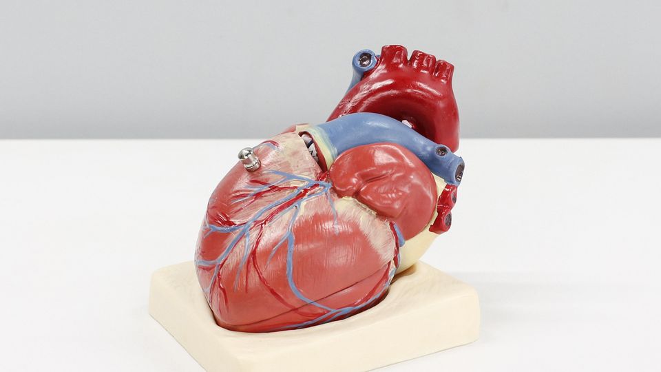
Key Highlights
- Innovative Biofabrication: Integration of iPSC cardiac cells and novel bioink formulations for creating realistic 3D printed heart tissues.
- Microfluidic Integration: Development of perfusable microfluidic channels to simulate physiological conditions and improve drug screening reliability.
- Advanced Pharmacokinetics: Application of pharmacokinetic-ADMET modeling to assess drug responses and toxicity in bioprinted cardiac models.
Introduction
The evolution of drug development has long been hindered by traditional in vitro culture systems and animal models that struggle to capture the complexity of human cardiac physiology. Recent advancements in 3D bioprinting offer a revolutionary approach by integrating induced pluripotent stem cell (iPSC) technology with state-of-the-art biofabrication methods. This proposal aims to advance the state-of-the-art in 3D printing of human heart models for enhanced drug screening. By emphasizing crucial skills such as iPSC-derived cardiac cell development, bioink optimization, advanced 3D bioprinting techniques, microfluidic system integration, and pharmacokinetic-ADMET analysis, the research design endeavors to create a model that accurately simulates the human heart's structure and function.
The envisioned platform will not only improve our understanding of cardiotoxicity and drug efficacy but will also mitigate the reliance on less reliable traditional methods. Specifically, the integration of microfluidics enables a precise simulation of blood flow and nutrient exchange, while advanced bioinks provide an environment conducive to cellular viability and natural tissue behavior. This holistic approach has the potential to transform the landscape of preclinical testing.
Objectives and Skills
Primary Objectives
The main goal is to develop an integrated platform that effectively marries 3D bioprinting with human cardiac tissue engineering and sophisticated drug screening protocols. The objectives include:
- Optimization of iPSC Cardiac Cells: Develop protocols to maintain high viability, proper differentiation, and enhanced maturation of iPSC-derived cardiomyocytes to closely mimic native heart tissue.
- Bioink Formulation: Engineer and synthesize bioinks that not only encapsulate the iPSC cardiac cells but also replicate the mechanical and electrical properties of heart tissue. This involves the integration of materials like collagen, alginate, fibrin, and extracellular matrix components.
- 3D Bioprinting Techniques: Utilize extrusion-based and stereolithography bioprinting methods to fabricate intricate heart models with multiple layers and complex cellular architectures.
- Microfluidics Integration: Design microfluidic devices that provide perfusion, mimicking blood flow to enhance nutrient supply and waste removal, thereby creating a dynamic in vitro environment.
- Pharmacokinetics-ADMET Analysis: Develop in vitro assays and computational models to assess drug absorption, distribution, metabolism, excretion, and toxicity in the printed heart models, ensuring a more accurate prediction of drug behavior and cardiotoxicity.
Essential Skills
- iPSC Cardiac Cells: Expertise in cell culture, differentiation protocols, and quality control measures to produce functional cardiomyocytes.
- Bioink Development: Familiarity with biomaterials, hydrogel mechanics, and the chemistry behind bioink synthesis to generate a supportive matrix for cells.
- 3D Bioprinting: Advanced skills in operating bioprinters, designing digital 3D models, and troubleshooting printing methodologies to create structurally sound tissues.
- Pharmacokinetics and ADMET: Understanding of drug metabolism, toxicology, and computational modeling to integrate drug screening into tissue engineering workflows.
- Microfluidics: Knowledge in microfabrication, fluid dynamics, and device integration to incorporate dynamic perfusion systems within printed models.
Proposed Methodology
Stage 1: iPSC Cardiac Cell Generation and Characterization
Cell Sourcing and Differentiation
Begin with the acquisition of iPSCs through established reprogramming techniques. Differentiate these iPSCs into cardiomyocytes using optimized protocols that ensure a high yield and purity. Regular assessments using immunohistochemistry and gene expression analysis will confirm the successful differentiation and maturation of these cells.
Quality Control
Employ viability assays and electrophysiological techniques to assess the function of the differentiated cardiomyocytes to validate their suitability for subsequent bioprinting.
Stage 2: Bioink Development and Optimization
Bioink Composition
Develop bioinks with properties that closely mimic the native cardiac extracellular matrix. Experiment with different combinations of natural polymers, such as collagen, alginate, and fibrin, assessing each for its rheological properties, biocompatibility, and electrical conductivity.
3D Printability and Stability
Characterize bioinks for viscoelasticity and printability. Optimize the crosslinking methods to ensure that the printed constructs remain stable under physiological conditions and support the maturation of iPSC cardiac cells.
Stage 3: 3D Bioprinting of Heart Models
Printing Strategy
Utilize extrusion-based bioprinting techniques for layering the cardiac cells along with the tailored bioink. Digital modeling tools will be used to design anatomically accurate heart tissues, taking into account the architecture of both myocardial and vascular structures.
Construct Complexity
Emphasize printing complexity by incorporating multiple cell types and gradients in bioink composition. This will facilitate the creation of constructs that better mimic the dynamic environment of the human heart, including contractility and electrophysiological properties.
Stage 4: Microfluidic Integration and Perfusion
Device Design
Develop microfluidic chips with channels that can be aligned with the printed heart tissues. These chips will be designed to simulate the mechanical shear forces and nutrient transport properties akin to the human circulatory system.
Perfusion and Functionality Assessment
Incorporate perfusion systems to continuously supply nutrients and remove metabolic waste from the printed tissues. Testing will include monitoring the contraction patterns and viability of the cardiac cells under dynamic flow conditions.
Stage 5: Pharmacokinetic and ADMET Evaluation
Drug Exposure Studies
Utilize the integrated platform to expose the 3D printed heart models to various pharmacological compounds. Perform assays to measure contractility, cell viability, and metabolic activity, comparing these results against known ADMET profiles.
Computational Modeling
Develop computational models to simulate drug absorption, distribution, metabolism, excretion, and toxicity. These models will be validated against experimental data obtained from the chip-integrated heart models, aiming for precise predictive power.
Project Timeline and Budget
| Phase | Timeline | Activities |
|---|---|---|
| Phase 1 | Months 1-6 | iPSC generation, differentiation, and initial quality control assessments. |
| Phase 2 | Months 7-12 | Development and testing of bioink formulations; preliminary 3D printing trials. |
| Phase 3 | Months 13-18 | Complex 3D bioprinting of heart models; refinement of printing protocols. |
| Phase 4 | Months 19-24 | Integration of microfluidic systems and functional perfusion testing. |
| Phase 5 | Months 25-30 | Pharmacokinetic and ADMET assays; computational modeling and validation studies. |
The preliminary budget is anticipated to be in the range of $950,000, allocated across personnel, equipment, reagents, and computational resources. Detailed budgeting will be refined as the project advances.
Interdisciplinary Integration and Expected Impact
Interdisciplinary Collaboration
This proposal brings together experts from several domains: tissue engineering, biomaterials science, microfluidics, pharmacology, and computational modeling. Through this interdisciplinary collaboration, each segment of the project is designed to synergize, ensuring that innovations in bioink formulation directly complement advances in bioprinting and microfluidic design. Such an approach guarantees the creation of robust and reliable in vitro models that are physiologically relevant and suitable for high-throughput drug screening.
Anticipated Results
The expected outcomes include the development of a highly functional 3D printed heart model that closely mimics native cardiac tissue. This will facilitate:
- Improved prediction of drug efficacy and safety, addressing intrinsic limitations of animal models.
- A reduction in time and cost associated with traditional pharmacokinetic studies and drug development cycles.
- A platform for personalized medicine, allowing for the customization of heart models to match patient-specific conditions.
- The establishment of enhanced ADMET profiles for new compounds, potentially reducing cardiotoxicity risks in clinical trials.
Validation and Testing Strategies
Analytical Techniques
The project will apply several analytical methods to validate the model's functionality:
- Immunohistochemistry: Used to confirm the expression of key cardiac markers in the printed tissues.
- Gene Expression Analysis: Monitoring gene profiles to ensure that the bioprinted tissues emulate the behavior of native heart cells.
- Electrophysiological Assessments: Recording the electrical activity of the cardiac cells to evaluate their functional maturity and response to drugs.
- Contractility Assays: Testing the dynamic response of the heart model by evaluating its contractile force in real-time under various pharmacological conditions.
- Computational Analysis: Implementing predictive models for ADMET parameters based on experimental data.
Iterative Refinement
Iterative loops between experimental results and computational predictions will be critical. This continuous feedback loop ensures that each subsystem – from bioink to microfluidics – is fine-tuned for maximum fidelity, thereby enhancing the model’s predictive capabilities in drug screening.
References
-
PMC Article - PubMed Central
-
Journal Article - ScienceDirect
-
Article - NCBI
- Frontiers in Bioengineering - Frontiers
- Nature Article - Nature
Recommended Related Queries
Last updated March 22, 2025