
Advancing 3D Printed Heart Models for Drug Screening
Exploring innovative strategies and breakthroughs in realistic heart model development
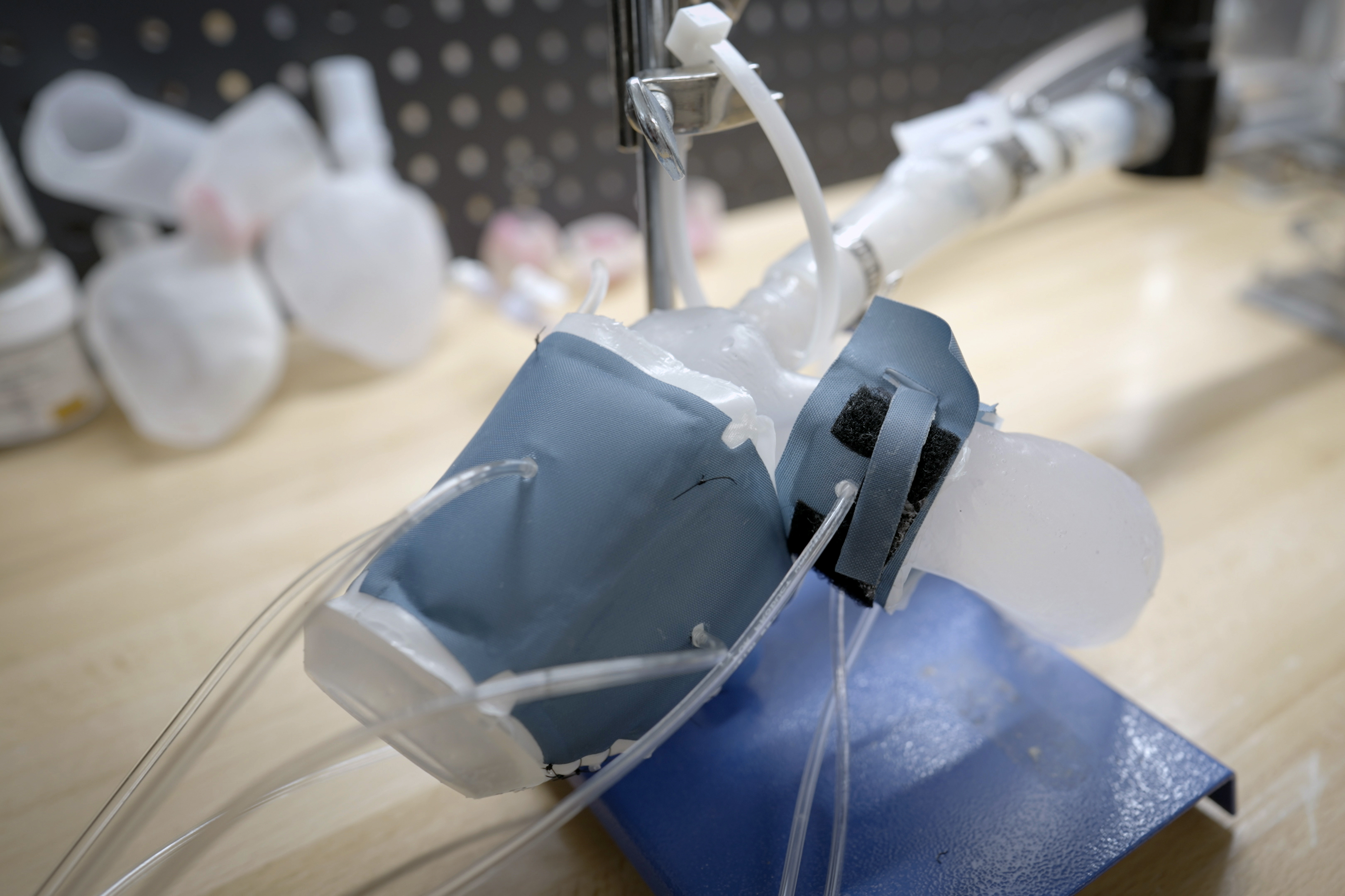
Key Highlights
- Advanced Biomaterials and Bioinks: Developing materials that replicate the physiological and mechanical properties of heart tissue.
- Integration of Cellular and Tissue Engineering: Employing techniques such as bioprinting to create functional, biologically accurate heart structures.
- High-Throughput and Personalized Screening: Leveraging automation, patient-specific data, and advanced techniques to enable precise drug evaluation.
Overview
The advancement of 3D printed human heart models for drug screening represents a major leap forward in both biomedical engineering and pharmacological testing. As traditional drug development often relies on animal models which do not fully recapitulate human cardiac physiology, researchers have increasingly turned to 3D printing technologies to create more realistic, accurate, and versatile cardiac models. These models not only allow for a better understanding of how drugs interact with human heart tissue but also pave the way towards personalized medicine.
Innovative Strategies in 3D Printing Heart Models
Advanced Biomaterials and Bioink Development
One of the key pillars in advancing 3D printed heart models is the development of novel biomaterials that closely mimic the native properties of heart tissue. These materials must fulfill several conditions: they need to be biocompatible, support cellular adhesion and proliferation, have mechanical integrity similar to human heart tissue, and ideally possess electrical conductivity to reproduce the heart’s electrophysiological characteristics.
Emerging Bioinks
Recent innovations have resulted in the creation of bioinks that incorporate living cells, extracellular matrix components, and growth factors. These bioinks can be fine-tuned to replicate the anisotropic properties of cardiac tissue, providing mechanical robustness while allowing for complex tissue growth. Advanced bioinks are designed to change viscosity under controlled conditions, allowing for high-definition printing of intricate structures that mimic the natural microarchitecture of the heart.
Moreover, the integration of conductive materials such as carbon nanotubes or graphene derivatives within the bioink is being explored to recreate the electrical conduction pathways of cardiac tissue. Such improvements are crucial in faithfully reproducing the dynamic electrical activity of the heart, thereby enhancing the fidelity of drug screening tests that evaluate electrophysiological responses.
Refined 3D Printing Techniques
Innovations in 3D printing techniques have significantly enhanced the resolution and complexity of printed heart models. Technologies such as stereolithography, selective laser sintering, and extrusion-based printing allow for the construction of layered architectures that replicate the heterogeneous structure of the heart.
Resolution and Accuracy
Precision is critical when modeling the intricate structures of the human heart. Enhanced printing resolution facilitates the accurate placement of different cell types and biomaterial components, ensuring that the printed model reproduces both the macro and microstructures found in live cardiac tissue. This precise layering leads to models that can demonstrate dynamic functions such as beating, contractility, and even vascularization.
Advanced printing techniques also enable the creation of patient-specific heart models utilizing data derived from medical imaging. Such precise replication not only supports personalized drug screening but also aids in surgical planning and cardiovascular training.
Cellular Integration and Tissue Engineering
To achieve functional heart models, recent research focuses on integrating living cells into the printed structures. The introduction of primary cardiomyocytes, cardiac fibroblasts, and endothelial cells into 3D printed scaffolds is essential for creating tissues that accurately replicate heart function.
Bioprinting and Cellularization
3D bioprinting techniques facilitate the simultaneous deposition of cells and biomaterials. This ensures that cells are precisely placed in a manner that reflects the natural organization of heart tissue. Post-printing, these cells can proliferate, differentiate, and begin to exhibit characteristic contractile and electrophysiological properties.
Techniques such as cell seeding can also be employed to enhance the viability and functionality of bioprinted tissues. By utilizing a combination of automated cell deposition and bioactive cues within the printed structure, researchers can promote the formation of a functional extracellular matrix that further supports tissue maturation.
Incorporation of Vascular Networks
For 3D printed heart models to be realistic in both function and longevity, they must include a complex network of vascular channels. Vascularization is vital not only for delivering oxygen and nutrients but also for removing metabolic wastes, mimicking the in vivo environment.
Engineering Perfusable Vascular Structures
Researchers are now employing techniques such as sacrificial bioinks and microfluidic chips to create interconnected vascular networks within heart models. These networks enhance the viability of the bioprinted tissues and also allow for the integration of perfusion systems. Such systems simulate blood flow, which is critical for testing drug-induced changes in vascular dynamics as well as cardiac function.
Understanding the interplay between vascular perfusion and tissue viability can also help in assessing cardiotoxic effects of various compounds, thereby providing a more comprehensive platform for drug safety evaluation.
High-Throughput Screening and Data Analytics
As the complexity of 3D printed heart models increases, so does the potential for high-throughput screening applications. The integration of automated imaging, robotics, and data analytics opens new horizons in drug screening efficiency.
Automated Platforms and Real-Time Monitoring
Leveraging high-throughput systems allows researchers to test a large number of drug candidates simultaneously. Automated platforms can provide real-time monitoring of key functional metrics such as heart rate, contractility, and electrophysiological parameters. Advanced imaging techniques and electrical sensors are being integrated into these systems to capture minute changes in tissue functionality.
Furthermore, the use of artificial intelligence and machine learning algorithms enables the rapid analysis of complex datasets, providing actionable insights into drug efficacy and toxicity. This data-driven approach not only speeds up the drug screening process but also facilitates the discovery of novel drug candidates by predicting outcomes based on previous experimental results.
Personalized Medicine and Patient-Specific Models
One of the most promising aspects of 3D printed heart models is their application in personalized medicine. By integrating patient-specific data, such as genetic information and imaging scans, researchers can create individualized heart models that closely reflect a person’s unique cardiac structure and function.
Tailoring Drug Therapies
Personalized heart models enable clinicians to test and predict patient-specific drug responses. This personalized approach can minimize adverse effects by ensuring that a given medication is tailored to the patient’s unique cardiac profile. As a result, potential therapies can be optimized before being clinically administered, significantly reducing risks associated with drug toxicity.
In addition, these models provide a valuable resource for understanding the progression of cardiovascular diseases in a patient-specific manner. This facilitates the development of targeted therapies and contributes to the broader field of regenerative medicine.
Collaborative and Interdisciplinary Approaches
The progress in 3D printing human heart models is not solely attributable to advancements in printing technologies or biomaterials. It is also the result of robust interdisciplinary collaborations. Biomedical engineers, material scientists, pharmacologists, and clinicians are working in tandem to solve complex challenges in tissue engineering and drug screening.
Interdisciplinary Research Initiatives
Interdisciplinary research initiatives are critical as they bring together diverse expertise and resources. Collaborative research facilitates the rapid translation of laboratory discoveries into clinical applications. For instance, a combined effort among experts in microfluidics, electrical engineering, and cellular biology has led to the development of perfusable heart-on-a-chip systems that simulate complex cardiac functions.
Additionally, international research partnerships and funding initiatives help standardize testing protocols and push for regulatory frameworks that will accelerate the adoption of these innovative technologies in mainstream drug development.
Data Integration and Computational Modeling
Another important dimension enhancing the refinement of 3D printed heart models is the integration of computational models and data analytics. Computational modeling can simulate the dynamic behavior of heart tissues under various pharmacological conditions. This in silico testing complements physical models by predicting cellular responses and potential off-target effects of drugs.
When paired with high-throughput screening data, computational models improve the predictive accuracy of drug testing. This not only optimizes the selection of candidate drugs, but it also provides insights into underlying mechanisms of drug action, paving the way for more innovative therapeutic strategies.
High-Throughput Screening Platforms and Standardization
To fully harness the potential of 3D printed heart models for drug screening, there is a growing need for developing standardized, high-throughput screening platforms. Such platforms should be capable of handling large-scale testing while delivering reliable, reproducible results.
Developing Robust Screening Systems
High-throughput screening systems integrate robotics, automation, and sophisticated imaging systems to test multiple drug candidates simultaneously. These platforms are designed to minimize human error and increase the reproducibility of experimental results. Automated platforms allow for the constant monitoring of heart model performance under various drug treatments, thereby providing immediate feedback on efficacy and toxicity.
Standardization in the fabrication process—from the selection of biomaterials and bioinks to the printing protocol and post-print processing—is also critical. Establishing benchmarking procedures and quality control measures ensures that all printed heart models exhibit consistent functional properties. This consistency not only supports their use in drug screening but also bolsters regulatory approval processes.
A Comprehensive View: Integration of Technologies
To illustrate the multidimensional nature of advancing 3D printed heart models, the table below summarizes the integration of various techniques and strategic approaches:
| Aspect | Technologies/Strategies | Benefits |
|---|---|---|
| Biomaterials | Advanced bioinks, hydrogels, conductive polymers, nanocomposites | Enhanced cell viability, mechanical integrity, electrical conductivity |
| Printing Techniques | Stereolithography, selective laser sintering, extrusion-based printing | High precision, accurate microarchitecture, patient-specific replication |
| Cellular Integration | Bioprinting, cell seeding, tissue engineering | Realistic tissue functionality, improved drug response accuracy |
| Vascularization | Sacrificial bioinks, microfluidic systems | Perfusable networks, mimicked hemodynamics, enhanced nutrient delivery |
| Screening Systems | High-throughput platforms, automated imaging, AI data analytics | Accelerated testing, consistent data, improved safety profiling |
| Personalization | Patient-specific imaging data, computational modeling | Customized therapy, reduced adverse drug reactions, improved outcomes |
| Collaborative Approaches | Interdisciplinary research, standardization protocols | Faster innovation, regulatory compliance, clinical translation |
Path Forward and Emerging Applications
The ongoing advancements in 3D printing of human heart models have set the groundwork for several emerging applications that could redefine drug screening and cardiovascular research:
Cardiotoxicity Assessment
One of the prime applications of these advanced models is in early cardiotoxicity testing. By exposing 3D printed heart tissues to new drug candidates, researchers can detect adverse effects with greater precision than conventional animal models, thereby reducing the risk of unforeseen side effects in clinical trials.
Disease Modeling and Therapeutic Optimization
In addition to drug screening, printed heart models facilitate the study of various cardiovascular diseases. For instance, models replicating the conditions of heart failure or myocardial infarction provide a platform to test regenerative therapies and stem cell treatments. This leads to more targeted therapeutic strategies, tailored to address specific pathological conditions.
Integration with Regenerative Medicine
With the convergence of tissue engineering and 3D printing, a future where functional cardiac tissue is engineered for transplantation becomes increasingly plausible. Such advancements not only promise improvements in drug screening but also offer new avenues for repairing damaged hearts through regenerative interventions.
Bridging Laboratory Research to Clinical Practice
Transitioning from research laboratories to real-world clinical practice is one of the significant challenges that these technologies face. To successfully translate these technologies, several critical factors must be addressed:
Validation and Standardization
Establishing standardized protocols for 3D printing and subsequent evaluation of heart models is vital. Consistent benchmarks and quality control frameworks enable reproducibility and reliability, ensuring that findings can be reliably interpreted and compared across different studies and laboratories.
This standardization is key for regulatory bodies and clinical institutions when considering the integration of these models into routine drug screening and surgical planning.
Regulatory and Commercial Pathways
In order to fully realize the clinical benefits of these advanced models, clear regulatory guidelines and commercial pathways must be established. Collaborative efforts between academic institutions, industry partners, and government agencies can help streamline the process for clinical trials and eventual market approval. Such collaborative models not only foster innovation but also facilitate broader adoption in the healthcare industry.
References
- Researchers Develop New Bioink to 3D Print Personalized Heart-on-a-Chip Devices - 3DPrintingIndustry
- 3D Printed Heart-on-a-Chip to be Sent to Space as Part of Belgian Astrocardia Project - 3DPrintingIndustry
- Researchers 3D Print New Ultra-Realistic Heart and Lung Models That Can Bleed, Beat, and Breathe - 3DPrintingIndustry
- 3D Printed Heart Models for Cardiovascular Training in Development - 3DPrint
- First 3D Engineered Vascularized Human Heart Is Bioprinted - GEN
Related Queries and Further Exploration
Last updated March 22, 2025