
Exploring Breakthrough Innovations in 3D Printed Heart Models
Innovative strategies to enhance drug screening with realistic human heart replicas
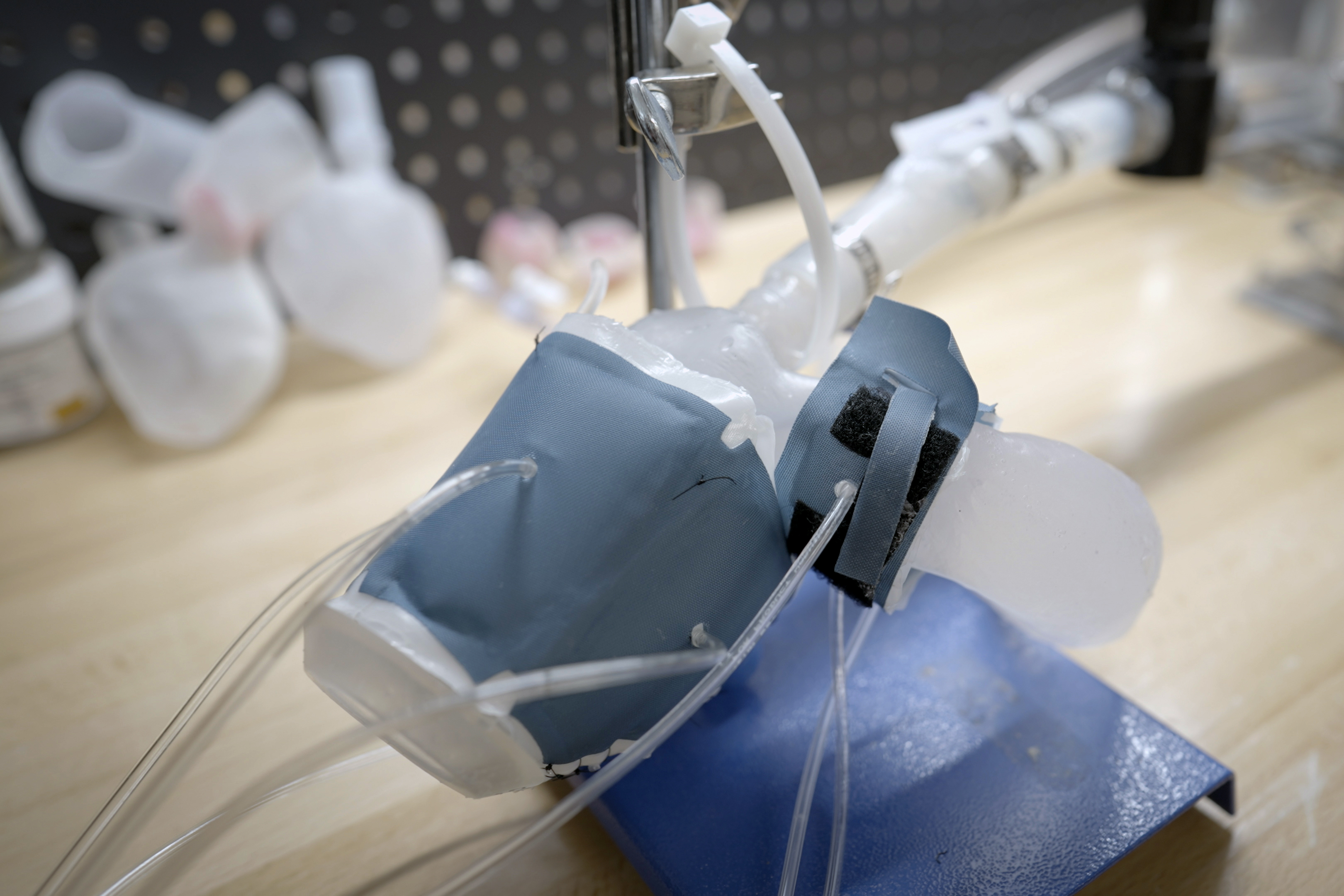
Key Highlights
- Advanced Bioinks & Biomaterials: Develop materials that precisely mimic native heart tissue's mechanical and biological properties.
- Functional and Vascular Integration: Integrate dynamic stimuli, perfusable vasculature, and microfluidic systems to simulate cardiac physiology.
- Personalized and High-Throughput Platforms: Leverage patient-derived data and scalable methods to create and analyze personalized heart models robustly.
Introduction to State-of-the-Art 3D Printed Heart Models for Drug Screening
Over the past decade, the emergence of 3D printing technology in biomedical engineering has opened up revolutionary opportunities in creating realistic human heart models tailored for drug screening. These models provide a much closer mimicry of the human heart’s anatomy and physiology than traditional 2D cultures or animal models. The integration of this technology into drug discovery processes promises to deliver more precise data on drug efficacy, toxicity, and overall safety, enhancing our understanding of cardiovascular pharmacology.
Enhanced Biomimicry and Material Innovation
Advanced Bioinks and Biomaterials
To replicate the natural complexities of heart tissue, the development of state-of-the-art bioinks is pivotal. Advanced bioinks must not only provide a supportive structure but also integrate biomimetic cues for cell attachment, proliferation, and differentiation.
Biomimetic Design
Using materials that combine collagen, alginate, and other extracellular matrix (ECM) components, scientists create scaffolds that closely mirror the architectural and functional attributes of native heart tissue. These biomaterials are designed to promote cell viability and can be tuned to match the electrical and mechanical properties of the actual myocardium.
Tailored Mechanical Properties
One major challenge is fabricating materials that can withstand the dynamic mechanical stresses of a beating heart. By incorporating nanocomposites and novel hydrogels, researchers are now able to create scaffolds that exhibit both robustness and the flexibility necessary for vascular and cellular integration.
Precision Bioprinting Techniques
The success of 3D printing human heart models hinges largely on the precision of bioprinting techniques. Breakthroughs in technologies like freeform reversible embedding of suspended hydrogels (FRESH) allow the creation of intricate cardiac structures with high resolution. Techniques that support coaxial printing and fiber-optic-assisted bioprinting are also pushing the boundaries by enabling the exact placement of multiple cell types within a complex 3D environment.
Enhanced precision not only improves structural fidelity but also ensures the functional integration of various elements like vascular networks, which are essential for nutrient and oxygen diffusion in a living tissue model.
Integrating Functional Complexity
Dynamic Stimulation Approaches
An evolving trend in the field is the incorporation of dynamic aspects into 3D printed heart models. Unlike static scaffolds, these models aim to mimic the living heart’s electrical, mechanical, and biochemical activity. Through multimodal stimulation such as electrical pulses and mechanical stretching, engineered heart tissues can demonstrate contractility and rhythmic beating patterns that are crucial for simulating real-life drug responses.
Electrical and Mechanical Stimulation
Application of external electrical impulses encourages synchronous contraction of cardiomyocytes, while mechanical stimulation fosters the development of physiologically relevant myocardial tissue. These stimulation protocols are invaluable in evaluating how drugs might affect heart rhythm and contractility.
Microfluidics and Vascular Integration
Integrating microfluidic systems into heart-on-a-chip devices significantly amplifies the physiological relevance of 3D printed models. Microfluidic channels mimic the heart’s natural vascular networks, enabling effective perfusion and real-time monitoring of drug interactions. Incorporating vascularization into the design not only improves the structural and functional viability of the model but also opens avenues for studying drug-induced vascular responses.
Perfusable Vascular Networks
Innovative techniques that embed perfusable channels within the printed tissues allow researchers to simulate blood flow and nutrient delivery, which is essential for maintaining cell viability during prolonged testing phases. This level of vascular integration mirrors the in vivo environment and produces more reliable drug response data.
Personalization and High-Throughput Screening
Personalized Heart Models
A transformative aspect of 3D printed heart technology is its potential for personalization. Utilizing patient-specific data—such as MRI scans and genomic information—to create tailor-made heart models can lead to the development of personalized medication regimens. By incorporating induced pluripotent stem cells (iPSCs) derived from patients, the printed models accurately reflect individual genetic profiles, thus predicting patient-specific responses to drug therapies.
Patient-Specific Data Integration
The use of patient-derived cells offers a dual benefit: it not only enhances the relevance of the 3D printed model as a platform for drug screening but also contributes to the burgeoning field of personalized medicine. These models allow researchers to delve into precision pharmacology, thereby reducing adverse reactions and improving therapeutic outcomes.
High-Throughput Drug Screening Platforms
To fully capitalize on the benefits of 3D printed heart models, scaling up for high-throughput screening is a must. Automated systems that combine robotics, real-time imaging, and sophisticated machine learning algorithms are being developed to accelerate drug testing. These platforms facilitate the simultaneous screening of multiple treatments, thereby expediting the drug discovery pipeline.
Data Integration and Analysis
Machine learning can be utilized to analyze complex data sets generated from these high-throughput platforms. The integration of advanced imaging techniques and computational models further refines the predictive capabilities regarding drug efficacy and potential side effects.
Multidisciplinary Approaches and Future Innovations
Collaborative Research and Standardization
The multidisciplinary nature of this field necessitates collaboration across biomaterials science, tissue engineering, cardiovascular biology, and pharmaceutical research. Establishing standard protocols for creating and validating 3D printed heart models is essential to ensure consistency and reproducibility across different laboratories and research facilities.
Interdisciplinary Collaboration
Partnerships between academic institutions, clinical researchers, and industry leaders can drive forward the standardized practices needed for model reliability. Collaborative platforms enable knowledge sharing and the pooling of resources, ultimately accelerating advancements in drug screening and personalized medicine.
Emerging Technologies and Multi-Scale Modeling
Beyond biomaterials and printing techniques, the integration of emerging technologies such as 4D printing and computational modeling represents the next frontier. 4D printing adds the element of time, enabling printed tissues to change their shape or properties in reaction to external stimuli. When paired with multi-scale computational models, researchers can simulate drug interactions from the molecular level to the entire organ, offering unprecedented insights into drug dynamics.
Integration with Computational Models
Combining in vitro experimental data with in silico simulations allows for a broad understanding of cardiac behavior under different drug regimens. This holistic approach is set to redefine drug efficacy assessments by linking detailed cellular responses with overall organ functionality.
A Comprehensive Overview Table
| Aspect | Strategy | Expected Outcome |
|---|---|---|
| Bioinks & Biomaterials | Development of ECM-mimicking hydrogels and nanocomposites | Enhanced cell viability and tissue functionality |
| Printing Precision | Utilization of FRESH and coaxial printing techniques | High-fidelity structures mimicking heart anatomy |
| Dynamic Functionality | Incorporation of electrical and mechanical stimulation | Synchronous beating and realistic cardiac responses |
| Vascularization | Integration of microfluidic channels | Perfusion and nutrient delivery enhancing tissue health |
| Personalization | Use of patient-specific iPSCs and imaging data | Personalized drug testing with reduced adverse effects |
| High-Throughput Platforms | Automated screening using robotics and ML algorithms | Accelerated drug discovery and improved data analytics |
| Multidisciplinary Integration | Collaborative research and standardization protocols | Consistent, reliable models approved across research centers |
| Computational Modeling | Multi-scale simulations integrated with experimental data | Comprehensive insights from molecular to organ level |
Pioneering Future Directions
Overcoming Current Limitations
Although substantial progress has been made, current challenges such as incomplete vascular integration and limited electrical conduction in tissue models remain. Future work should aim to refine the microarchitecture of printed tissues and introduce more complex cellular interplays. Addressing these areas will further bridge the gap between in vitro models and the native human heart environment.
Improving Cellular Heterogeneity
Incorporating a mix of cardiomyocytes, fibroblasts, endothelial cells, and even immune cells can provide a more complete model of heart tissue. This cellular diversity is crucial for replicating not only the mechanical and electrical properties of the heart but also the myriad biochemical interactions that occur during drug exposure.
Adopting Real-Time Monitoring and Automated Analyses
Innovations in sensor technology and imaging techniques are enabling real-time monitoring of cellular behavior and tissue dynamics. The coupling of these approaches with automated data pipelines and machine learning algorithms will allow researchers to extract actionable insights quickly and more reliably. Such digital integration is a game changer that will allow for adaptive experimentation and continuous improvement of model accuracy.
Real-Time Data Integration
Continuous monitoring of drug effects using real-time imaging and sensor data can allow researchers to detect subtle shifts in cardiac tissue response, ensuring more precise adjustments in experimental procedures and improved drug efficacy evaluations.
References
- Researchers Develop New Bioink to 3D Print Personalized “Heart on a Chip” Devices - 3D Printing Industry
- 3D Bioprinting for the Construction of Drug Testing Models - Frontiers
- Fiber-Infused Ink Enables 3D-Printed Heart Muscle to Beat - Harvard SEAS
- Advances in 3D Bioprinted Cardiac Tissue Using Stem Cell-Derived Cardiomyocytes - Oxford Academic
-
Recent Advances in 3D Bioprinting for Regenerative Medicine and Drug Development
Recommended Queries for Future Exploration
Last updated March 23, 2025