
Recommended Workup for Newly Diagnosed Acute Myeloid Leukemia (AML)
A Comprehensive Approach to Diagnosing and Stratifying AML
Key Takeaways
- Comprehensive Clinical Evaluation: A thorough history and physical examination are essential to identify symptoms, risk factors, and potential comorbidities associated with AML.
- Extensive Laboratory and Molecular Testing: Detailed laboratory studies, including CBC, metabolic panels, and advanced molecular genetic testing, are crucial for accurate diagnosis and risk stratification.
- Bone Marrow Examination: Bone marrow aspirate and biopsy, complemented by cytogenetic and molecular analyses, are pivotal in confirming AML and guiding treatment strategies.
1. Initial Clinical Evaluation
History and Physical Examination
A detailed medical history and comprehensive physical examination are foundational in the workup of newly diagnosed AML. The clinician should assess for common symptoms such as fatigue, frequent infections, easy bruising, and bleeding, which may indicate cytopenias resulting from bone marrow infiltration by leukemic cells.
Key aspects of the history include:
- Risk Factors: Previous chemotherapy or radiation therapy, history of myelodysplastic syndromes, exposure to specific chemicals (e.g., benzene), and a family history of hematologic malignancies.
- Comorbidities: Pre-existing medical conditions that may affect treatment options and prognosis, such as cardiovascular disease, liver or kidney dysfunction.
The physical examination should focus on identifying signs indicative of AML, including:
- Pallor: Suggestive of anemia.
- Petechiae and Ecchymoses: Indicative of thrombocytopenia.
- Organomegaly: Enlargement of the liver or spleen (hepatosplenomegaly).
- Gingival Hypertrophy: Swelling of the gums, which can occur in certain AML subtypes.
- Extramedullary Manifestations: Presence of skin lesions (leukemia cutis) or chloromas (granulocytic sarcomas).
2. Laboratory Tests
Complete Blood Count (CBC) with Differential and Peripheral Blood Smear
The CBC is a critical initial test in the evaluation of AML. It provides information on the levels of various blood cells and can reveal abnormalities suggestive of leukemia.
- Leukocytosis or Leukopenia: Elevated or decreased white blood cell counts.
- Anemia: Low hemoglobin levels leading to fatigue and pallor.
- Thrombocytopenia: Reduced platelet counts resulting in increased bleeding and bruising.
- Circumstances Blasts: Presence of immature white blood cells (blasts) in the peripheral blood.
- Peripheral Blood Smear: Evaluation of cell morphology to identify abnormal or immature cells characteristic of AML.
Comprehensive Metabolic Panel
This panel assesses organ function and electrolyte balance, which are crucial for determining a patient's ability to tolerate specific AML treatments.
- Liver Function Tests: Elevated liver enzymes may indicate hepatic involvement or treatment-related toxicity.
- Renal Function: Assessment of creatinine and blood urea nitrogen (BUN) levels is vital before initiating chemotherapy.
- Tumor Lysis Syndrome Indicators: Elevated uric acid, lactate dehydrogenase (LDH), phosphorus, and potassium levels should be monitored, especially in patients with high tumor burden.
- Vitamin B12 and Folate Levels: These may be measured to rule out other causes of macrocytic anemia.
Coagulation Studies
Coagulation profiles are particularly important in cases of suspected Acute Promyelocytic Leukemia (APL), a subtype of AML associated with a high risk of disseminated intravascular coagulation (DIC).
- Prothrombin Time (PT) and Partial Thromboplastin Time (PTT): Prolonged times can indicate coagulation abnormalities.
- Fibrinogen Levels: Low levels may suggest ongoing consumption in DIC.
Lactate Dehydrogenase (LDH) and Uric Acid Levels
Elevated LDH is a marker of cellular turnover and can be indicative of disease burden in AML. Elevated uric acid levels may suggest tumor lysis syndrome.
Flow Cytometry (Immunophenotyping)
Flow cytometry is essential for confirming the diagnosis of AML by identifying specific cell surface markers that distinguish AML from other leukemias.
- Cell Surface Markers: Common markers include CD13, CD33, CD34, and HLA-DR.
- Lineage Confirmation: Helps in determining the myeloid origin of the blasts.
Cytogenetic and Molecular Genetic Testing
Genetic analysis is crucial for classifying AML, determining prognosis, and guiding targeted therapy options.
- Cytogenetic Analysis: Karyotyping and fluorescence in situ hybridization (FISH) are used to detect chromosomal abnormalities such as t(8;21), inv(16), and t(15;17) translocations.
- Molecular Genetic Testing: Identification of mutations in genes like NPM1, FLT3-ITD, CEBPA, IDH1/2, and TP53 using techniques like next-generation sequencing (NGS).
3. Bone Marrow Examination
Bone Marrow Aspirate and Biopsy
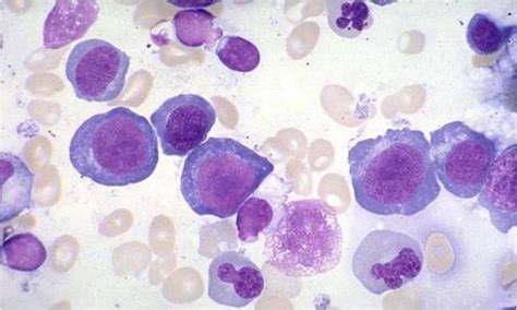
A bone marrow aspirate and biopsy provide definitive evidence for AML diagnosis by assessing the cellularity, morphology, and proportion of blasts in the bone marrow.
- Blast Percentage: A diagnosis of AML is confirmed if the bone marrow contains ≥20% blasts, or if specific genetic abnormalities associated with AML are present.
- Cellularity Assessment: Determines the proportion of hematopoietic cells to fat cells in the marrow.
- Morphological Evaluation: Identifies the appearance and differentiation stage of leukemic cells.
Cytochemistry
Cytochemical staining differentiates AML subtypes based on enzyme activity within the leukemic cells.
- Myeloperoxidase (MPO) Staining: Positive staining supports the diagnosis of myeloid lineage.
- Non-Specific Esterase (NSE) Staining: Helps differentiate between myeloid and monocytic subtypes.
Immunophenotyping by Flow Cytometry
Further analysis of cell surface markers provides detailed information about the differentiation state and lineage of leukemia cells.
Cytogenetic and Molecular Genetic Testing
Comprehensive genetic profiling guides risk stratification and therapeutic decisions.
4. Additional Tests
Minimal Residual Disease (MRD) Assessment
Post-treatment MRD evaluation using flow cytometry or molecular techniques helps in assessing the effectiveness of therapy and the likelihood of relapse.
Cardiac Assessment
Before initiating anthracycline-based chemotherapy, it's essential to evaluate cardiac function to mitigate the risk of cardiotoxicity.
- Electrocardiography (ECG): Assesses heart rhythm and detects any electrical abnormalities.
- Echocardiography or MUGA Scan: Evaluates the ejection fraction and overall cardiac function.
Infectious Disease Screening
Screening for infectious agents such as hepatitis B, hepatitis C, and HIV is crucial as these infections can influence treatment choices and outcomes.
Lumbar Puncture (If Indicated)
In cases where there are neurological symptoms, high white blood cell counts, or specific AML subtypes (e.g., monocytic), a lumbar puncture may be necessary to evaluate central nervous system involvement.
Imaging Studies
Imaging may be required to assess for extramedullary disease or complications related to AML.
- Chest X-ray or CT Scan: Evaluates for mediastinal masses or infections.
- Brain MRI or PET/CT: Used if there is suspicion of extramedullary involvement such as granulocytic sarcomas.
5. Risk Stratification
Risk stratification categorizes AML patients based on genetic, molecular, and clinical factors to guide treatment decisions and predict prognosis. Common stratification systems include the European LeukemiaNet (ELN) and the World Health Organization (WHO) classifications.
Cytogenetic and Molecular Findings
Patients are grouped into favorable, intermediate, or adverse risk categories based on specific cytogenetic abnormalities and molecular mutations.
- Favorable Risk: Includes patients with favorable cytogenetics such as t(8;21), inv(16), or mutated NPM1 without FLT3-ITD.
- Intermediate Risk: Patients with normal cytogenetics or incomplete cytogenetic information.
- Adverse Risk: Includes unfavorable cytogenetic abnormalities like -7, complex karyotypes, or mutations in TP53.
Clinical Factors
- Age and Performance Status: Older age and poorer performance status are associated with worse prognosis.
- Comorbidities: Assessed using tools like the Hematopoietic Cell Transplantation-Comorbidity Index (HCT-CI).
6. Subtype-Specific Workup
Identification of specific AML subtypes is critical for appropriate management. Acute Promyelocytic Leukemia (APL) is a distinct subtype that requires immediate initiation of targeted therapy.
- APL Identification: Rapid testing for the PML-RARA fusion gene using techniques like FISH or RT-PCR is essential due to the high risk of coagulopathy and the need for prompt treatment with all-trans retinoic acid (ATRA) and arsenic trioxide (ATO).
7. Treatment Planning
Based on the comprehensive workup, an individualized treatment plan is formulated, which may include:
-
Induction Chemotherapy: Common regimens include the "7+3" protocol, which consists of cytarabine for seven days combined with an anthracycline (e.g., daunorubicin) for three days, aimed at achieving complete remission.
-
Targeted Therapies: For patients with specific genetic mutations, targeted agents such as FLT3 inhibitors (e.g., midostaurin), IDH inhibitors (e.g., enasidenib, ivosidenib), or BCL-2 inhibitors (e.g., venetoclax) may be incorporated.
-
Hematopoietic Stem Cell Transplantation: Recommended for patients in the intermediate or adverse risk categories, especially those who achieve remission but are at high risk of relapse.
-
Supportive Care: Includes transfusions, antimicrobial prophylaxis, and growth factor support to manage complications and improve quality of life during treatment.
Multidisciplinary Approach
Collaboration with a hematologist/oncologist, transplant specialist, and other healthcare professionals is essential to optimize treatment outcomes and manage side effects effectively.
Conclusion
The recommended workup for newly diagnosed Acute Myeloid Leukemia involves a multifaceted approach encompassing clinical evaluation, extensive laboratory and molecular testing, bone marrow examination, and additional assessments tailored to individual patient needs. Accurate diagnosis and risk stratification are paramount in guiding effective treatment strategies, improving prognosis, and enhancing the quality of life for patients with AML.
References
Last updated January 17, 2025