
Unveiling the Mystery of Angle Kappa: Normal Values Every Eye Specialist Should Know
Decoding the crucial measurement between visual and pupillary axes that can make or break refractive surgery outcomes
Essential Angle Kappa Insights
- Definition: Angle kappa is the angle between the visual axis and the pupillary axis of the eye, critical for proper centration in refractive procedures.
- Normal Range: Generally, values between 3-5° are considered physiologically normal, with values >5° potentially leading to pseudostrabismus.
- Clinical Significance: Hyperopic eyes typically have larger angle kappa values than myopic eyes, directly impacting surgical planning and outcomes.
Understanding Angle Kappa
Angle kappa represents the angular difference between the visual axis (the line connecting the fovea to the object of regard) and the pupillary axis (the line perpendicular to the cornea that passes through the center of the pupil). This measurement is crucial in ophthalmology, particularly for refractive and cataract surgeries where precise centration determines visual outcomes.
Proper assessment of angle kappa helps surgeons avoid decentered treatments that could lead to unwanted visual phenomena such as glare, halos, and decreased visual quality. Ignoring angle kappa during planning stages may result in suboptimal surgical outcomes, especially in procedures like LASIK, PRK, and multifocal intraocular lens implantation.
Explanation of Angle Kappa Mindmap
The mindmap above illustrates the comprehensive framework of angle kappa, showcasing its anatomical basis, various measurement methods, clinical significance in different procedures, and the factors that influence its value. Understanding these interconnected elements is essential for proper surgical planning and interpreting measurement variations across patients.
Normative Values By Measurement Device
The normative values for angle kappa vary depending on the measurement device used. This variation stems from differences in technology, methodology, and calibration across instruments. Understanding device-specific norms is crucial for accurate clinical interpretation.
| Measurement Device | Right Eye (Mean ± SD) | Left Eye (Mean ± SD) | Population |
|---|---|---|---|
| Synoptophore | 2.78° ± 0.12° | 3.32° ± 0.13° | Emmetropic |
| Orbscan II | 5.55° ± 0.13° | 5.62° ± 0.10° | Emmetropic |
| Orbscan II (alternate study) | 3.98° ± 1.12° | General | |
| UBM with Corneal Topography | 3.19° ± 1.15° | General | |
| Displacement Measurement | 0.41mm (IQR: 0.30-0.53mm) | General | |
| Mesopic Conditions | 0.33mm ± 0.15mm | Caucasian | |
| Photopic Conditions | 0.31mm ± 0.15mm | Caucasian | |
Population-Specific Normative Values
Research shows that angle kappa values may vary across different populations and demographic groups. A study on healthy Iranian adults (ages 18-45) found an average angle kappa of approximately 5.00° ± 1.36° measured using Orbscan II. These findings highlight the importance of considering ethnic and regional variations when interpreting angle kappa measurements in clinical settings.
Angle Kappa Values by Refractive Error
Refractive error significantly influences angle kappa measurements. Hyperopic patients consistently demonstrate larger angle kappa values compared to myopic patients. This correlation has important clinical implications, particularly in refractive surgery planning where hyperopic patients may require special consideration for treatment centration.
Visualizing Angle Kappa Variations
The radar chart above visually represents how angle kappa measurements vary across different refractive conditions, their clinical significance, and potential surgical impact. Note how hyperopic eyes consistently show higher values across all metrics, highlighting why surgeons must pay particular attention to angle kappa when planning procedures for hyperopic patients.
Factors Influencing Angle Kappa Measurements
Several factors can influence angle kappa measurements, making standardization important for accurate clinical assessment:
Lighting Conditions
Pupil size changes due to varying illumination directly affect the location of the pupil center, influencing angle kappa measurements. Studies have documented different values under mesopic (0.33mm ± 0.15mm) versus photopic (0.31mm ± 0.15mm) conditions.
Measurement Technique
The specific technology and methodology used for measurement significantly impact the values obtained. Direct comparison between different devices should be made cautiously, as demonstrated by the variation between Synoptophore and Orbscan II readings.
Eye Dominance
Some studies suggest that eye dominance may influence angle kappa measurements, though the exact relationship remains under investigation. This factor should be considered when interpreting bilateral asymmetries in measurements.
Age and Gender
While less pronounced, some studies have noted potential variations in angle kappa related to age and gender, though most differences are statistically insignificant in larger population samples.
Clinical Significance in Refractive Surgery
Understanding angle kappa's normative values has profound implications for refractive surgery outcomes. A large angle kappa can lead to decentration during procedures like LASIK, PRK, or multifocal IOL implantation. This decentration may cause:
- Induced higher-order aberrations
- Decreased visual quality
- Glare and halos
- Reduced contrast sensitivity
- Unsatisfactory refractive outcomes
For patients with angle kappa values greater than 5°, surgeons often consider modified treatment approaches such as shifting the ablation center or selecting different IOL technologies to compensate for the offset between axes.
This informative video explains the concept of angle kappa and its critical importance in refractive surgery planning. It details how proper understanding of this measurement can help avoid complications and optimize visual outcomes after procedures like LASIK and cataract surgery with premium intraocular lenses.
Visual Representation of Angle Kappa
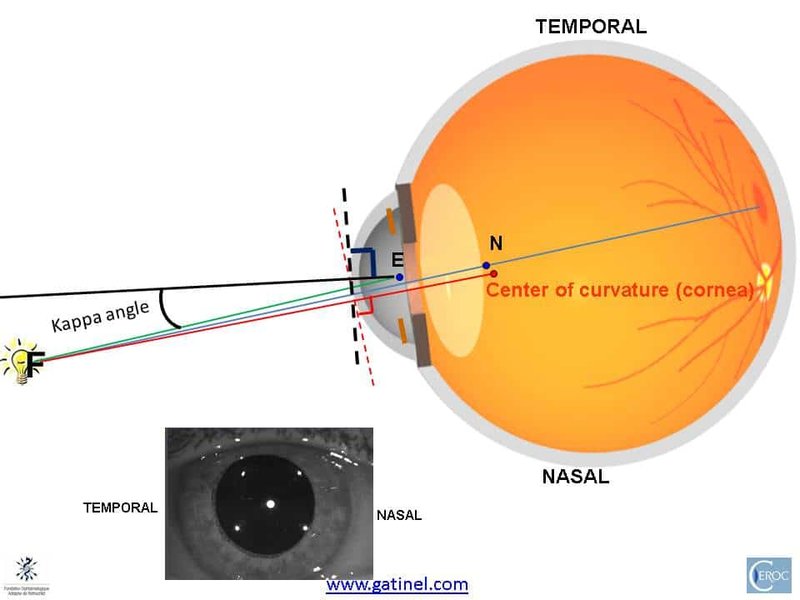
The image above illustrates the anatomical basis of angle kappa, showing the relationship between the visual axis and the pupillary axis. Understanding this spatial relationship is fundamental to interpreting clinical measurements and planning centered surgical treatments.
Frequently Asked Questions
References
-
Angle Kappa and its Importance in Refractive Surgery - PMC
- Angle Kappa Measurements: Normal Values in Healthy Iranian Population - PubMed
-
A Method to Measure Angle Kappa Using UBM and Corneal Topography - PMC
-
Normative Values for Pupil Size, Angle Kappa, and Aberrations in a Caucasian Population - PMC
- Measurement of Angle Kappa With Synoptophore and Orbscan II in a Normal Population - PubMed
- Optical Axes and Angle Kappa - EyeWiki
Recommended Searches
Last updated April 8, 2025