
Comprehensive Analysis of Cervical Spine MRI Findings
Insights into Degenerative Changes and CSF Collection in the Cervical Spine
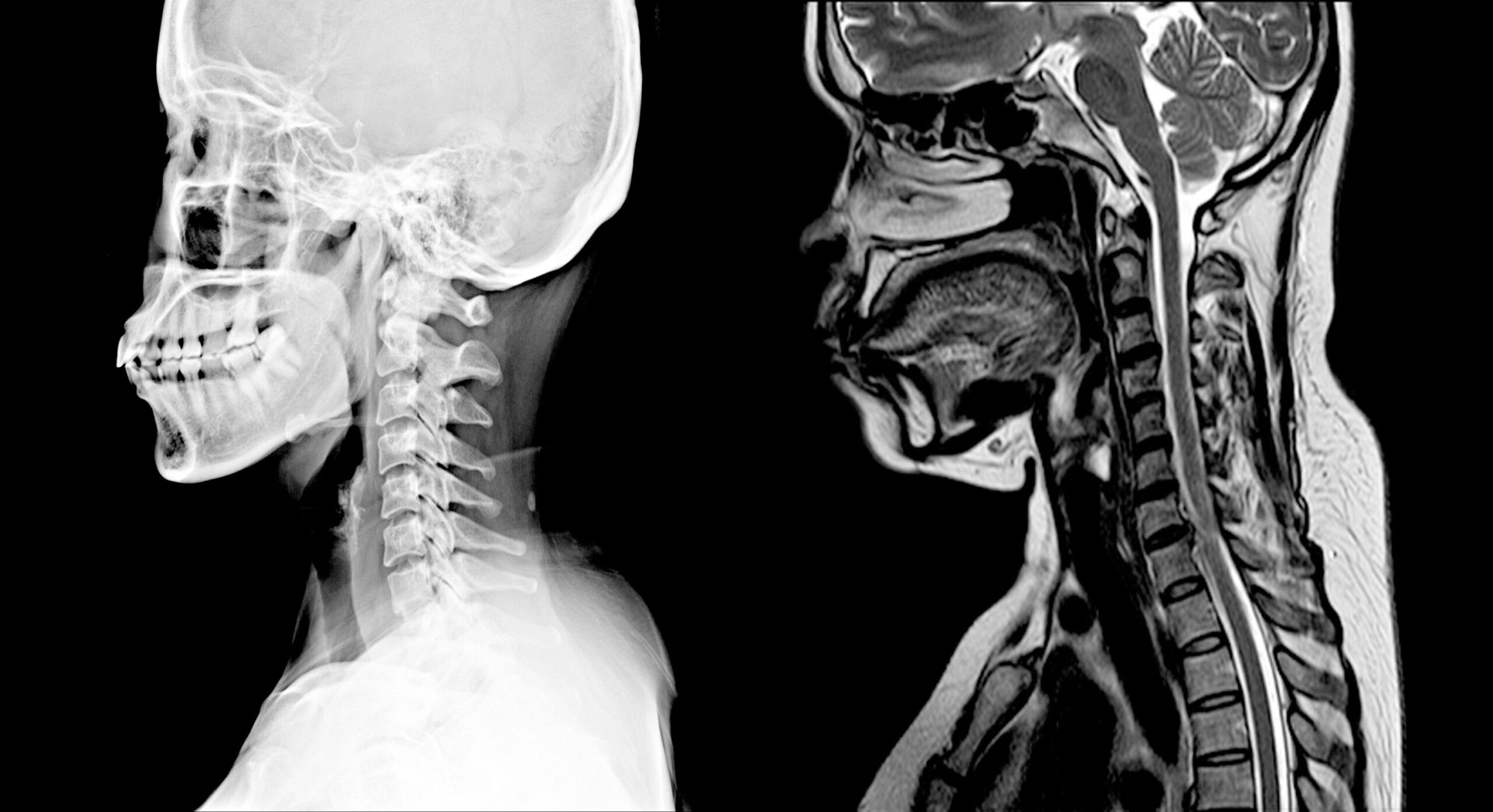
Key Highlights
- Straightening of Cervical Curvature and Disk Desiccation: Indicative of degenerative changes with muscle spasm or early aging signs.
- Ventral Epidural CSF Fluid Collection: A focal CSF fluid signal collection whose clinical significance may require further evaluation.
- Minor Disk Protrusion at C2-3: A small central disk protrusion noted without causing neural impingement or canal stenosis.
Detailed Breakdown of the MRI Findings
1. Cervical Curvature and Alignment
Straightening of Cervical Curvature
The MRI report indicates that there is a straightening of the natural cervical curvature. Ordinarily, the cervical spine exhibits a natural lordotic curve (an inward curve) that aids in load distribution and absorbs shock. Straightening may suggest underlying issues such as muscle spasm, tension, or chronic postural adaptations that can arise due to degenerative changes or prolonged discomfort. Notably, the absence of anteroposterior (AP) malalignment implies that there is no significant lateral or misdirection of the vertebral bodies despite the alteration in curvature.
2. Vertebral Body and Disk Evaluation
Disk Desiccation with Maintained Disk Height
A prominent portion of the findings is the presence of disk desiccation throughout the cervical spine. Desiccation refers to the dehydration or reduction in water content of the intervertebral discs. In many cases, disk desiccation is a typical degenerative change related to aging or chronic wear and tear. Despite this dehydration, the maintained disk height is significant; it implies that while the nutritional status and hydration of the disc may be compromised, there is no gross collapse of the disc space. This is an important finding because maintained disk height typically suggests that the mechanical function of the intervertebral discs remains relatively preserved, and there is no evidence of acute disc herniation or severe degenerative loss.
Tiny Central Disk Protrusion at C2-3
Additionally, the report notes a tiny central disk protrusion at the C2-3 level. A disk protrusion is when the disc’s soft inner material extends slightly beyond its normal margins. In this case, the protrusion is described as tiny and centrally located. Importantly, it does not create central canal or foraminal stenosis, meaning that it does not compress the spaces where spinal nerves exit. Hence, while it is a deviation from the norm, it does not appear to be currently affecting neural structures or leading to symptoms like radiculopathy by narrowing the canal for nerve roots.
3. Muscular and Ligamentous Integrity
No Evidence of Muscle or Ligament Damage
The MRI findings specifically mention the absence of paraspinal muscular strain and interspinous ligamentous sprain. Paraspinal muscles support and stabilize the vertebral column, and their integrity is essential for normal spinal function. Similarly, the interspinous ligaments, which connect adjacent spinous processes, contribute to the stability of the spine. The absence of injury in these areas strongly indicates that the changes observed in the spine – including the altered curvature and disk desiccation – are primarily degenerative rather than acute traumatic injuries. This is a reassuring aspect of the report as it rules out recent muscle or ligament injuries, and instead indicates a chronic process.
4. Ventral Epidural CSF Fluid Collection
Description and Localization
One of the more notable and potentially significant findings is the ventral epidural collection of cerebrospinal fluid (CSF). This collection begins at the level of the superior endplate of C7 and extends caudally (downward) to at least the level of the superior endplate of T2. CSF collections in this area may be suggestive of several underlying conditions, including a CSF leak, an epidural cyst, or an incidental finding that requires further clinical correlation. The reported measurement of the collection is 0.4 cm in maximum depth at the mid-T1 vertebral body, as seen on series 6/image 46.
Clinical Implications of CSF Collection
The clinical significance of a ventral epidural CSF fluid collection can be variable. In some instances, it may be an incidental finding without symptoms. However, it can also be associated with conditions like a dural tear, especially in the presence of trauma or degenerative changes that compromise the dura. Given that the CSF collection is not described as causing compression of the spinal cord (and the spinal cord signal remains normal), its current presentation might be subclinical. However, further clinical evaluation is recommended. Monitoring for any symptoms such as headache, neurologic deficits, or changes in posture and balance would be essential to rule out a more serious condition.
5. Spinal Cord and Craniocervical Junction Integrity
Spinal Cord Signal
An important aspect of any cervical spine MRI analysis is the assessment of the spinal cord. The findings report that there is no abnormal signal within the spinal cord. This is a positive outcome, indicating that, despite the degenerative changes and the presence of CSF fluid collection, there is no direct impact on the spinal cord tissue, such as edema, demyelination, or compression-related changes. Preservation of normal spinal cord signal is critical for ensuring that neural functions related to both motor and sensory activities remain intact.
Craniocervical Junction
The integrity of the craniocervical junction – the region where the skull meets the cervical spine – is also noted to be intact. This is significant because any disruption in this area can result in serious neurological deficits due to the proximity of the brainstem and cerebellum. The intact status here reassures that there are no anatomical disruptions, fractures, or misalignments at this critical juncture.
Integrated Analysis and Clinical Relevance
Chronic Degenerative Changes Versus Acute Findings
The MRI report provides clear evidence of chronic degenerative changes in the cervical spine, predominantly reflected by the disk desiccation and straightening of the normal lordotic curvature. Such degenerative changes are common with age or chronic mechanical stress and usually do not constitute an immediate emergency. Nonetheless, they are vital in understanding the natural progression of spinal aging and may predispose individuals to further issues if left unmonitored.
On the other hand, the ventral epidural CSF fluid collection represents a finding that may have different clinical implications. Its presence raises questions about the potential for a CSF leak or a cystic pathology that might contribute to neck discomfort or even cerebrospinal hypotension in some cases. In clinical practice, additional investigations, such as a contrast-enhanced study or CT myelography, may be used to further delineate this finding and understand its cause. Importantly, the measurement of 0.4 cm suggests a small, localized fluid accumulation, yet its extension over several vertebral levels warrants careful observation.
Comparative Summary Table
| Finding | Description | Clinical Relevance |
|---|---|---|
| Straightening of Cervical Curvature | Loss of the natural lordotic curve without AP malalignment | May indicate muscle spasm or chronic degenerative changes; no acute misalignment |
| Disk Desiccation | Dehydration process in discs with maintained overall height | Sign of chronic degeneration; maintained height is favorable |
| Ventral Epidural CSF Collection | Fluid signal collection from C7 to T2, 0.4 cm depth | Potential CSF leak or cyst; requires further evaluation if symptomatic |
| Central Disk Protrusion at C2-3 | Small bulge without canal or foraminal stenosis | Minor finding unlikely to cause neural compromise |
| Spinal Cord Signal & Craniocervical Junction | No abnormal signal; intact junction | Reassures that neural elements are not affected |
Implications for Management and Follow-Up
Given the composite findings, the patient may be experiencing symptoms related to chronic degenerative changes of the cervical spine. The presence of disk desiccation and loss of normal curvature is common in individuals experiencing age-related wear and postural stress. In such cases, a conservative approach often includes physical therapy, posture modification, and possibly the use of non-steroidal anti-inflammatory medications (NSAIDs) to alleviate pain and improve function.
However, the ventral epidural CSF fluid collection merits a more cautious approach. Although the small size (0.4 cm) and lack of abnormal spinal cord signal are reassuring, clinicians might consider longitudinal monitoring. If there are any evolving clinical symptoms—such as headache, radicular pain, or neurological deficits—a repeat imaging study or further investigations like a CT myelogram could be warranted to ascertain whether there has been any progression or if intervention is needed.
Similarly, while the minor disk protrusion at C2-3 is not causing canal stenosis or interfering with neural structures, it serves as a marker of ongoing degenerative processes. This reinforces the importance of a comprehensive approach to spinal health which may involve lifestyle adjustments, regular physical activity, and possibly ergonomically optimized environments to reduce stress on the cervical spine over time.
Additional Considerations and Future Directions
Non-invasive Therapies and Conservative Management
In light of the MRI findings, managing the underlying chronic changes typically focuses on non-invasive methods. Treatments such as physiotherapy, chiropractic care, and ergonomically guided educational programs have shown benefit for patients with similar cervical spine complaints. For many patients, targeted exercises to strengthen neck muscles, improve flexibility, and correct posture play pivotal roles in alleviating symptoms and preventing further spinal degeneration.
Moreover, monitoring the CSF fluid collection is essential since its presence might evolve. Regular follow-up imaging allows physicians to detect any subtle changes early on. In the absence of acute worsening, a patient can often be managed conservatively with periodic reassessment.
Potential Role of Advanced Imaging Techniques
While the current MRI provides comprehensive insight into the structural integrity of the cervical spine, there are circumstances where additional imaging modalities may be beneficial. Advanced imaging studies such as CT scans or dynamic MRI evaluations can offer further clarity, particularly regarding the nature of the CSF collection and any potential ventral dural disruption. CT myelography, for example, might be employed if there is persistent concern about a CSF leak or if clinical symptoms do not correlate with the MRI findings. Utilizing these tools can significantly aid in fine-tuning the diagnosis and ensuring tailored management strategies.
Integration with Clinical History and Symptoms
It is paramount to integrate these imaging findings with the patient’s clinical history and present symptoms. Imaging serves as a critical component of diagnosis, but it functions best when correlated with physical examination, history of symptoms, and any prior incidents of trauma or chronic pain. A multidisciplinary approach involving a neurologist, orthopedic specialist, and physical therapist can provide comprehensive care and ensure that the findings are contextualized within the broader clinical picture.
Additionally, consideration of the patient’s lifestyle, occupational demands, and any history of previous cervical interventions can influence the overall management plan. For example, ergonomic adjustments at work or modifications in daily activities may mitigate further degenerative change and enhance overall spinal health.
Summary Table of Findings and Clinical Insights
| Aspect | Observation | Clinical Insight |
|---|---|---|
| Cervical Curvature | Straightening without AP malalignment | Suggests chronic muscle spasm or degenerative adaptation |
| Disk Desiccation | Present throughout with maintained height | Indicative of age-related degenerative changes; mechanical disc function preserved |
| Ventral Epidural CSF | Collection from C7 to T2; 0.4 cm at mid-T1 | Cautionary sign that may require further evaluation if symptomatic |
| Disk Protrusion | Tiny central protrusion at C2-3 | Minor finding without evidence of neural compression |
| Spinal Cord & Junction Integrity | Normal signal and intact craniocervical junction | Reassuring findings indicating no immediate neurological compromise |
References
- Cervical Spine Degeneration - Hindawi
-
MRI Analysis of Cervical Spine - NCBI
-
CSF Collections in Spine - Radiopaedia
- Cervical MRI Scan - Mount Sinai
-
Cervical Disc Protrusion - Spine Universe
Recommended Further Queries
Last updated March 19, 2025