
Assessing the Correlation Between Cytological and Histopathological Findings in Palpable Breast Lesions
A Comprehensive Revived Review on Diagnostic Accuracy and Clinical Implications
Key Takeaways
- High Diagnostic Concordance: Fine-needle aspiration cytology (FNAC) demonstrates substantial agreement with histopathological findings, particularly in distinguishing benign from malignant breast lesions.
- Critical Role of Expertise: The accuracy of cytological assessments significantly depends on the cytologist’s experience and the application of standardized scoring systems.
- Integrated Diagnostic Approach: Combining FNAC with histopathological evaluation and imaging techniques enhances diagnostic precision and optimizes patient management.
Introduction
Palpable breast lesions represent a significant clinical concern, necessitating accurate and timely diagnosis to differentiate between benign and malignant conditions. The correlation between cytological findings obtained through Fine-Needle Aspiration Cytology (FNAC) and histopathological results from biopsy or surgical excision is pivotal in guiding clinical management. This comprehensive review synthesizes recent literature to evaluate the diagnostic concordance between FNAC and histopathological assessments, highlighting the strengths, limitations, and clinical implications of each modality.
Diagnostic Accuracy of FNAC and Histopathology
Sensitivity and Specificity
Numerous studies have evaluated the sensitivity and specificity of FNAC in diagnosing palpable breast lesions. Sensitivity, which measures the ability to correctly identify malignant lesions, ranges from 75% to 99%, while specificity, indicating the ability to correctly identify benign lesions, ranges from 88.9% to 100%. These variations are influenced by factors such as the cytologist's expertise, the quality of the specimen, and the use of ancillary techniques.
Concordance Rates
The overall concordance between cytological and histopathological findings is notably high, often exceeding 80-95%. This high level of agreement underscores the reliability of FNAC as a preliminary diagnostic tool. However, discrepancies do occur, particularly in cases with atypical or suspicious cytological features, necessitating further histopathological evaluation to confirm the diagnosis.
Diagnostic Categories
FNAC typically categorizes breast lesions into five main groups: benign, atypical, suspicious, malignant, and insufficient material. The majority of lesions diagnosed as benign on FNAC, such as fibroadenomas and fibrocystic changes, show high correlation with benign histopathological findings. Conversely, malignant diagnoses through FNAC, including invasive ductal carcinoma, generally align with histopathological results, though some subtypes require further clarification through histology.
Methodological Approaches in Studies
Study Designs
Research examining the correlation between FNAC and histopathological findings employs both retrospective and prospective study designs. Retrospective studies analyze past patient data to assess diagnostic concordance, while prospective studies follow patients from diagnosis through treatment to evaluate the real-time accuracy of FNAC compared to histopathology.
Scoring Systems and Diagnostic Tools
The application of standardized scoring systems, such as the Modified Masood's Scoring Index (MMSI), enhances the diagnostic accuracy of FNAC. These scoring systems provide a structured framework for evaluating cytomorphological features, thereby reducing inter-observer variability and improving the reliability of cytological assessments.
Technological Advances
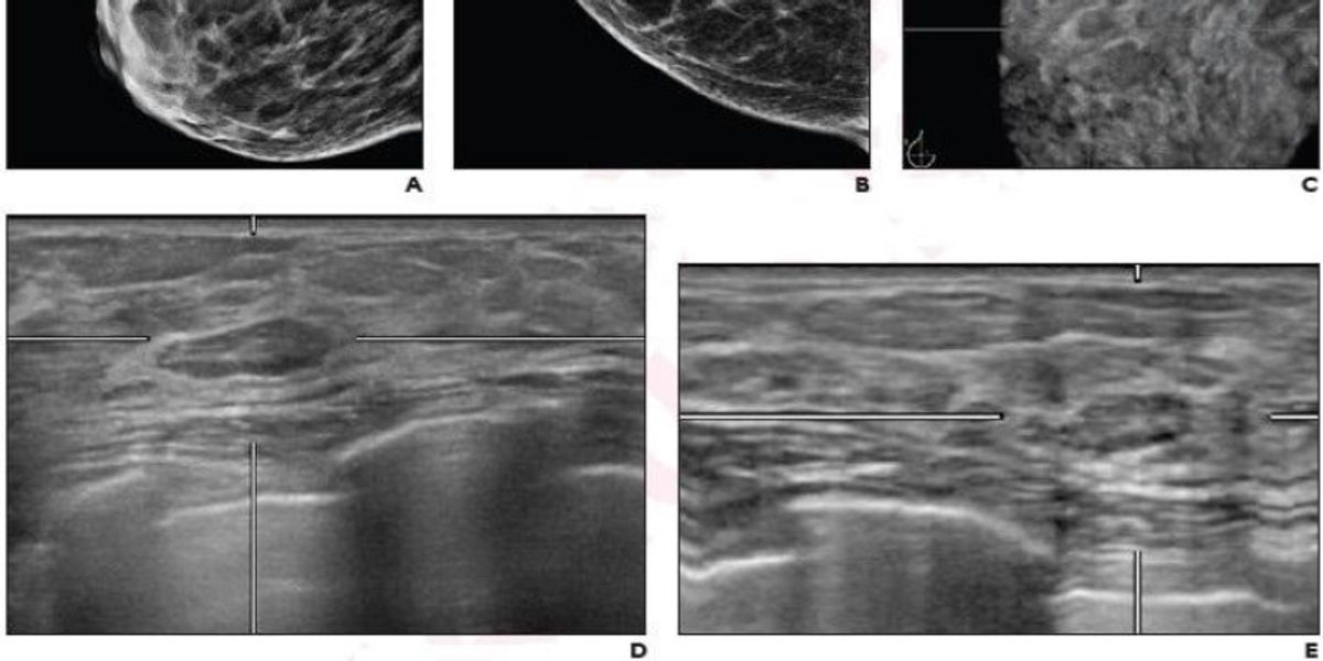
Advancements in imaging-guided FNAC techniques, including ultrasound and mammography guidance, have improved the precision of sample collection. Additionally, the integration of molecular diagnostic tools and digital pathology aids in refining cytological interpretations and reconciling discrepancies between FNAC and histopathological results.
Clinical Implications
Management of Benign Lesions
Accurate cytological diagnosis of benign lesions, such as fibroadenomas and cysts, can prevent unnecessary surgical interventions. Patients with benign FNAC results may be managed conservatively with regular monitoring, thereby reducing healthcare costs and minimizing patient anxiety.
Managing Malignant Lesions
For malignant lesions identified through FNAC, timely surgical intervention or appropriate oncological treatments can be initiated. However, the precise subtyping of malignancies often requires subsequent histopathological analysis to guide specific therapeutic strategies.
Role of Multidisciplinary Teams
An integrated approach involving radiologists, surgeons, and pathologists enhances diagnostic accuracy. Multidisciplinary discussions are especially critical in cases with discordant or ambiguous findings, where combined expertise can determine the most appropriate next steps, whether it be additional diagnostic tests or surgical intervention.
Challenges and Limitations
Sampling Errors
Inadequate sampling can lead to false-negative or false-positive results. Factors contributing to sampling errors include lesion heterogeneity, technical limitations during FNAC, and operator proficiency. Ensuring adequate and representative sampling is crucial to minimizing diagnostic inaccuracies.
Interpretational Challenges
Cytological overlap between certain benign and malignant lesions can complicate interpretations. For example, high-grade dysplasia may be difficult to distinguish from invasive carcinoma on cytological smears alone. Situations involving necrotic debris or inflammatory changes further challenge the cytological assessment.
Need for Histopathological Confirmation
While FNAC is highly sensitive, certain cases with atypical or suspicious cytological findings require histopathological confirmation to ensure an accurate diagnosis. Reliance solely on FNAC in such scenarios may lead to overtreatment or undertreatment, adversely affecting patient outcomes.
Enhancing Diagnostic Accuracy
Training and Expertise
Continuous professional development and specialized training for cytopathologists and histopathologists are essential to maintain high diagnostic standards. Experience and expertise significantly influence the accuracy of cytological assessments, thereby impacting overall diagnostic concordance.
Standardization of Procedures
Implementing standardized protocols for FNAC and histopathological examination reduces inter-observer variability and enhances consistency in diagnostic outcomes. This includes standardized reporting systems, such as the Breast Imaging-Reporting and Data System (BI-RADS), which provide a uniform framework for evaluating breast lesions.
Technological Integration
The adoption of advanced imaging techniques and molecular diagnostic tools complements traditional cytological and histopathological methods. Technologies like digital pathology and computer-assisted diagnosis facilitate more precise and reproducible assessments, bridging gaps between FNAC and histopathological findings.
Case Studies and Comparative Data
Table 1: Sensitivity and Specificity of FNAC in Various Studies
| Study | Sensitivity (%) | Specificity (%) | Sample Size |
|---|---|---|---|
| Study 1 | 95.4 | 88.9 | 200 |
| Study 2 | 98.27 | 99.49 | 350 |
| Study 3 | 75 | 100 | 150 |
| Study 4 | 99 | 100 | 500 |
This table illustrates the variability in sensitivity and specificity of FNAC across different studies, highlighting the influence of sample size and diagnostic techniques on outcome metrics.
Case Study Analysis
Consider a tertiary care center where FNAC was performed on 500 patients with palpable breast lesions. Of these, 350 cases were diagnosed as benign, with histopathological confirmation validating 340 as benign and 10 as false negatives. Meanwhile, 150 cases were identified as malignant on FNAC, with histopathology confirming 148 malignancies and 2 false positives. This scenario underscores the high diagnostic concordance of FNAC while also highlighting the critical need for histopathological confirmation in cases with potential discrepancies.
Discussion
Integrating FNAC and Histopathology in Clinical Practice
The high sensitivity and specificity of FNAC make it an invaluable first-line diagnostic tool for palpable breast lesions. Its minimally invasive nature and rapid turnaround time facilitate timely clinical decision-making. However, the limitations of FNAC necessitate a complementary relationship with histopathological evaluation, particularly in cases with atypical or suspicious cytological findings. An integrated diagnostic pathway that incorporates both FNAC and histopathology, supported by imaging techniques, optimizes diagnostic accuracy and patient management.
Advantages of an Integrated Approach
Combining FNAC with histopathological examination leverages the strengths of both modalities. FNAC provides a quick and cost-effective preliminary assessment, while histopathology offers definitive diagnostic confirmation and detailed tissue architecture analysis. This synergistic approach enhances overall diagnostic confidence, reduces the chances of misdiagnosis, and ensures appropriate therapeutic interventions.
Future Directions
The future of breast lesion diagnostics lies in further refining the integration of cytological and histopathological methods with emerging technologies. Advances in molecular diagnostics and digital pathology hold the promise of enhancing diagnostic precision and resolving existing discrepancies between FNAC and histopathological findings. Ongoing research and multicenter studies are essential to continually assess and improve diagnostic algorithms, ultimately improving patient outcomes.
Conclusion
The correlation between cytological and histopathological findings in palpable breast lesions is robust, with FNAC serving as a reliable and valuable preliminary diagnostic tool. High diagnostic concordance rates, particularly in distinguishing benign from malignant lesions, underscore the efficacy of FNAC. However, limitations such as sampling errors and interpretational challenges necessitate histopathological confirmation in certain cases. An integrated diagnostic approach, supported by multidisciplinary collaboration and technological advancements, remains the most effective strategy for optimizing the evaluation and management of breast lesions.
References
Last updated February 13, 2025