
Unraveling Medical Imaging: CT Scans, CAT Scans, and MRIs Explained
Demystifying the distinctions between these vital diagnostic tools for a clearer understanding of your health.
Key Insights into Advanced Medical Imaging
- CAT Scan and CT Scan are synonymous terms: The terms "CAT scan" (Computed Axial Tomography) and "CT scan" (Computed Tomography) refer to the exact same X-ray-based imaging procedure. "CT scan" is the more current and universally used term.
- Fundamental difference in technology: CT scans utilize X-rays and ionizing radiation to generate images, primarily excelling in visualizing bones, acute trauma, and certain lung/abdominal conditions. Conversely, MRIs employ powerful magnetic fields and radio waves, producing highly detailed images of soft tissues without any radiation exposure.
- Clinical applications and diagnostic strengths vary: CT scans are faster and often preferred in emergencies for rapid diagnoses of fractures, internal bleeding, and organ damage. MRIs, while slower and more expensive, offer superior contrast and detail for assessing the brain, spinal cord, ligaments, muscles, and subtle tissue abnormalities, making them crucial for neurological and musculoskeletal diagnostics.
In the realm of modern medicine, diagnostic imaging plays a pivotal role in identifying and understanding a vast array of health conditions. Among the most commonly employed techniques are Computed Tomography (CT) scans, often historically referred to as CAT scans, and Magnetic Resonance Imaging (MRI). While both provide invaluable insights into the body's internal structures, they operate on entirely different scientific principles, offering distinct advantages and applications in clinical practice. Understanding these differences is crucial for comprehending why a healthcare provider might choose one over the other for a specific diagnostic purpose.
Decoding the Terminology: CAT Scan vs. CT Scan
The first point of clarification is often the most straightforward: the terms "CAT scan" and "CT scan" refer to the same medical imaging procedure. Historically, when computed tomography technology first emerged, the term "CAT scan" was prevalent, standing for Computed Axial Tomography. The "axial" component referred to the cross-sectional slices of the body that the scanner captured. As technology advanced and scanning capabilities evolved to include helical (spiral) data acquisition, the term was simplified to "CT scan," or Computed Tomography. Therefore, if you hear either term, rest assured they denote the same diagnostic test that uses X-rays and computer processing to create detailed images of the body's interior.
A CT scanner (left) and an MRI machine (right) illustrate their unique appearances, reflecting their differing technologies.
Fundamental Differences in Technology and Physics
The core distinction between a CT scan and an MRI lies in the underlying physics and technology they employ to generate images:
How CT Scans Work: The Power of X-Rays
A CT scan utilizes X-rays, a form of ionizing radiation, to produce images. During a CT scan, an X-ray tube rotates around the patient's body, emitting a narrow beam of X-rays. As these X-rays pass through different tissues, they are attenuated (weakened) to varying degrees depending on the tissue density. Denser structures, like bone, absorb more X-rays and appear white on the images, while less dense tissues, like air in the lungs, appear black. Detectors on the opposite side of the X-ray tube measure the attenuated beams. A computer then processes this data from multiple angles to create detailed cross-sectional (slice) images of the bones, organs, and soft tissues. These slices can be reconstructed into 3D models for a comprehensive view.
How MRI Works: Magnetism and Radio Waves
In contrast, MRI operates without ionizing radiation. It harnesses the power of a strong magnetic field and radio waves. The patient is placed inside a large, powerful magnet, which causes the hydrogen atoms (abundant in water molecules within the body's tissues) to align with the magnetic field. Short bursts of radio waves are then emitted, temporarily knocking these aligned hydrogen atoms out of alignment. When the radio waves are turned off, the hydrogen atoms rapidly realign with the main magnetic field, releasing energy signals. Different tissues release these signals at varying rates and intensities, which are detected by the MRI scanner. A computer then processes these signals to create incredibly detailed images, particularly of soft tissues, distinguishing subtle differences between normal and abnormal structures.
Diagnostic Applications and Imaging Strengths
The choice between a CT scan and an MRI is largely dictated by the specific diagnostic question and the type of tissue or condition being investigated. Each modality excels in different areas:
CT Scan's Diagnostic Prowess
CT scans are often the preferred choice in emergency situations due to their speed and ability to rapidly image a large area of the body. They are exceptionally good for:
- Bone Injuries: Clearly visualizing fractures, dislocations, and degenerative bone conditions.
- Acute Trauma: Quickly detecting internal bleeding, organ damage, and other injuries following accidents.
- Lungs and Chest: Diagnosing conditions like pneumonia, emphysema, lung nodules, and pulmonary embolisms.
- Abdominal Issues: Identifying appendicitis, kidney stones, tumors, and other abdominal pathologies.
- Cancer Staging: Used to determine the size and location of tumors and whether cancer has spread to other parts of the body.
- Vascular Imaging: With contrast, CT angiography (CTA) can visualize blood vessels to detect aneurysms, blockages, or dissections.
MRI's Unmatched Soft Tissue Detail
MRIs are superior for providing detailed images of soft tissues, making them invaluable for:
- Brain and Spinal Cord: Detecting brain tumors, strokes, multiple sclerosis, aneurysms, and spinal cord injuries.
- Joints and Musculoskeletal System: Visualizing torn ligaments, tendons, cartilage damage, muscle injuries, and complex joint pathologies (e.g., knee, shoulder, wrist).
- Soft Tissue Tumors: Offering better differentiation between cancerous and non-cancerous soft tissue masses than CT.
- Organ-Specific Imaging: Providing highly detailed views of organs like the liver, kidneys, prostate, and uterus for detecting specific diseases or abnormalities that might be missed on CT.
- Neurological Conditions: Diagnosing conditions affecting nerves and the central nervous system.
Procedural Aspects, Patient Experience, and Safety Considerations
Beyond the technological differences, the patient experience, procedure duration, and safety profiles also vary significantly between CT and MRI:
The CT Scan Experience
CT scans are generally very quick, often completed within a few minutes. The patient lies on a motorized table that slides into a doughnut-shaped scanner. The machine is relatively open and quiet. Due to the use of X-rays, there is exposure to ionizing radiation. While the dose is typically low, especially with modern scanners and protocols, cumulative exposure is a consideration, particularly for patients requiring multiple scans or in vulnerable populations like children and pregnant women. Contrast agents, if used, are typically iodine-based and administered intravenously or orally, with a small risk of allergic reactions or kidney impact.

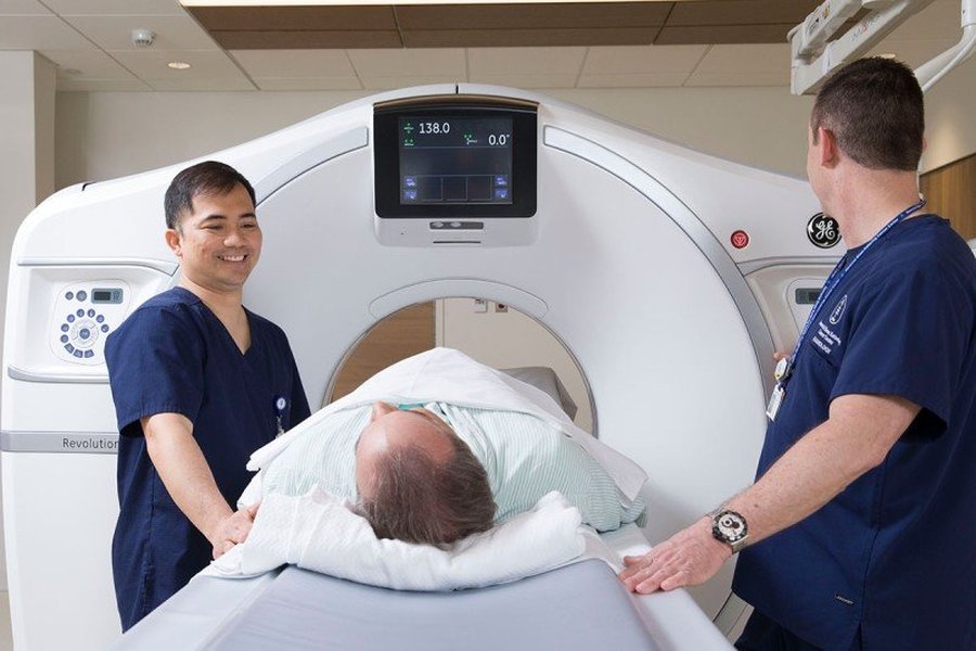
A healthcare professional discusses the benefits of a CT scan with a patient, highlighting its efficiency.
The MRI Experience
MRI scans take significantly longer, typically ranging from 15 minutes to over an hour, depending on the complexity of the exam. The patient lies inside a long, narrow, tunnel-like machine. The machine produces loud banging, clanging, and whirring noises during the scan, so earplugs or headphones are usually provided. Claustrophobia can be a concern for some patients due to the enclosed space. A major advantage of MRI is the absence of ionizing radiation, making it safer for repeated imaging and for pregnant women (though typically avoided in the first trimester unless medically necessary). Contrast agents, if used, are usually gadolinium-based, which are generally associated with a lower risk of allergic reactions and kidney issues compared to iodine-based contrasts used in CT.

A patient prepares to enter an MRI scanner, illustrating the machine's enclosed design.
Visualizing Key Distinctions: CT vs. MRI Performance
To further illustrate the multifaceted differences between CT and MRI, the radar chart below provides a comparative overview of their performance across several key operational and diagnostic attributes. This chart is based on general clinical consensus and highlights typical strengths and weaknesses for each modality.
This radar chart illustrates the comparative strengths of CT and MRI. For example, CT scans rank highly in 'Speed of Scan' and 'Emergency Use Suitability' due to their rapid image acquisition, making them indispensable in critical situations. They also excel in 'Bone Imaging Detail' given their X-ray technology. However, their primary drawback is 'Radiation Exposure', where MRI has a significant advantage with 'None'. Conversely, MRI shines in 'Soft Tissue Contrast', providing superior detail for soft tissues, and has no radiation risk. Its main limitations are 'Speed of Scan' and potential 'Claustrophobia Risk' due to the longer, enclosed procedure. 'Cost-Effectiveness' and 'Emergency Use Suitability' are generally lower for MRI compared to CT. This visualization helps to understand why a doctor might choose one over the other based on specific patient needs and diagnostic requirements.
Understanding When Each Scan is Recommended
The decision to use a CT scan or an MRI is a clinical one, made by a healthcare provider based on a patient's symptoms, medical history, and the specific information needed for diagnosis. The following mindmap visually summarizes the typical clinical scenarios that favor one imaging modality over the other:
This mindmap effectively illustrates the primary factors influencing the choice between a CT scan and an MRI. It categorizes each modality by its core technology, typical procedure duration, and most effective diagnostic applications. For instance, the branches under "CT/CAT Scan" highlight its utility in urgent scenarios and for skeletal imaging, while "MRI" branches emphasize its detailed soft tissue capabilities and lack of radiation. This clear structural representation helps in understanding the distinct roles each imaging technique plays in medical diagnosis.
Comparative Overview Table
For a quick reference, the table below summarizes the key differences between CT/CAT scans and MRIs, providing a comprehensive side-by-side comparison:
| Feature | CAT Scan / CT Scan | MRI (Magnetic Resonance Imaging) |
|---|---|---|
| Technology Used | X-rays (ionizing radiation) | Strong magnetic fields and radio waves (no radiation) |
| Primary Use Cases | Bones, acute trauma, internal bleeding, lung conditions, cancer staging, fast emergency diagnosis | Soft tissues (brain, spinal cord, ligaments, muscles, tendons), tumors, joint problems, detailed organ assessment |
| Image Detail Strengths | Excellent for bone definition and detecting internal bleeding/trauma. Good spatial resolution. | Superior contrast resolution for soft tissues, better at differentiating subtle tissue types. |
| Procedure Time | Fast (typically 5-15 minutes) | Slower (typically 15-60+ minutes) |
| Radiation Exposure | Yes, involves low doses of ionizing radiation | None |
| Patient Experience | Relatively quiet, open machine, quicker scan. | Noisy (requires earplugs/headphones), enclosed tunnel, longer scan, potential for claustrophobia. |
| Contrast Agents Used | Iodine-based (oral or IV), higher volume, slight risk of kidney damage/allergic reaction. | Gadolinium-based (IV), lower volume, generally safer for kidneys and fewer allergic reactions. |
| Suitability for Implants | Generally safe with most metal implants (check with doctor) | Contraindicated for many metal implants (pacemakers, certain surgical clips) due to magnetic fields. |
| Cost (General) | Lower | Higher |
Delving Deeper: Visualizing the Differences
To further contextualize the distinctions between these imaging modalities, consider this informative video. It visually explains the core concepts and applications of CT and MRI, helping bridge the gap between technical explanations and practical understanding in a medical setting. This video succinctly captures why medical professionals choose between these powerful tools based on specific patient needs and diagnostic goals.
A comprehensive overview explaining the differences between MRI and CT scans, highlighting their respective uses and safety considerations.
The video above titled "MRI vs CT Scan In Under 2 Minutes (Radiation, Cancer, and ...)" effectively distills the core differences between MRI and CT scans, focusing on their distinct technologies, the presence or absence of radiation, and their primary diagnostic applications. It reinforces that while both are invaluable diagnostic tools, their underlying mechanisms – X-rays for CT and magnetic fields/radio waves for MRI – dictate their strengths. This visual aid complements the detailed text by providing a quick and clear summary, helping to solidify the understanding of when and why each scan is utilized in healthcare.
Frequently Asked Questions (FAQ)
Conclusion
In conclusion, while CAT scans and CT scans are synonymous, representing the same X-ray-based imaging technology, an MRI is a fundamentally different diagnostic tool. The choice between a CT scan and an MRI hinges on several critical factors: the suspected condition, the specific body part requiring examination, the need for speed, and patient-specific considerations such as metal implants or concerns about radiation exposure. CT scans excel in rapid diagnostics for bone injuries and acute trauma, whereas MRIs provide unparalleled detail for soft tissues, making them indispensable for neurological and musculoskeletal conditions. Understanding these distinctions empowers patients to have more informed conversations with their healthcare providers about the most appropriate imaging strategy for their diagnostic needs.
Recommended Further Exploration
- Explore the detailed applications of CT scans in emergency medicine.
- Understand the specific advantages of MRI for diagnosing neurological conditions.
- Learn more about radiation safety protocols and guidelines in medical imaging.
- Investigate preparation guidelines and patient experience variations for CT vs. MRI scans.