
Common Dermatology Tests
Essential diagnostic procedures in skin care
Key Highlights
- Skin Biopsy Variations – Different biopsy techniques tailored for various conditions.
- Allergy and Patch Testing – Critical for diagnosing contact and environmental allergies.
- Non-Invasive Examinations – Procedures such as dermoscopy and Wood’s lamp exam enhance diagnosis without discomfort.
Overview of Dermatological Diagnostic Tests
Dermatology encompasses a variety of tests designed to assess skin conditions ranging from allergic reactions to malignancies. These diagnostic procedures provide invaluable information regarding the structure, function, and any pathological anomalies associated with the skin. In the following sections, we delve into the numerous tests utilized in the discipline, their purposes, methodologies, and the contexts in which they are applied.
Skin Biopsy Techniques
A skin biopsy remains one of the most vital diagnostic procedures in dermatology. It involves the removal of a small skin sample which is subsequently analyzed histologically. The information garnered can be pivotal in diagnosing conditions such as skin cancers, inflammatory diseases, and infections. The procedure is not only diagnostic but may also serve a therapeutic role in certain cases.
Types of Biopsies
The choice of biopsy depends on the nature of the lesion, its size, location, and the suspected diagnosis. Here are the primary types:
- Punch Biopsy: Utilizes a circular blade to remove a cylindrical core of skin tissue, including the epidermis, dermis, and upper subcutis. It is highly effective for sampling inflammatory dermatoses and melanocytic lesions.
- Shave Biopsy: Involves slicing the top layers of the skin. It is frequently chosen for lesions presumed to be superficial and benign.
- Excisional Biopsy: The entire lesion is removed. This is the preferred method for suspected melanoma or other lesions that require complete removal for definitive diagnosis.
- Wedge Excision: Used when deeper tissue sampling is necessary, particularly for larger or more complex lesions.
| Test | Purpose | Procedure Overview |
|---|---|---|
| Skin Biopsy | Diagnose skin cancers, infections, and inflammatory conditions | Removal of a skin sample using punch, shave, or excisional techniques; histopathological examination. |
| Patch Testing | Identify contact dermatitis and allergens | Application of allergens under adhesive patches on the skin; evaluation after 48-96 hours. |
| Skin Scraping | Diagnose fungal infections and scabies | Gentle scraping of the skin’s surface to gather scales for microscopic evaluation. |
| Wood's Lamp Examination | Detect pigment disorders, fungal infections, and bacterial infections | Examination using UV light to highlight abnormalities in pigmentation or the presence of infections. |
| Tzanck Test | Diagnose viral infections like herpes simplex and zoster | Examination of cells scraped from blister bases to identify characteristic cytologic features. |
| Dermatoscopy | Evaluate pigmented lesions and monitor moles | Use of a specialized magnifying device to assess skin lesions in detail. |
Allergy and Immunologically Focused Tests
Allergies can profoundly impact skin health, leading to conditions such as contact dermatitis and atopic dermatitis. Dermatologists often employ both patch testing and skin prick testing to pinpoint specific allergens. These procedures are crucial in determining the cause of immune-mediated reactions.
Patch Testing
Patch testing involves the application of various allergens directly to the skin using adhesive patches. This test is typically conducted on the back of a patient since the area is relatively uniform and well-covered. The patches are left in place for an extended period, generally between 48 and 96 hours, after which the skin is examined for signs of allergic reaction.
Procedure and Considerations
The patient’s clinical history is significant in determining which allergens to test for, such as preservatives, fragrances, dyes, metals (like nickel), and latex. The reactions range from mild redness and swelling to more pronounced blistering. The results can guide effective avoidance strategies and tailored treatment plans.
Skin Prick Testing
Used primarily for identifying immediate hypersensitivity reactions, the skin prick test is another common method. In this test, a drop of allergen extract is placed on the skin, which is then lightly pricked to allow the substance to enter the dermis. The test is interpreted within 15-20 minutes based on the formation of a wheal (raised, red area) or flare.
Non-Invasive and Specialized Examinations
Several non-invasive techniques allow dermatologists to examine skin lesions with minimal discomfort for the patient. These methods augment physical examination and provide additional insights necessary for an accurate diagnosis.
Dermoscopy
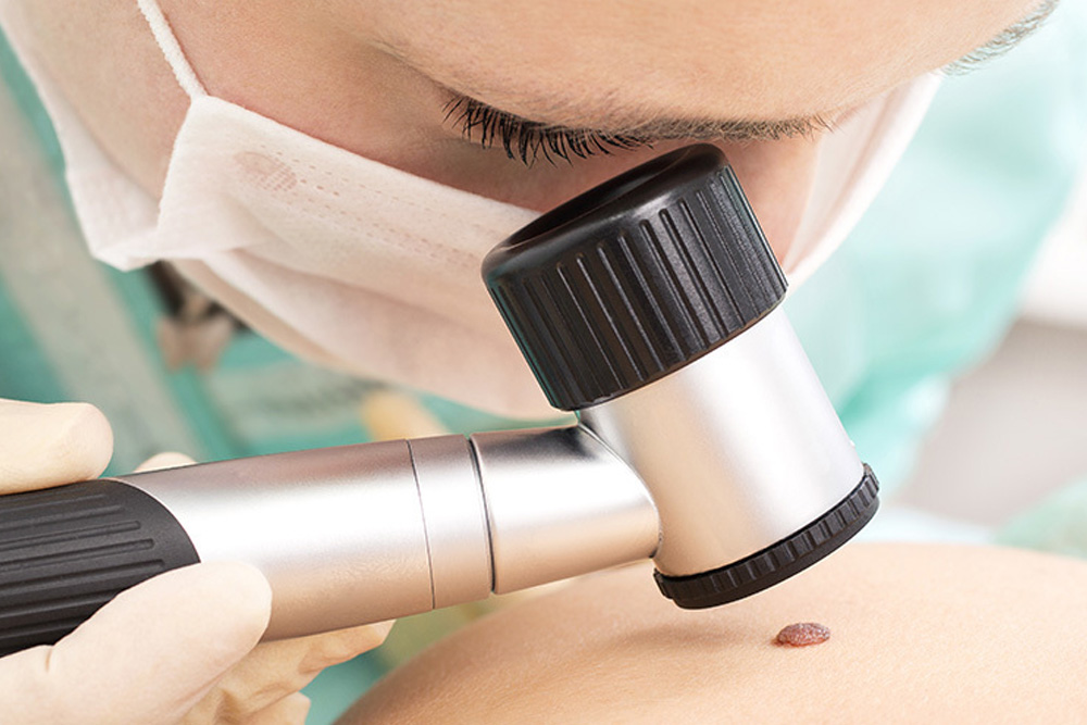
Dermoscopy is an advanced, non-invasive imaging method that employs a dermatoscope—a specialized magnifying device—to closely inspect skin lesions, particularly pigmented ones. This magnification coupled with polarized light aids in distinguishing between benign moles and malignant lesions, including melanoma. It allows dermatologists to evaluate structures and patterns that are otherwise invisible to the naked eye.
Wood's Lamp Examination
The Wood's lamp examination is a diagnostic procedure that uses ultraviolet light to evaluate skin conditions. Under this light, certain skin pathogens, pigment disorders such as vitiligo, and fungal infections can become more apparent. For instance, a fungal infection might fluoresce under the lamp, aiding in pinpointing the affected area and extending the diagnostic accuracy.
Skin Scrapings and Tzanck Test
Skin Scraping
A skin scraping involves gently removing a layer of scales from the surface of the skin. It is especially useful for diagnosing fungal infections like tinea versicolor and scabies. The scraping is then examined under a microscope, sometimes after being treated with a chemical stain to highlight fungal elements.
Tzanck Test
The Tzanck test is a cytological examination used mainly to diagnose viral infections such as herpes simplex and zoster. A sample is taken from the base of a blister and stained. The presence of multinucleated giant cells is a hallmark diagnostic feature in these viral infections.
Additional Diagnostic Considerations
Beyond the aforementioned procedures, other diagnostic methods are occasionally employed to unravel complex dermatologic cases. For instance, blood tests may be ordered to assess immunological or endocrine components related to skin manifestations. These tests might include:
- Complete Blood Count (CBC): To detect systemic inflammation or infection.
- Immunoglobulin E (IgE) Levels: Often elevated in atopic conditions.
- Hormonal and Thyroid Function Tests: To identify endocrine disorders impacting skin health.
- Antinuclear Antibody (ANA) Tests: Used in the evaluation of autoimmune conditions manifesting in the skin.
Although these blood tests are not direct skin examinations, they contribute to a more complete understanding of the patient’s overall condition and can direct further dermatologic evaluation.
Comparative Overview Table
The table below summarizes key dermatological tests, providing a quick reference to their purposes and procedural aspects.
| Diagnostic Test | Common Purpose | Procedure Details |
|---|---|---|
| Skin Biopsy | Diagnose cancers, inflammatory, and infectious conditions | Various methods including punch, shave, and excision to obtain tissue for histopathological analysis |
| Patch Testing | Identify allergens causing contact dermatitis | Application of allergen patches on the skin with assessments after 48-96 hours |
| Skin Prick Testing | Detect IgE mediated allergies | Introduction of allergen extracts via pricking; evaluation of immediate wheal reaction |
| Skin Scraping | Diagnose fungal infections and scabies | Gentle removal of skin scales for microscopic examination often aided by special stains |
| Wood's Lamp Examination | Identify pigment disorders and certain infections | Use of ultraviolet light to reveal abnormalities unnoticeable in the regular light spectrum |
| Tzanck Test | Detect viral infections | Cytological examination of cells from blister bases to identify multinucleated giant cells |
| Dermoscopy | Enhanced visualization of pigmented lesions | Utilizes a dermatoscope to provide magnification and illumination for accurate lesion assessment |
Integration and Application in Clinical Practice
The selection of a diagnostic test in dermatology is often dictated by patient history, clinical presentation, and the need to clarify uncertain diagnoses. Dermatologists utilize a combination of these tests to form a comprehensive clinical picture. For example, a patient presenting with an evolving pigmented lesion might undergo a dermoscopic evaluation followed by an excisional biopsy if malignancy is suspected. Similarly, a patient with a suspected fungal infection might first be evaluated with a Wood's lamp exam and confirmed with a skin scraping.
Modern dermatologic practices also incorporate molecular testing and advanced imaging techniques to further refine diagnoses, especially in ambiguous cases. The evolving field of dermatology continues to integrate innovative technology and procedural refinements, ensuring that physicians can deliver personalized treatment based on accurate and timely diagnostic results.
Role of Complementary Diagnostic Procedures
Complementary to direct dermatologic examinations, blood tests and serological markers play an essential role in uncovering systemic issues that may have cutaneous manifestations. Conditions like autoimmune diseases, hormonal imbalances, or infections can manifest in the skin, requiring a holistic approach to diagnosis. By combining information from skin evaluations and systemic tests, dermatologists are better equipped to design treatment regimens that address both local and systemic pathology.
Balancing Invasiveness and Diagnostic Yield
A critical aspect in the field of dermatology is the balance between invasiveness and diagnostic yield. Non-invasive exams such as dermoscopy and Wood's lamp testing offer quick insights without discomfort, making them ideal first-line assessments. In contrast, more invasive procedures like biopsies, while slightly uncomfortable, provide definitive histological evidence that can be crucial in managing chronic or life-threatening conditions.
Patient-Centric Diagnostic Approach
Patient comfort and clinical efficiency are paramount. Dermatologists routinely tailor testing protocols according to the patient’s age, overall health, and the specific dermatologic concern. For instance, when evaluating a child, less invasive procedures such as patch testing or dermoscopy might be preferred over procedures that require anesthesia, thus making the patient’s experience more positive while still ensuring accurate diagnosis.
References
- Skinsight - Common Dermatology Procedures
- WebMD - Diagnosing Skin Problems
- Merck Manuals - Diagnostic Tests for Skin Disorders
- Rabkin Dermatopathology - Common Tests and Procedures
- DermNet NZ - Dermatological Investigations and Tests
Recommended Topics for Further Exploration
Last updated March 6, 2025