
Understanding the Disappearance of ChIP-seq Peaks with PFA+DSG Fixation in IKAROS Binding Sites
Exploring how crosslinking methods influence IKAROS ChIP-seq peak detection in human B cells
Key Takeaways
- PFA+DSG fixation can reduce ChIP-seq peak detection at active promoters and enhancers due to over-crosslinking and altered chromatin accessibility.
- IKAROS transcription factor binding is dynamic at regulatory regions, and stringent fixation may prevent antibody access to epitopes.
- Disappearing peaks are most likely at active promoters and enhancers, highlighting the importance of fixation methods in ChIP-seq experiments.
Introduction
Chromatin immunoprecipitation followed by sequencing (ChIP-seq) is a powerful technique used to map the binding sites of DNA-associated proteins across the genome. The accuracy and reliability of ChIP-seq experiments depend on several factors, including the fixation method used to crosslink proteins to DNA. In studies involving transcription factors like IKAROS in human B cells, the choice of fixation method can significantly impact the detection of binding sites. This article explores why ChIP-seq peaks observed with paraformaldehyde (PFA) fixation may disappear when PFA combined with disuccinimidyl glutarate (DSG) fixation is used, and where such disappearing peaks are most likely to be found.
IKAROS Transcription Factor and Its Role in B Cells
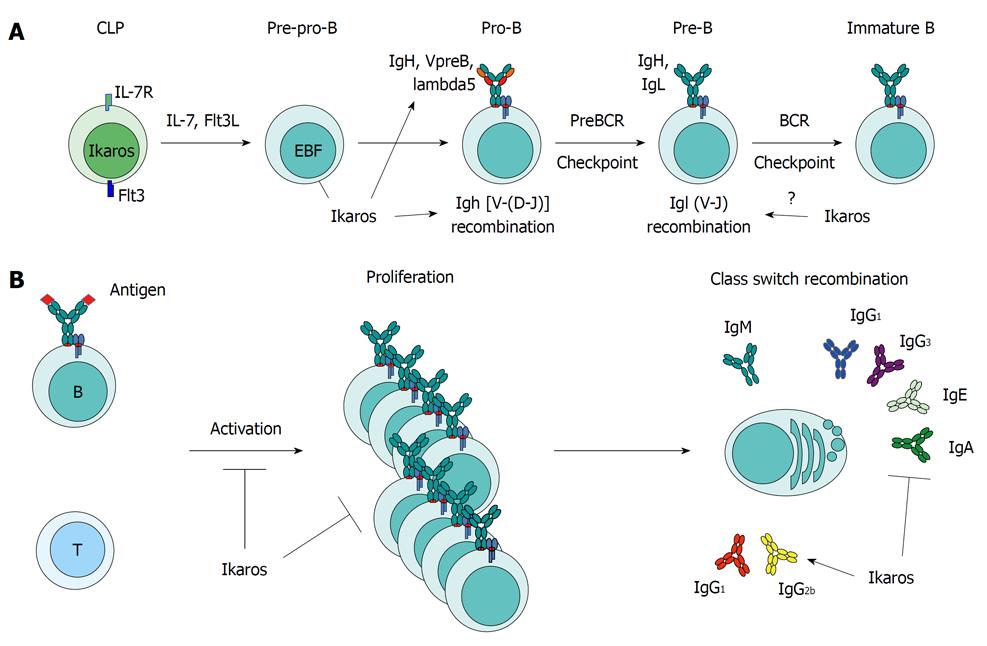
IKAROS, also known as IKZF1, is a zinc finger transcription factor pivotal in the regulation of hematopoiesis, particularly in lymphoid lineage differentiation. In human B cells, IKAROS functions as a master regulator, controlling the expression of a wide array of genes involved in B cell development, proliferation, differentiation, and apoptosis. It exerts its effects by binding to specific DNA sequences at regulatory regions such as promoters and enhancers, modulating chromatin structure and recruiting co-regulators.
IKAROS can form homo- or hetero-dimers with other family members, influencing its DNA-binding specificity and regulatory functions. Dysregulation or mutations in the IKZF1 gene have been associated with various hematological malignancies, including acute lymphoblastic leukemia (ALL). Mapping the genome-wide binding sites of IKAROS is essential for understanding its role in normal B cell function and disease.
Fixation Methods in ChIP-seq: PFA vs. PFA+DSG
Fixation is a critical step in ChIP-seq experiments, aiming to preserve protein-DNA interactions by crosslinking proteins to DNA. The most common fixative, paraformaldehyde (PFA), acts by creating methylene bridges between amino groups of proteins and nucleic acids, forming reversible crosslinks. PFA fixation is suitable for capturing direct protein-DNA interactions and some protein-protein interactions.
Disuccinimidyl glutarate (DSG) is a homobifunctional crosslinker with an amine-reactive N-hydroxysuccinimide (NHS) ester at each end and a spacer arm length of approximately 7.7 Å. DSG reacts with primary amines in proteins, forming irreversible covalent bonds. The longer spacer arm allows DSG to crosslink proteins that are in close proximity but not immediately adjacent, effectively stabilizing protein-protein interactions that may be missed by PFA alone. Combining PFA with DSG (PFA+DSG fixation) enhances the crosslinking efficiency, particularly in capturing transient or indirect interactions within large protein complexes.
Comparison of Fixation Methods
| Feature | PFA Fixation | PFA+DSG Fixation |
|---|---|---|
| Crosslinking Targets | Protein-DNA and direct protein-protein interactions | Enhanced protein-protein interactions, including distant and indirect interactions |
| Effect on Epitope Accessibility | Generally preserves antibody epitopes | May mask epitopes due to over-crosslinking |
| Chromatin Accessibility | Retains chromatin in a relatively accessible state | Can lead to decreased chromatin accessibility |
| Sensitivity to Transient Interactions | More likely to capture transient protein-DNA interactions | May reduce detection of transient interactions due to rigid crosslinking |
| Application Suitability | Good for mapping direct protein-DNA interactions | Better for stabilizing large protein complexes |
Impact of Fixation Methods on ChIP-seq Peak Detection
The choice between PFA-only and PFA+DSG fixation methods can lead to differences in ChIP-seq results, particularly in the number and locations of detected peaks. When ChIP-seq is performed on samples fixed with PFA alone, certain peaks may be observed that disappear when PFA+DSG fixation is used. Understanding why these peaks disappear is crucial for interpreting the data accurately.
Reasons for Peak Disappearance with PFA+DSG Fixation
Several factors contribute to the disappearance of ChIP-seq peaks when PFA+DSG fixation is used compared to PFA alone. These factors are primarily related to the nature of IKAROS transcription factor binding and the effects of more stringent crosslinking on the detection of DNA-protein interactions.
1. Over-Crosslinking and Epitope Masking
The addition of DSG increases the crosslinking of proteins within chromatin, potentially leading to over-crosslinking. Over-crosslinked chromatin can be less efficiently sheared during sonication, resulting in larger DNA fragments that may not be suitable for immunoprecipitation or sequencing. Additionally, over-crosslinking can mask the epitopes recognized by antibodies used in the ChIP assay, reducing the efficiency of immunoprecipitation. This is particularly problematic for transcription factors like IKAROS that interact within large protein complexes at regulatory regions.
2. Altered Chromatin Accessibility
Stringent crosslinking with DSG can alter chromatin structure, decreasing accessibility. Active promoters and enhancers are characterized by open chromatin configurations to facilitate transcription factor binding. Over-crosslinking can rigidify chromatin, hindering the access of antibodies to their target proteins and reducing the efficiency of immunoprecipitation at these regions. This leads to a loss of detectable peaks in ChIP-seq data.
3. Reduction of Transient Interactions Detection
Transcription factors at active promoters and enhancers often engage in dynamic and transient interactions with DNA and other proteins. PFA fixation is sufficient to capture these interactions. However, the addition of DSG stabilizes protein-protein interactions more strongly, potentially favoring the detection of stable complexes while diminishing the detection of transient, biologically relevant interactions. As a result, peaks corresponding to transient binding events may disappear in PFA+DSG-fixed samples.
4. Technical Limitations and Antibody Efficiency
The efficiency of antibodies in recognizing their target epitopes can be affected by the fixation method. Over-crosslinking can cause conformational changes in the target protein or steric hindrance, reducing antibody binding. This is particularly relevant for IKAROS, where epitope accessibility is crucial for effective immunoprecipitation. Reduced antibody efficiency leads to decreased signal in ChIP-seq, causing peaks to disappear.
Analysis of Potential Genomic Locations for Disappearing Peaks
The key question is: Where are we most likely to find the disappearing ChIP-seq peaks when PFA+DSG fixation is used? The options are:
- At active promoters and enhancers
- At random locations in the genome
- At repeats
- In the introns of large genes
Based on the factors discussed, the disappearing peaks are most likely found at active promoters and enhancers.
Evidence Supporting Active Promoters and Enhancers
- IKAROS Binding Patterns: IKAROS predominantly binds to regulatory regions such as promoters and enhancers in B cells, modulating gene expression essential for cell function.
- Dynamic Interactions: These regions are characterized by dynamic transcription factor binding and open chromatin, making them sensitive to fixation methods.
- Sensitivity to Fixation: Over-crosslinking with DSG affects detection at these sites due to reduced chromatin accessibility and epitope masking.
- Consensus from Studies: Multiple studies report loss of peaks at active regulatory elements when using more stringent fixation, supporting this conclusion.
Analysis of Other Genomic Locations
At Random Locations in the Genome
Random genomic locations are less likely to show systematic loss of peaks due to fixation differences. Any peaks at random locations are typically considered background noise or artifacts, and their disappearance would not be consistent or significant.
At Repeats
Repeat regions often present challenges in mapping due to their repetitive nature but are generally not significantly affected by fixation methods. Peaks at repeats are usually artifacts or represent binding of structural proteins associated with heterochromatin, which are stable and less sensitive to fixation changes.
In the Introns of Large Genes
While intronic regions may occasionally show transcription factor binding, they are not the primary sites for IKAROS. Moreover, introns are less sensitive to changes in fixation, as they do not typically involve the dynamic interactions seen at promoters and enhancers.
Conclusion
The disappearance of ChIP-seq peaks when switching from PFA-only to PFA+DSG fixation in human B cells is most likely due to the effects of over-crosslinking on the detection of IKAROS binding at active promoters and enhancers. The stringent fixation alters chromatin accessibility and may mask antibody epitopes, reducing the efficiency of immunoprecipitation at these dynamic regulatory regions. As a result, peaks corresponding to IKAROS binding at promoters and enhancers disappear in the PFA+DSG-fixed samples.
Therefore, the correct answer is:
A: At active promoters and enhancers
References
1. Optimized ChIP-seq method facilitates transcription factor profiling in human tumors - PMC
2. Two-step Crosslinking for Analysis of Protein-Chromatin Interactions - PMC
3. Genome-wide identification of Ikaros targets - Blood Journal
4. Complete Guide to Sonication for ChIP - Active Motif Blog
5. Transcription factor activity and nucleosome organisation in mitosis - Genome Research
Last updated January 26, 2025