
Unraveling the Double Helix: The Iconic Structure of DNA
A Deep Dive into the Architecture of Life's Genetic Blueprint
The double helix structure of Deoxyribonucleic Acid (DNA) stands as one of the most profound discoveries in the history of science. First accurately described by James Watson and Francis Crick in 1953, building upon the crucial work of scientists like Rosalind Franklin, Maurice Wilkins, and Erwin Chargaff, this elegant molecular architecture provides the fundamental basis for heredity and life itself. It’s a sophisticated yet remarkably simple design that allows DNA to store, replicate, and transmit genetic information with extraordinary precision across generations.
Key Insights into the Double Helix
- Twisted Ladder Configuration: DNA is structured like a twisted ladder or spiral staircase, with two long strands winding around each other in a helical shape. This iconic "double helix" is a right-handed coil, meaning it twists clockwise.
- Sugar-Phosphate Backbone: The 'sides' of this twisted ladder are formed by alternating sugar (deoxyribose) and phosphate groups. This sugar-phosphate backbone is on the exterior of the helix, providing structural integrity.
- Complementary Base Pairing: The 'rungs' of the ladder are made of specific pairs of nitrogenous bases. Adenine (A) always pairs with Thymine (T) via two hydrogen bonds, and Guanine (G) always pairs with Cytosine (C) via three hydrogen bonds. This precise pairing is fundamental to DNA's ability to replicate and carry genetic information.
The Fundamental Components of DNA
At its core, DNA is a polymer, meaning it's composed of many repeating molecular units. These small units are called nucleotides. Each nucleotide itself is a tripartite structure, essential for the overall integrity and function of the DNA molecule.
The Nucleotide Building Blocks
Every single nucleotide is made up of three distinct components:
- A Nitrogenous Base: These are the information-carrying parts of DNA. There are four types:
- Adenine (A)
- Thymine (T)
- Cytosine (C)
- Guanine (G)
- A Pentose (Five-Carbon) Sugar: In DNA, this sugar is deoxyribose. It forms a crucial part of the backbone and links to both the phosphate group and the nitrogenous base.
- A Phosphate Group: This group provides the link between successive nucleotides in a single strand, forming the sugar-phosphate backbone. It carries a negative charge, which contributes to the overall stability of the DNA molecule.
These nucleotides link together to form long polynucleotide strands, and it's the pairing of two such strands that creates the double helix.
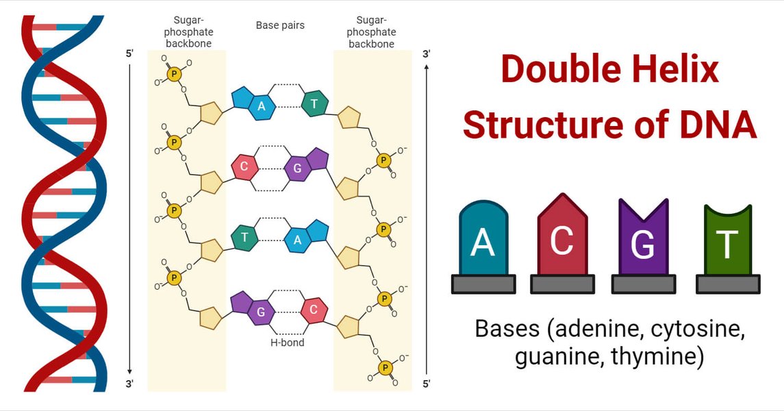
An illustrative representation of the DNA double helix, highlighting its key components.
The Architecture of the Double Helix
The double helix isn't just a simple twist; it's a precisely organized structure with specific characteristics that enable its biological functions.
The Sugar-Phosphate Backbone
The "sides" of the DNA ladder are robust sugar-phosphate backbones. The phosphate groups are on the outside of the helix, while the pentose sugars provide the linkage points for the nitrogenous bases. This external positioning of the sugar-phosphate backbone provides structural stability and protection to the genetic information housed within the bases.
Antiparallel Strands: A Crucial Orientation
One of the critical features of the double helix is that its two strands run in opposite directions, a characteristic known as antiparallel orientation. This means that the 5' end of one DNA strand aligns with the 3' end of the other, and vice versa. This specific arrangement is vital for DNA replication and repair, as well as for how other molecules interact with DNA.
Base Pairing and Hydrogen Bonds
The nitrogenous bases face inwards, forming the "rungs" of the ladder. They are held together by hydrogen bonds, which are relatively weak individually but collectively provide significant strength to the entire molecule due to their sheer number across the length of the DNA. The specificity of base pairing—Adenine with Thymine (A-T) and Guanine with Cytosine (G-C)—is known as Chargaff's rules and is a cornerstone of DNA's function. A-T pairs form two hydrogen bonds, while G-C pairs form three, contributing to the differing stability of DNA regions rich in G-C pairs.
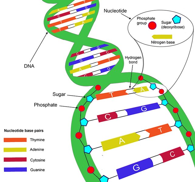
Illustration of hydrogen bonds between complementary base pairs, highlighting their specificity.
Major and Minor Grooves
As the two DNA strands twist around each other, they create two distinct types of grooves along the surface of the helix: a wider major groove and a narrower minor groove. These grooves are important because they expose the edges of the base pairs, allowing various proteins to bind to specific DNA sequences without having to unwrap the helix. This interaction is crucial for processes like gene regulation and DNA replication.
The Significance of the Double Helix Discovery
The elucidation of the DNA double helix was more than just a scientific breakthrough; it revolutionized biology and medicine, providing answers to long-standing questions about heredity and the fundamental mechanisms of life.
Implications for Genetic Information Storage and Replication
The most profound implication of the double helix structure was its immediate suggestion of a copying mechanism for genetic material. If the two strands could separate, each could then serve as a template for synthesizing a new complementary strand, resulting in two identical daughter DNA molecules. This semiconservative replication mechanism explains how genetic information is faithfully passed from one generation of cells to the next, and from parent to offspring.
This video from Khan Academy provides a concise overview of the James Watson and Francis Crick model of DNA and how the double helix structure was discovered. It delves into the historical context and the collaborative scientific efforts that led to this monumental understanding, emphasizing the roles of key researchers and the elegance of the structure itself.
The "Twisted Ladder" Analogy in Action
The analogy of a "twisted ladder" perfectly captures the essence of the DNA double helix. The ladder's side rails are the sugar-phosphate backbones, strong and consistent. The rungs are the base pairs, forming the informational steps. This simple yet accurate analogy helps visualize how the molecule is structured and how its components interact to form a stable yet functional entity capable of carrying complex biological instructions.
Evolution of Understanding: Beyond Watson and Crick
While Watson and Crick's model was groundbreaking, scientific understanding of DNA has continued to evolve. Researchers have identified different conformations of the DNA double helix, such as A-DNA, B-DNA (the most common form described by Watson and Crick), and Z-DNA. These variations highlight the dynamic nature of DNA and its ability to adapt its structure under different physiological conditions or when interacting with various proteins.
Contributions of Other Scientists
It is crucial to acknowledge the extensive work of other scientists that preceded and informed Watson and Crick's discovery. Rosalind Franklin's exceptional X-ray diffraction images, particularly "Photo 51," provided critical evidence of DNA's helical nature and key dimensions. Maurice Wilkins also played a significant role in collecting and sharing X-ray data. Erwin Chargaff's rules of base composition, which stated that the amount of adenine equals thymine and guanine equals cytosine, were instrumental in postulating the specific base pairing. These collective contributions underscore the collaborative nature of scientific progress.
Detailed Structural Characteristics of B-DNA
B-DNA, the most prevalent form in living cells, exhibits specific dimensions and features:
- It is a right-handed helix.
- Each complete turn of the helix contains approximately 10-10.5 base pairs.
- The helical pitch (the distance per turn) is approximately 3.4 nanometers.
- The diameter of the helix is about 2 nanometers.
These precise measurements contribute to the stability and functionality of the DNA molecule within biological systems.
A Comparative Look at DNA Structure
To further appreciate the Watson-Crick model, let's compare its key features and implications:
| Feature | Description in Double Helix | Biological Significance |
|---|---|---|
| Overall Shape | Double helix (twisted ladder), right-handed. | Compact storage of vast genetic information within the cell. |
| Backbone Composition | Alternating sugar (deoxyribose) and phosphate groups on the exterior. | Structural integrity, protection of internal bases, negatively charged for interaction with proteins. |
| Strand Orientation | Antiparallel (one strand 5' to 3', other 3' to 5'). | Essential for DNA replication (template for new strands) and protein binding. |
| Internal Components | Nitrogenous bases (A, T, C, G) forming rungs. | Carry the genetic code sequence. |
| Base Pairing Rules | A pairs with T (2 H-bonds); G pairs with C (3 H-bonds). | Ensures accurate replication and repair, maintains consistent helix width. |
| Forces Holding Strands | Hydrogen bonds between bases; base stacking interactions. | Provides stability while allowing for unwinding during replication/transcription. |
| Grooves | Major and minor grooves. | Binding sites for DNA-binding proteins involved in gene expression and regulation. |
Exploring the Functional Impact of DNA's Structure
The double helical structure is not merely an aesthetic marvel; it directly dictates DNA's fundamental biological roles. The organization of base pairs within the helix allows for the encoding of complex biological instructions, while the ability of the strands to separate provides a straightforward mechanism for copying these instructions. This elegant design enables DNA to serve as the blueprint for all living organisms.
The radar chart above illustrates the perceived strengths of the double helix structure of DNA across several critical biological functions, compared to a hypothetical single-stranded genetic molecule. The values are qualitative, representing the enhanced capabilities afforded by the double-helical arrangement. For instance, the double helix significantly boosts 'Replication Fidelity' due to complementary base pairing and provides superior 'Structural Stability' compared to a lone strand. Its complex surface, with major and minor grooves, also offers specialized 'Protein Interaction' sites crucial for gene regulation.
Frequently Asked Questions (FAQ)
Conclusion
The double helix structure of DNA, first proposed by Watson and Crick, is a marvel of biological engineering. Its elegant and deceptively simple design—a twisted ladder composed of sugar-phosphate backbones and complementary base pairs—provides the molecular framework for life's most fundamental processes. This structure not only accounts for how genetic information is stored and expressed but, crucially, also immediately suggested a mechanism for its faithful replication, thereby solving the riddle of heredity. The discovery of the double helix cemented DNA's role as the genetic material and laid the groundwork for modern molecular biology, continuing to inspire and enable scientific advancements today.
Recommended Further Exploration
- Explore the fascinating history and key figures behind the discovery of DNA's structure.
- Delve deeper into how the double helix structure enables DNA to accurately copy itself.
- Learn about the different conformational forms of the DNA double helix and their biological significance.
- Understand how the double helix influences the process of gene expression and protein synthesis.