
Unlocking Molecular Secrets: A Deep Dive into ELISA and PCR Techniques
Explore the powerful principles, diverse applications, and critical precautions of these essential laboratory tools.
Key Insights at a Glance
- ELISA relies on antigen-antibody specificity coupled with enzymatic reactions to detect and quantify proteins, antibodies, and other molecules, crucial for diagnostics and research.
- PCR exponentially amplifies specific DNA/RNA sequences through thermal cycling, enabling sensitive detection vital for genetics, forensics, and infectious disease identification.
- Strict precautions are paramount for both techniques to ensure accuracy, prevent contamination (especially critical for PCR's sensitivity), and guarantee reliable results.
ELISA: The Immunological Detective
Decoding the Enzyme-Linked Immunosorbent Assay
The Enzyme-Linked Immunosorbent Assay, universally known as ELISA, stands as a cornerstone technique in modern biology and medicine. Developed in the 1970s, it's a highly versatile plate-based assay designed primarily for detecting and quantifying substances such as peptides, proteins, antibodies, and hormones. Think of it as a highly specific molecular detection system that leverages the immune system's own targeting mechanism – the antibody-antigen interaction.

A typical 96-well microplate used in ELISA, showing varying color intensities indicating different concentrations of the target molecule.
The Core Principle: Specificity Meets Signal Amplification
At its heart, ELISA hinges on the highly specific binding affinity between an antibody and its corresponding antigen (ScienceVivid, 2020; R&D Systems, 2023). This interaction forms the basis for capturing the target molecule within the wells of a microplate. To make this binding event detectable, an enzyme is chemically linked (conjugated) to either an antibody or an antigen involved in the assay. When the appropriate chemical substrate is added, this enzyme catalyzes a reaction, typically producing a visible color change or fluorescent signal (PraxiLabs, 2021; NCBI Bookshelf, 2020). The intensity of this signal is directly proportional to the amount of the target analyte present in the sample, allowing for quantitative measurement (Slideshare, 2022).
Diverse Flavors: Types of ELISA and Their Methods
ELISA is not a monolithic technique; several variations exist, each tailored for specific analytical needs (R&D Systems, 2023):
- Direct ELISA: This is the simplest format. The antigen from the sample is directly adsorbed onto the microplate well surface. An enzyme-conjugated primary antibody, specific to the antigen, is then added. After washing away unbound antibody, the substrate is added, and the signal is measured. While quick, its sensitivity can be limited, and it requires labeling the primary antibody for each specific antigen (Danaher Life Sciences, 2023; Bio-Rad, 2025).
- Indirect ELISA: Similar to direct ELISA, the antigen is first immobilized on the plate. However, an unlabeled primary antibody binds to the antigen. Then, an enzyme-conjugated secondary antibody, which recognizes the primary antibody, is added. This secondary antibody binding amplifies the signal because multiple secondary antibodies can bind to a single primary antibody, increasing overall sensitivity compared to the direct method (R&D Systems, 2023; Microbiology Notes, 2020). This is commonly used for determining antibody concentrations in samples (e.g., serum screening).
- Sandwich ELISA: Often considered the most sensitive and specific format for antigen detection, this method uses matched antibody pairs. A "capture" antibody is coated onto the plate. The sample containing the antigen is added, and the antigen binds to the capture antibody. After washing, a second "detection" antibody (which can be directly enzyme-conjugated or detected via an enzyme-conjugated secondary antibody) is added, binding to a different site (epitope) on the captured antigen. This "sandwiches" the antigen between the two antibodies (Microbiology Notes, 2020; Excedr, 2023).
- Competitive ELISA: This type measures the concentration of an antigen in a sample by detecting signal inhibition. An antibody is coated onto the plate. The sample (containing the unknown antigen) is pre-incubated with a known amount of enzyme-conjugated antigen and then added to the wells. The sample antigen competes with the conjugated antigen for binding to the limited antibody sites. Higher concentrations of antigen in the sample result in less conjugated antigen binding, leading to a weaker signal. The signal intensity is thus inversely proportional to the sample antigen concentration (Slideshare, 2017; Bio-Rad, 2025). It's particularly useful for small molecules with limited antibody binding sites.
Color development in ELISA wells using TMB substrate, indicating positive results.
Broad Spectrum: Applications of ELISA
The sensitivity, specificity, and relatively straightforward nature of ELISA have led to its widespread adoption across numerous fields:
- Clinical Diagnostics: Screening blood donations for viral antigens (e.g., HIV, Hepatitis B/C), detecting infections (like Lyme disease, COVID-19 antibodies), diagnosing allergies, measuring hormone levels (e.g., pregnancy tests, thyroid function), and identifying biomarkers for various diseases (Cleveland Clinic, 2025; PraxiLabs, 2021).
- Pharmaceutical Industry: Used in drug discovery and development, monitoring drug efficacy, quality control of biological drugs, and vaccine development (testing immunogenicity) (PraxiLabs, 2021; Slideshare, 2022).
- Food Safety and Quality Control: Detecting food allergens (like peanuts or gluten), identifying contaminants (e.g., toxins, bacterial contamination), and verifying food labeling (PraxiLabs, 2021).
- Research: Quantifying cytokines, growth factors, signaling molecules in cell culture or biological samples; studying protein-protein interactions; basic immunology research (ConductScience, 2023).
- Environmental Monitoring: Detecting pollutants or toxins in water and soil samples.
Handle with Care: Essential Precautions for Reliable ELISA
Achieving accurate and reproducible ELISA results requires meticulous attention to detail and adherence to best practices:
- Reagent Quality: Use high-quality, validated antibodies with high specificity and affinity for the target analyte to minimize cross-reactivity and false positives (Jackson ImmunoResearch, 2025). Ensure buffers are correctly prepared (pH, composition) and stored (NCBI Bookshelf, 2020).
- Plate Handling: Use appropriate microplates and ensure consistent coating. Avoid letting plates dry out during incubation steps, as this can denature proteins and affect binding.
- Washing Steps: Thorough and consistent washing between steps is critical to remove unbound reagents and reduce background noise. Inadequate washing is a common source of error (ThermoFisher Scientific).
- Blocking: Proper blocking of non-specific binding sites on the plate surface (usually with bovine serum albumin or non-fat dry milk) is essential to prevent antibodies from sticking non-specifically and causing high background (Jackson ImmunoResearch, 2025).
- Incubation Conditions: Maintain consistent incubation times and temperatures as specified in the protocol, as variations can significantly impact reaction rates and binding kinetics (NCBI Bookshelf, 2020).
- Sample Handling: Avoid repeated freeze-thaw cycles of samples, which can degrade target proteins. Be mindful of potential matrix effects from complex sample types (e.g., serum, plasma) that might interfere with the assay (NCBI Bookshelf, 2020).
- Controls: Always include appropriate controls (positive, negative, standard curve for quantitative assays) in every experiment to validate the assay performance and ensure results are reliable (NCBI, 2023).
- Pipetting Technique: Accurate and consistent pipetting is crucial, especially when preparing serial dilutions for standard curves or adding small volumes of reagents.
PCR: The Molecular Photocopier
Amplifying DNA with Polymerase Chain Reaction
Polymerase Chain Reaction (PCR) is a revolutionary technique that allows scientists to amplify a specific segment of DNA exponentially, creating millions to billions of copies from a very small starting amount. Invented by Kary Mullis in the 1980s (for which he received the Nobel Prize), PCR has become indispensable in molecular biology, genetics, diagnostics, and forensics.
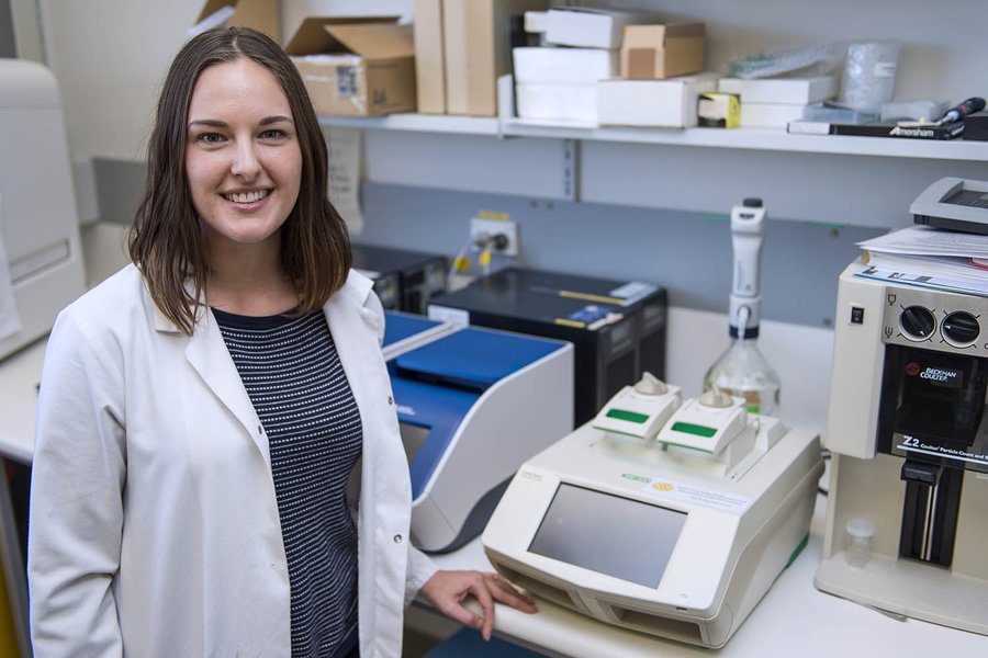
A scientist using a thermal cycler, the machine essential for performing PCR by precisely controlling temperature cycles.
The Core Principle: Cycling Through DNA Replication
PCR mimics the natural process of DNA replication in a test tube but focuses on a specific target region. It involves repeated cycles of temperature changes, facilitated by a machine called a thermal cycler (NCBI Bookshelf, 2025). Each cycle typically consists of three key steps:
- Denaturation: The reaction mixture is heated to a high temperature (usually around 94-98°C) to separate the two strands of the target DNA template.
- Annealing: The temperature is lowered (typically 50-65°C, depending on the primers) to allow short synthetic DNA sequences, called primers, to bind (anneal) specifically to the complementary sequences flanking the target region on each single strand.
- Extension (or Elongation): The temperature is raised again (usually to 72°C, the optimal temperature for the enzyme) to allow a heat-stable DNA polymerase enzyme (most famously Taq polymerase, isolated from the thermophilic bacterium *Thermus aquaticus*) to synthesize new DNA strands, starting from the primers and using the template strands as a guide (Byju’s, 2025; Thermo Fisher Scientific, 2025).
These three steps constitute one cycle. Repeating this cycle 25-40 times results in an exponential amplification of the target DNA sequence, as the newly synthesized strands also serve as templates in subsequent cycles (Microbe Notes, 2025).
Variations on a Theme: Types of PCR and Their Methods
While the basic principle remains the same, several variations of PCR have been developed to suit different applications:
- Conventional PCR: The original method, where amplification occurs over a set number of cycles. The final product (amplicon) is typically visualized and analyzed after the reaction is complete, often using gel electrophoresis to determine its size and approximate quantity.
- Real-Time PCR (qPCR): This technique allows for the detection and quantification of the amplified DNA *during* the reaction (in real-time). It uses fluorescent reporters (either DNA-binding dyes like SYBR Green or sequence-specific probes like TaqMan probes) whose signal increases proportionally to the amount of amplified product. This provides quantitative data much faster and with higher precision than conventional PCR (IntechOpen, 2023).
- Reverse Transcription PCR (RT-PCR): Used to amplify RNA targets (like mRNA for gene expression studies or RNA viruses like influenza or HIV). It involves an initial step where an enzyme called reverse transcriptase converts the RNA template into complementary DNA (cDNA). This cDNA then serves as the template for standard PCR amplification (Microbe Notes, 2025; Thermo Fisher Scientific, n.d.-b). Note: RT-PCR (Reverse Transcription PCR) is distinct from qPCR (quantitative PCR), although RT-qPCR combines both techniques.
- Multiplex PCR: Allows the simultaneous amplification of multiple different DNA targets in a single reaction tube by using multiple primer pairs.
- Nested PCR: Increases the specificity and sensitivity of amplification by using two sequential rounds of PCR. The second round uses primers designed to bind *within* the product amplified in the first round.
- Hot-Start PCR: A modification designed to reduce non-specific amplification that can occur at lower temperatures during reaction setup. It uses components (like a modified polymerase or sequestered magnesium) that prevent the reaction from starting until the high denaturation temperature is reached in the first cycle (Takara Bio, 2018).
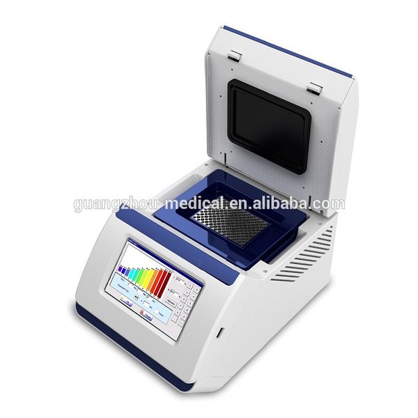
Real-Time PCR (qPCR) machines allow for quantification of DNA amplification as it happens.
Transformative Power: Applications of PCR
PCR's ability to amplify minute amounts of specific nucleic acid sequences has revolutionized countless areas:
- Clinical Diagnostics: Detecting infectious agents (viruses like SARS-CoV-2, HIV, HPV; bacteria like *Mycobacterium tuberculosis*), identifying genetic mutations associated with inherited diseases (e.g., cystic fibrosis, sickle cell anemia), cancer diagnosis and monitoring (NCBI Bookshelf, 2025; Assay Genie, 2025).
- Forensic Science: DNA fingerprinting for identifying individuals from trace biological samples (blood, hair, saliva) found at crime scenes or in paternity testing (NCBI Bookshelf, 2025).
- Molecular Biology Research: Gene cloning and sequencing, studying gene expression (using RT-qPCR), site-directed mutagenesis, genetic mapping, and evolutionary studies (PubMed, 2008).
- Agriculture and Food Science: Detecting genetically modified organisms (GMOs), identifying plant and animal pathogens, species identification.
- Environmental Science: Detecting microbial contamination in water or soil, monitoring biodiversity.
Guard Against Errors: Critical Precautions for Accurate PCR
The extreme sensitivity of PCR means that contamination is a major concern, potentially leading to false positive results. Rigorous precautions are essential:
- Prevent Contamination: This is the cardinal rule. Use separate, dedicated areas and equipment (pipettes, tube racks) for pre-PCR (reagent preparation, sample addition) and post-PCR (amplicon analysis) steps. Use aerosol-resistant filter tips for all pipetting. Wear gloves and change them frequently (Microbe Notes, 2025; Laboratory Equipment, 2015).
- Workflow: Maintain a unidirectional workflow, moving from pre-PCR to post-PCR areas, never the reverse (World Health Organization, 2018).
- Reagent Handling: Aliquot reagents into smaller volumes to avoid contaminating stock solutions. Keep enzymes and dNTPs (building blocks of DNA) stored correctly (usually frozen) and minimize freeze-thaw cycles. Prepare master mixes (containing buffer, dNTPs, polymerase, primers, water) on ice to maintain enzyme stability and reduce non-specific primer binding before the reaction starts (Takara Bio, 2018).
- Primer Design: Design primers carefully to be specific to the target sequence and avoid sequences that could lead to primer-dimers (primers annealing to each other) or non-specific binding elsewhere in the genome (Byju’s, 2025).
- Controls: Always include appropriate controls:
- Negative Control (No Template Control - NTC): Contains all reaction components except the DNA template. Amplification here indicates contamination.
- Positive Control: Contains a known template that should amplify, confirming the reaction components and conditions are working correctly.
- Internal Control (sometimes): A non-target sequence added to the sample and amplified with separate primers to check for PCR inhibition.
- Workstation Cleaning: Regularly clean benchtops, pipettes, and equipment with solutions like 10% bleach followed by 70% ethanol or specialized DNA-decontaminating solutions. UV irradiation of PCR hoods can also help sterilize surfaces (Takara Bio, 2018).
- Hot-Start Techniques: Employing hot-start PCR methods can significantly reduce non-specific amplification products formed at lower temperatures during setup (Takara Bio, 2018).
Comparing ELISA and PCR Techniques
A Visual and Tabular Overview
While both ELISA and PCR are powerful detection techniques, they target different molecules and operate on fundamentally distinct principles. The following radar chart provides a comparative visualization of different ELISA and PCR formats based on key performance characteristics. Note that these are generalized comparisons; actual performance can vary based on specific assay design and optimization.
This chart highlights relative strengths: Sandwich ELISA and qPCR generally offer the highest sensitivity and specificity. Direct ELISA is often faster and simpler but less sensitive. Conventional PCR provides good sensitivity but typically requires post-reaction analysis for results, whereas qPCR offers real-time quantitative data. Costs and throughput can vary significantly depending on the specific assay and scale of operation.
Key Differences Summarized
The table below provides a concise comparison of the fundamental aspects of ELISA and PCR.
| Feature | ELISA (Enzyme-Linked Immunosorbent Assay) | PCR (Polymerase Chain Reaction) |
|---|---|---|
| Primary Target | Proteins, Peptides, Antibodies, Hormones (Antigens/Antibodies) | DNA or RNA (Nucleic Acids) |
| Core Principle | Antigen-Antibody Binding + Enzymatic Signal Detection | Enzymatic Amplification of Target Nucleic Acid Sequence via Thermal Cycling |
| Detection Method | Colorimetric, Fluorometric, or Chemiluminescent Signal (proportional to amount) | Gel Electrophoresis (Conventional) or Real-Time Fluorescence Detection (qPCR) |
| Key Enzyme(s) | Enzyme Conjugate (e.g., HRP, AP) + Substrate | Heat-Stable DNA Polymerase (e.g., Taq), Reverse Transcriptase (for RT-PCR) |
| Sensitivity | High (pg/mL to ng/mL range typically) | Extremely High (can detect single copies of DNA/RNA) |
| Primary Use | Quantification/Detection of Proteins/Antibodies (Diagnostics, Research) | Detection/Quantification of specific DNA/RNA sequences (Genetics, Diagnostics, Forensics) |
| Major Concern | Cross-reactivity, Background Signal, Matrix Effects | Contamination (leading to false positives), Primer Specificity |
Visualizing the Concepts: ELISA & PCR Mindmap
Connecting the Key Ideas
To further illustrate the core components of each technique, the following mindmap provides a hierarchical overview of the principles, types, applications, and essential precautions associated with both ELISA and PCR.
(Colorimetric/Fluorometric)"] Types Direct Indirect Sandwich Competitive Applications Diagnostics["(HIV, Hormones, Allergens)"] Research["(Protein Quantification)"] Food Safety["(Allergen Detection)"] Pharma["(Drug Development)"] Precautions ["Reagent Quality"] ["Washing Steps"] ["Blocking"] ["Controls"] ["Sample Handling"] PCR Principle ["DNA/RNA Amplification"] ["Thermal Cycling"] Denaturation Annealing Extension ["Heat-Stable Polymerase"] Types Conventional Real-Time (qPCR) RT-PCR Multiplex Nested Applications Diagnostics["(Infectious Diseases, Genetic Disorders)"] Forensics["(DNA Fingerprinting)"] Research["(Gene Cloning, Expression)"] Agriculture["(GMO Detection)"] Precautions ["Contamination Control
(Strict Separation, Filter Tips)"] ["Primer Design"] ["Reagent Handling
(Master Mix, Storage)"] ["Controls (NTC, Positive)"] ["Workflow (Unidirectional)"]
This mindmap visually organizes the information presented earlier, showing how the fundamental principles lead to different variations (types), which enable diverse applications, and highlighting the critical precautions needed for reliable outcomes in both ELISA and PCR methodologies.
Watch and Learn: ELISA Principles Explained
A Video Overview of the Technique
For a visual explanation of how ELISA works, the following video provides a clear overview of the principle behind this widely used immunoassay. It demonstrates the key steps involved in detecting the presence of antigens or antibodies using enzyme-linked antibodies and a color-changing substrate, reinforcing the concepts discussed earlier.
This video illustrates the interaction between antigens and antibodies on the microplate surface and how the enzyme conjugate facilitates signal generation. Understanding these steps visually can help solidify comprehension of the ELISA process and its reliance on specific molecular interactions for detection.
Frequently Asked Questions (FAQ)
Common Queries about ELISA and PCR
Recommended Further Exploration
- What are the differences between Sandwich ELISA and Competitive ELISA sensitivity?
- How is Real-Time PCR (qPCR) used to quantify gene expression?
- Explain common troubleshooting steps for ELISA assays.
- Describe the role of primers and probes in qPCR.
References

 assets.thermofisher.com
Last updated April 27, 2025