
Unlocking Neural Secrets: The Frontier of Flexible Microelectrode Arrays
Exploring advancements, challenges, and future horizons in brain interface technology for recording and stimulation.
Key Insights
- Flexible probes minimize tissue damage: Ultra-thin, polymer-based probes offer better biocompatibility and long-term stability compared to rigid predecessors by reducing mechanical mismatch with brain tissue.
- Multifunctionality drives innovation: Integrating electrical recording/stimulation with optical (optogenetics) and microfluidic (drug delivery) capabilities in single probes enhances research and therapeutic potential.
- Overcoming key challenges is crucial: Addressing the foreign body response, ensuring signal fidelity amidst artifacts, improving insertion techniques for soft probes, and scalable manufacturing remain critical hurdles.
The Dawn of Neuro-Interfacing: MEAs and Neural Probes
Bridging Electronics and Biology
Microelectrode arrays (MEAs) and neural probes are sophisticated tools designed to interact directly with the nervous system. Implanted into specific brain regions or peripheral nerves, these devices allow researchers and clinicians to record electrical signals from individual neurons (spikes) or larger neural populations (local field potentials - LFPs) and to electrically stimulate neural tissue. This capability provides unparalleled insights into brain function and offers potential therapeutic avenues for neurological disorders. While rigid implantable stimulating electrodes have been available for years, recording microelectrode technology, especially for long-term, high-fidelity applications, is undergoing rapid evolution.
The Implantation Process
The implantation of MEAs and neural probes is a delicate surgical procedure. It involves accessing the target neural tissue and carefully inserting the probe. The design, materials, and surgical technique are critical to minimize tissue damage and ensure the probe's functionality. Traditionally, rigid materials like silicon were used, offering ease of insertion but often causing significant tissue reaction due to mechanical mismatch. The procedure requires precision to place electrodes accurately within target structures like the cortex or spinal cord.
The Rise of Flexibility: Revolutionizing Neural Interfaces
Next-Generation Probe Design
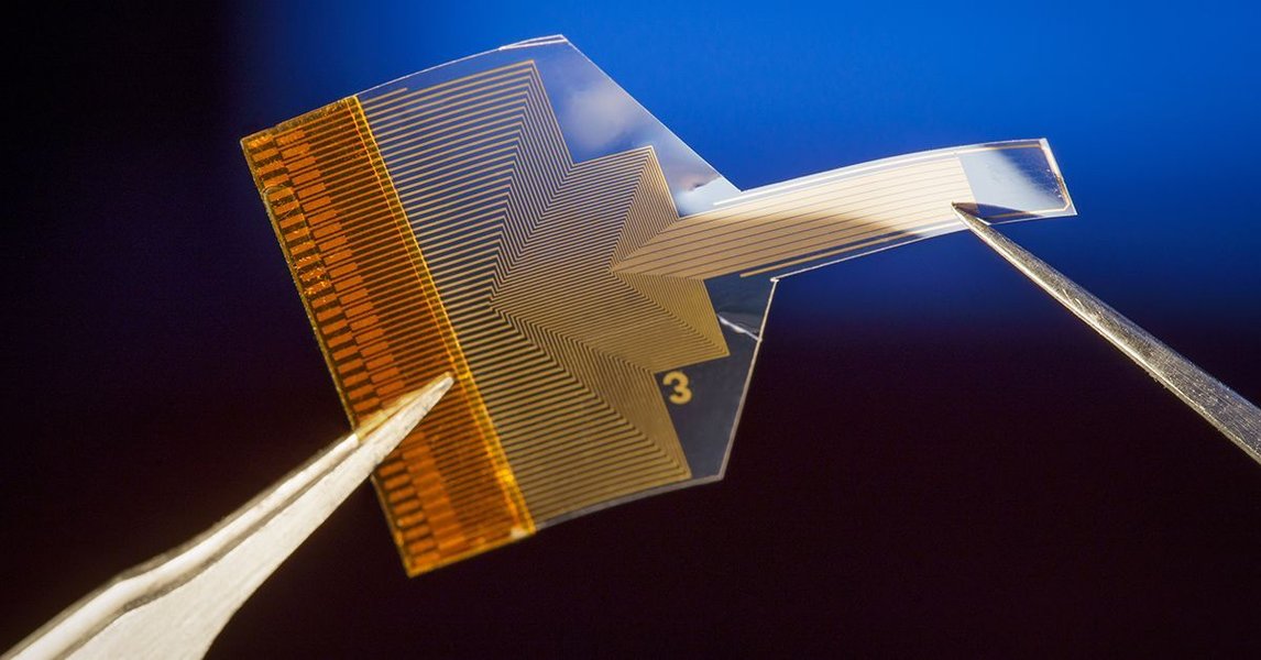
A major advancement in the field is the development of flexible neural probes. These probes, often fabricated from biocompatible polymers like polyimide, parylene-C, or polydimethylsiloxane (PDMS), offer significant advantages over their rigid counterparts. Their mechanical properties more closely match those of soft neural tissue, reducing chronic inflammation, glial scarring (the foreign body response), and tissue damage over long implantation periods. This improved biocompatibility leads to more stable and reliable recordings.

Design Innovations in Flexible Probes
- Ultra-thin Dimensions: Probes are becoming incredibly thin, sometimes just 8-14 micrometers in diameter (about one-fifth the width of a human hair), further minimizing invasiveness.
- High-Density Arrays: Advanced fabrication allows for packing numerous electrode sites (e.g., 32 or more) into small areas with very close spacing (down to 5 μm), enabling high-resolution mapping of neural activity.
- Multifunctional Integration: Modern probes often combine electrical recording/stimulation with other modalities. Optical fibers can be integrated for optogenetics (controlling neuron activity with light), and microfluidic channels allow for targeted drug delivery or sampling.
- Advanced Materials: Beyond base polymers, coatings like platinum (Pt), platinum-black (Pt-Black), or conductive polymers like PEDOT (poly(3,4-ethylenedioxythiophene)), sometimes doped with graphene oxide (GO), are used to lower electrode impedance, enhance charge transfer, and improve signal quality.
- Low-Cost Fabrication: Techniques like using standardized flexible printed circuit boards (PCBs) or 3D printing are being explored to make these sophisticated devices more accessible and scalable.
Listening and Talking to Neurons: Stimulation and Recording
Capturing and Modulating Neural Signals
The core functions of neural probes are electrical recording and stimulation. Recording involves detecting the minute voltage changes associated with neuronal firing (action potentials or spikes) and collective synaptic activity (LFPs). High signal-to-noise ratio (SNR) and stability over time are crucial for meaningful data. Electrical stimulation involves delivering controlled current pulses through the electrodes to activate or inhibit nearby neurons, aiming to modulate neural circuits or evoke specific responses.
Advancements focus on improving both aspects. Lowering electrode impedance through materials science (e.g., Pt-Black, PEDOT coatings) significantly enhances the ability to record faint neural signals. For stimulation, precision is key – delivering charge effectively to targeted neurons without causing tissue damage or eliciting widespread, non-specific activation. Multifunctional probes allow for sophisticated experiments, such as recording neural responses immediately following optogenetic stimulation.
Mapping the Neural Interface Landscape
Key Concepts in MEA Technology
The following mindmap provides a conceptual overview of the key areas involved in microelectrode array and neural probe technology, highlighting the interplay between device design, biological interaction, functional capabilities, and research challenges.
(Foreign Body Response)"] id4b["Signal Quality & Stability"] id4c["Stimulation Artifacts"] id4d["Mechanical Mismatch"] id4e["Insertion Difficulty (Flexible)"] id4f["Fabrication & Cost"] id4g["Long-Term Performance"] id5["Advancements & Future"] id5a["Novel Materials (PEDOT, Hydrogels)"] id5b["Advanced Fabrication (3D Printing)"] id5c["Artifact Reduction Techniques"] id5d["Wireless & Biodegradable Probes"] id5e["AI Integration"] id5f["Closed-Loop Systems"]
Navigating the Hurdles: Challenges in Neural Interfacing
Obstacles on the Path to Seamless Integration
Despite significant progress, the field faces substantial challenges that researchers are actively working to overcome. These issues impact the reliability, longevity, and effectiveness of neural probes.
Biocompatibility and the Foreign Body Response (FBR)
Perhaps the most significant hurdle is the biological response to the implanted device. The mechanical mismatch between even flexible probes and the ultra-soft brain tissue, along with the chemical nature of the implant materials, triggers an inflammatory response. This leads to the formation of glial scars—dense layers of astrocytes and microglia—around the probe. This encapsulation increases the distance between electrodes and neurons, raises impedance, degrades signal quality (lower SNR), and can eventually lead to device failure. Minimizing FBR is critical for long-term functionality.
Signal Quality and Stability
Maintaining high-quality, stable recordings over months or years remains difficult. Issues include:
- Low SNR: Faint neural signals can be easily lost in background noise, especially as FBR progresses.
- Electrode Impedance: High or unstable impedance hinders effective signal capture and charge injection for stimulation. Material degradation or biofouling can alter impedance over time.
- Motion Artifacts: Movement of the brain relative to the probe (e.g., due to breathing, heartbeat, or head movement) can introduce noise or cause signal loss, particularly problematic in dynamic areas like the spinal cord or peripheral nerves.
Stimulation Issues and Artifacts
Electrical stimulation presents its own set of challenges:
- Artifacts: The large electrical currents used for stimulation can overwhelm the sensitive recording amplifiers, creating large artifacts that obscure nearby neural activity. In optogenetics, light itself can induce photoelectrochemical (PEC) and photovoltaic (PV) effects at the electrode interface, generating artifacts that contaminate electrical recordings.
- Specificity: Precisely stimulating only the desired neuronal population without affecting neighboring cells is difficult.
- Tissue Damage: High stimulation currents can potentially damage surrounding tissue through electrochemical reactions or heating.
- Electrode Degradation: Repeated stimulation can degrade electrode materials, altering their properties and performance.
Mechanical and Insertion Challenges
While flexibility is advantageous for long-term implantation, it poses challenges during insertion. Flexible probes lack the rigidity to penetrate brain tissue easily and can buckle or bend. This necessitates temporary stiffeners (e.g., dissolvable coatings like PEG, or removable shuttles), adding complexity to the implantation procedure and potentially causing more initial tissue disruption.
Fabrication, Scalability, and Cost
Manufacturing high-density, multifunctional, flexible probes involves complex microfabrication processes. Ensuring consistent quality, high yield, and reducing production costs are essential for wider adoption in research and clinical settings.
Comparative Performance Analysis of Neural Probes
Visualizing Strengths and Weaknesses
Different types of neural probes offer varying performance characteristics. The radar chart below provides a comparative visualization based on key metrics. "Rigid Silicon" represents traditional probes. "Basic Flexible Polymer" refers to early-generation flexible probes. "Advanced Multifunctional Flexible" represents the cutting edge, incorporating features like high density, low-impedance coatings, and potentially optical/fluidic integration. Scores are relative estimates (higher is generally better, except for Insertion Difficulty and Cost where lower is better).
This chart highlights the trade-offs: while advanced flexible probes excel in biocompatibility, signal quality, and functionality, they currently face greater challenges in ease of insertion and fabrication cost compared to older technologies. Research aims to push the boundaries further, improving all aspects simultaneously.
Strategies for Advancing Neural Probe Research
Paving the Way for Better Brain Interfaces
Overcoming the aforementioned challenges requires a multi-pronged approach, integrating materials science, engineering, neuroscience, and advanced fabrication techniques. Key strategies include:
Material Innovations
- Developing novel polymers and composites with enhanced biocompatibility and mechanical properties that closely mimic brain tissue.
- Using advanced coating materials (e.g., PEDOT, Pt-Black, nanostructured surfaces) to lower impedance, improve charge injection capacity, and reduce FBR. Surface modification strategies aim to reduce microglial adhesion and neuronal cell death.
- Exploring bioactive coatings or drug-eluting materials to actively modulate the tissue response.
- Investigating biodegradable materials (e.g., silk fibroin) that provide initial support but dissolve over time, potentially reducing chronic FBR.
- Incorporating adaptive materials like hydrogels that change properties (e.g., soften upon hydration) after implantation.
Design and Fabrication Enhancements
- Optimizing electrode size, shape, and spacing for specific applications (e.g., single-unit vs. LFP recording).
- Developing sophisticated probe geometries (e.g., 3D structures, tissue-like scaffolds) using techniques like 3D printing or thermal drawing to improve tissue integration.
- Miniaturizing probes and increasing electrode density without compromising mechanical integrity or signal quality.
- Refining methods for integrating multiple modalities (electrical, optical, fluidic) seamlessly onto flexible substrates.
Improving Insertion Techniques
- Designing improved temporary stiffeners or bioresorbable insertion aids.
- Developing robotic-assisted or micro-actuated insertion systems for greater precision and minimal tissue damage.
- Utilizing transient lubricating coatings to ease insertion.
Signal Processing and Artifact Reduction
- Employing advanced algorithms for real-time noise cancellation and artifact removal (especially stimulation artifacts).
- Developing electrochemical modifications or specific electrode materials (e.g., PEDOT-GO) designed to suppress PEC/PV artifacts during optogenetic experiments.
- Implementing closed-loop systems where recorded neural activity informs and adjusts stimulation parameters in real time.
Scalability and Cost Reduction
- Leveraging scalable manufacturing processes like flexible PCB technology or roll-to-roll processing.
- Standardizing designs and materials to reduce variability and cost.
Summary of Improvement Strategies
The table below summarizes key areas and strategies for improving flexible neural probe design and performance:
| Area of Improvement | Strategies | Expected Outcome |
|---|---|---|
| Biocompatibility / FBR | Softer materials, bioactive coatings, biodegradable components, hydrogels | Reduced inflammation, less scarring, longer device lifespan |
| Signal Quality (SNR) | Low-impedance coatings (PEDOT, Pt-Black), optimized electrode design, noise-cancelling algorithms | Clearer neural signals, ability to detect weaker signals |
| Stimulation Fidelity | Artifact reduction techniques (material/algorithmic), precise electrode placement, closed-loop control | Accurate recording during stimulation, targeted neural modulation |
| Insertion Reliability | Temporary stiffeners, robotic assistance, lubricated coatings | Reduced tissue damage during implantation, successful placement of highly flexible probes |
| Functionality | Integration of optical/fluidic channels, high-density arrays | Multimodal interrogation/manipulation of neural circuits |
| Accessibility | Scalable fabrication (PCB, 3D printing), design standardization | Lower cost, wider availability for research and clinical use |
Future Frontiers: Project Ideas and Hypotheses
Pushing the Boundaries of Neurotechnology
Building on current knowledge, several exciting research directions and project ideas emerge:
-
AI-Powered Adaptive Probes
Idea: Develop probes with integrated low-power processing or wireless connection to AI algorithms for real-time analysis of neural signals and adaptive adjustment of stimulation parameters. Hypothesis: AI-driven closed-loop systems can optimize therapeutic stimulation for conditions like epilepsy or Parkinson's disease more effectively than pre-set parameters by responding dynamically to brain states. -
Fully Biodegradable Neural Interfaces
Idea: Design probes entirely from bioresorbable materials that function for a predetermined period (e.g., for post-operative monitoring or temporary therapy) and then safely dissolve, eliminating the need for removal surgery. Hypothesis: Transient biodegradable probes will minimize long-term FBR and reduce risks associated with permanent implants, making them suitable for short-term diagnostic or therapeutic applications. -
Wireless and Battery-less Operation
Idea: Create highly miniaturized probes powered wirelessly (e.g., via inductive coupling or ultrasound) and transmitting data wirelessly, eliminating tethered connections and bulky headstages. Hypothesis: Fully wireless probes will improve long-term biocompatibility by reducing device weight and strain, and enhance user comfort and mobility, making chronic monitoring more feasible. -
Hybrid Hydrogel-Polymer Probes
Idea: Fabricate probes combining a flexible polymer backbone for structural integrity and electrode routing with a hydrogel coating or matrix that swells in situ to better match tissue mechanics and potentially elute therapeutic agents. Hypothesis: Hybrid probes will offer superior tissue integration and reduced FBR compared to purely polymer probes due to the hydrogel's tissue-like properties. -
Artifact-Resistant Optoelectronic Probes
Idea: Engineer probes with novel electrode materials (e.g., specific transparent conductors or tailored coatings) and geometries specifically designed to minimize photoelectrochemical and photovoltaic artifacts during simultaneous optogenetic stimulation and electrical recording. Hypothesis: Optimized optoelectronic probes will enable artifact-free recording of neural activity immediately adjacent to sites of optical stimulation, providing clearer insights into optogenetically controlled circuits.
Visualizing Flexible Probe Technology in Action
A Glimpse into Ultra-Flexible Neural Interfaces
The following video showcases the development and potential of tiny, ultra-flexible neural probes. It highlights their minimal invasiveness and ability to interface with delicate neural tissues, representing a significant step forward in brain research capabilities. Understanding the physical characteristics and handling of these probes is essential for appreciating both their advantages and the associated implantation challenges.
As demonstrated, the extreme flexibility allows these probes to conform to the brain's natural movements, potentially reducing chronic inflammation and enabling longer-lasting, more stable recordings compared to rigid implants. This technology opens doors for studying neural circuits involved in learning, memory, and disease with unprecedented detail and duration.
Frequently Asked Questions (FAQ)
What is the 'foreign body response' (FBR) to neural implants?
The foreign body response is the natural reaction of biological tissue to any implanted foreign material. In the brain, this involves the activation of immune cells like microglia and astrocytes, leading to inflammation and the formation of a dense glial scar encapsulating the implant. This scar isolates the probe from the surrounding neurons, increasing electrical impedance and degrading the quality of recorded signals over time. It's a major challenge for the long-term stability of neural interfaces.
Why are flexible neural probes considered better than rigid ones?
Flexible probes, typically made from polymers like polyimide or parylene, have mechanical properties (stiffness) much closer to soft brain tissue than traditional rigid probes (often silicon-based). This reduced mechanical mismatch leads to less chronic tissue irritation, inflammation, and glial scarring (FBR). Consequently, flexible probes generally offer better long-term biocompatibility, leading to more stable and reliable neural recordings over extended periods (months to years).
What are stimulation artifacts and why are they a problem?
Stimulation artifacts are large, unwanted electrical signals that contaminate recordings during or immediately after electrical or optical stimulation. Electrical stimulation pulses themselves are much larger than neural signals and can saturate recording amplifiers. Optical stimulation (optogenetics) can cause photoelectrochemical or photovoltaic effects at the electrode-tissue interface, generating voltage artifacts. These artifacts can obscure or mimic real neural activity (spikes or LFPs), making it difficult or impossible to accurately study the direct effects of the stimulation on nearby neurons. Reducing or removing these artifacts is crucial for closed-loop systems and detailed circuit analysis.
How can researchers insert very flexible probes into the brain without them buckling?
Inserting highly flexible probes requires strategies to temporarily increase their stiffness during penetration of the brain tissue. Common methods include:
- Biodegradable Coatings: Coating the probe with a stiff, biocompatible material (like polyethylene glycol - PEG, or silk fibroin) that dissolves after insertion.
- Removable Stiffeners/Shuttles: Attaching the flexible probe to a rigid carrier (shuttle) that is withdrawn after the probe is in place.
- Guide Cannulas: Using a rigid tube (cannula) to guide the flexible probe to the target depth.
- High-Speed Insertion: Inserting the probe very quickly can sometimes overcome buckling forces, although this carries risks.
References
-
Design, fabrication, and implantation of invasive microelectrode arrays (MEAs) as in vivo brain-machine interfaces: A comprehensive review - ScienceDirect
- Double-Layer Flexible Neural Probe With Closely Spaced Microelectrode Sites for High-Density In Vivo Brain Recordings - Frontiers in Neuroscience
-
Flexible neural probes: current advantages, drawbacks, and future demands - NCBI PMC
-
Microelectrode arrays (MEAs): A critical review on neuronal interface systems and strategies to alleviate the foreign body response - NCBI PMC
- Implantable intracortical microelectrodes for recording neuronal action potentials: challenges and future directions - Nature Microsystems & Nanoengineering
-
Ultra-thin, flexible probe provides neural interface that’s minimally invasive and long-lasting - UC San Diego Today
- Flexible Neural Probes With Electrochemical Modified Microelectrodes for Artifact-Free Optogenetic Application - PubMed
Recommended Reading
Last updated April 14, 2025