
Uncovering the Precise Location of the Fovea: How Our Retina's Most Critical Region is Positioned
Understanding the exact horizontal and vertical coordinates of the eye's high-definition center and why it matters for your vision
Key Insights About Foveal Positioning
- Horizontal Placement: The fovea is positioned approximately 4 mm temporal (toward the temple) from the center of the optic disc.
- Vertical Alignment: It sits about 0.8 mm inferior (below) the center of the optic disc.
- Anatomical Significance: This precise positioning maximizes visual acuity by ensuring light falls directly on the area with the highest concentration of cone photoreceptors.
Anatomical Coordinates of the Fovea Centralis
The fovea centralis represents the most critical area for high-acuity vision in the human eye. Located within the center of the macula lutea in the retina, its precise positioning is essential for sharp central vision and color perception. Understanding its exact coordinates relative to other retinal landmarks provides valuable insights into visual processing and potential vision disorders.
Horizontal Position Relative to the Optic Disc
The fovea centralis is positioned temporally (toward the side of the head) from the optic disc. This horizontal displacement has been measured with remarkable consistency across multiple studies:
Linear Measurements
When measured in millimeters or micrometers, research consistently shows the fovea is positioned:
- Approximately 4 mm temporal to the center of the optic disc
- Specific studies have measured this distance as 4,147-4,357 μm (4.15-4.36 mm)
- The fovea is typically located at a distance of approximately 2.5 optic nerve diameters away from the optic disc
Angular Measurements
When expressed as an angular measurement (which accounts for variations in eye size):
- The horizontal angle typically ranges from 13.0° to 17.9° relative to the optic disc
- The mean angular displacement is approximately 15.5°
- This angular position remains fairly consistent between right and left eyes of the same individual
Vertical Position Relative to the Optic Disc
The vertical positioning of the fovea shows it is slightly inferior (below) the level of the optic disc:
Linear Measurements
- Approximately 0.8 mm inferior to the center of the optic disc
- Some studies report a narrower range of 552-568 μm (0.55-0.57 mm)
- Interestingly, gender differences have been observed, with men having the fovea positioned on average 57 μm more inferior than women
Angular Measurements
- Vertically, the angular position typically ranges from -3.65° to 0.65°
- The negative values indicate an inferior position relative to the optic disc
- The angle between the fovea and the center of the optic disc versus the horizon averages approximately -5.6° ± 3.3°
Factors Influencing Foveal Position
The exact position of the fovea can vary due to several factors, though it remains relatively consistent across healthy individuals:
Individual Variations
While the measurements above represent averages, individual variations do exist. These variations can be attributed to:
- Genetic factors influencing retinal development
- Age-related changes, with older individuals showing potentially greater vertical distances
- Axial length of the eye (the distance from cornea to retina)
- Refractive errors such as myopia (nearsightedness) or hyperopia (farsightedness)
Influence of Parapapillary Gamma Zone
The parapapillary gamma zone—an area adjacent to the optic disc where Bruch's membrane is absent—can influence foveal positioning:
- A wider temporal gamma zone may increase the horizontal disc-fovea distance
- An inferior gamma zone is associated with a more inferior vertical position of the fovea
- These correlations suggest developmental relationships between the optic disc, gamma zone, and foveal positioning during eye formation
Gender Differences
Interesting gender-related differences have been observed in foveal positioning:
- Men tend to have the fovea positioned approximately 57 μm more inferiorly than women
- This difference persists even after accounting for axial length differences
- The clinical significance of this gender difference is still being investigated
Visualizing Foveal Position Characteristics
Comparative Analysis of Foveal Position Attributes
The radar chart below illustrates the relationship between various aspects of foveal positioning, highlighting the relative importance and variability of each factor:
This chart demonstrates how horizontal positioning is more consistent than vertical positioning across individuals, and how both measurements have significant clinical relevance. The influence of parapapillary regions shows moderate impact on both horizontal and vertical positioning.
Structural Relationships of the Fovea
Anatomical Connections and Relationships
The mindmap below illustrates the key relationships between the fovea and surrounding ocular structures, highlighting its central role in vision and anatomical connections:
This mindmap demonstrates how the fovea's position relates to its structural features and function, as well as potential pathologies that can affect this critical region of the retina.
Visual Guide to Foveal Positioning
Anatomical Visualization of the Retina and Fovea
These images provide visual context for understanding the fovea's position within the eye:
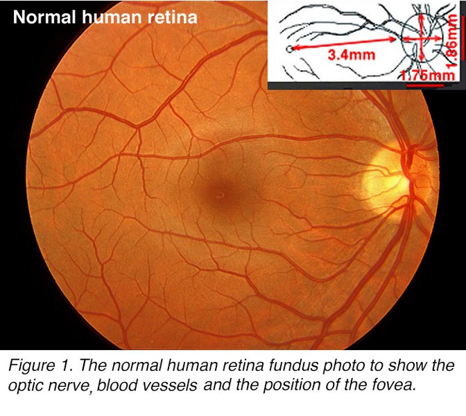


These images clearly demonstrate the fovea's position within the retina and its relationship to the optic disc and other ocular structures. Note how the fovea is located temporal (to the side) and slightly inferior (below) the optic disc, exactly matching the measurements discussed above.
Comparative Measurements of Foveal Position
Summary of Key Position Measurements
The following table summarizes the key measurements of foveal position from multiple research studies:
| Measurement Type | Horizontal Position | Vertical Position | Measurement Method | Notable Variations |
|---|---|---|---|---|
| Linear Distance | 4.0-4.4 mm temporal | 0.55-0.8 mm inferior | OCT/SLO Imaging | Slight variations between imaging devices |
| Angular Distance | 13.0°-17.9° (mean ~15.5°) | -3.65° to 0.65° | Fundus Photography | Minimal intraindividual difference between eyes |
| Relative to Optic Disc | ~2.5 optic disc diameters | ~0.5 optic disc diameters | Clinical Observation | Used as clinical reference point |
| Gender Comparison | Similar between genders | Men: ~57 μm more inferior | Comparative Studies | Difference persists after accounting for axial length |
| Age-Related Changes | Relatively stable | May increase with age | Cross-sectional Studies | Potentially related to retinal thinning with age |
Visual Understanding of the Fovea
Video Explanation of Foveal Anatomy and Position
This video provides an excellent visual explanation of the fovea's position within the retina and its critical role in vision:
This informative video explains the location of the optic disc, macula, and fovea, demonstrating their relative positions and the critical role the fovea plays in central vision due to its unique cellular composition and precise positioning.
Clinical Significance of Foveal Position
The precise position of the fovea has significant clinical implications:
Diagnostic Applications
- Abnormal foveal position (foveal ectopia) can indicate developmental anomalies or pathological conditions
- Changes in the disc-to-fovea distance may be associated with glaucomatous damage
- Precise knowledge of foveal position is essential for accurate interpretation of visual field tests
- Modern imaging techniques like OCT rely on accurate foveal localization for standardized measurements
Therapeutic Considerations
- Retinal laser treatments must account for precise foveal positioning to avoid damaging central vision
- Surgical interventions for macular pathology require detailed understanding of foveal position relative to surrounding structures
- Visual rehabilitation strategies for macular disease take into account the relationship between the fovea and preferred retinal locus
Research Implications
Understanding normal variations in foveal position helps researchers:
- Establish normative databases for retinal imaging
- Study developmental processes in the visual system
- Investigate potential genetic influences on foveal position
- Analyze the relationship between structural positioning and functional visual outcomes
Frequently Asked Questions
References
- Anatomy, Head and Neck, Eye Fovea - StatPearls - NCBI Bookshelf
- Fovea Centralis - Structure, Function, Anatomy, Location - Kenhub
- The Architecture of the Human Fovea - Webvision
-
Determining the Location of the Fovea Centralis via En-face SLO and Cross-sectional OCT Imaging - PMC
-
Location of Parapapillary Gamma Zone and Vertical Fovea Location - PMC
- Determination of the Location of the Fovea on the Fundus - ResearchGate
-
Fovea Centralis - Science Direct
Recommended Searches
Last updated April 8, 2025