
The Hidden Architecture of Vision: How Foveal Positioning Shapes What We See
Understanding the precise location of the eye's most critical region reveals why our vision works the way it does
Key Insights About Foveal Positioning
- Horizontal Offset: The fovea is positioned temporally from the optical axis, creating angle kappa which averages 5°
- Vertical Displacement: The fovea sits approximately 6.11° below the horizontal midline bisecting the optic disc
- Clinical Significance: This precise positioning affects visual acuity, measurement of eye conditions, and surgical outcomes
Understanding the Fovea and Optical Axis
The fovea centralis is a small depression in the retina responsible for our sharpest vision. At just 1.5mm wide with a central 0.5mm avascular zone, this tiny region houses the highest concentration of cone photoreceptors in the eye. The optical axis, meanwhile, is an imaginary line passing through the center of the pupil and the optic disc. Understanding the spatial relationship between these structures is crucial for ophthalmologists, vision scientists, and anyone studying visual perception.
The positioning of the fovea relative to the optical axis is not random but precisely calibrated for optimal visual function. This positioning creates multiple axes in the eye: the optical axis, the visual axis (connecting the object of regard to the fovea), and the pupillary axis (perpendicular to the cornea through the pupil center). These relationships form the foundation of how we perceive our visual world.
Anatomical Relationship in the Human Eye
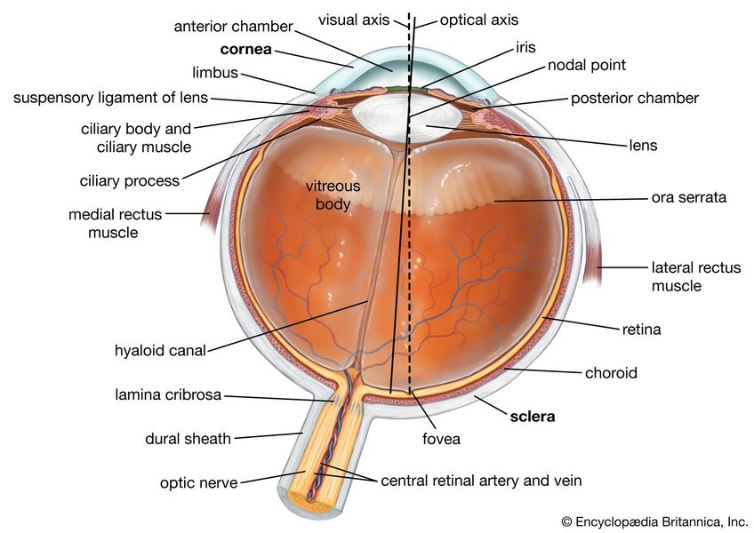
Horizontal Position of the Fovea
The fovea is positioned nasally from the center of the optic disc but temporally relative to the optical axis of the eye. This horizontal displacement creates what ophthalmologists call angle kappa.
Angle Kappa: The Key to Horizontal Positioning
Angle kappa is defined as the angle formed between the visual axis (which passes through the fovea and the nodal point of the eye) and the pupillary axis (which is essentially aligned with the optical axis). Due to the temporal positioning of the fovea relative to the optical axis, angle kappa is typically positive in most individuals.
Measurements and Variations
Research indicates that the horizontal angular distance between the fovea and the center of the optic disc generally averages around 15.5 ± 1.1 degrees. This positioning places the fovea nasal to the optic disc but temporal to the optical axis. Studies using spectral domain optical coherence tomography (SD-OCT) have examined this angle to assess interindividual variability, finding significant variations among individuals.
The horizontal displacement can be affected by various factors including:
- Axial length of the eye (particularly in myopia)
- Age-related changes in ocular structures
- Individual anatomical variations
- Ethnic differences in ocular anatomy
Vertical Position of the Fovea
In terms of vertical alignment, the fovea is typically positioned below the horizontal line that bisects the optic nerve head. This vertical positioning is critical for understanding visual field mapping and processing.
Vertical Displacement Measurements
Studies have consistently found that the fovea is positioned approximately 6.11 degrees (±3.32 degrees) below the horizontal midline passing through the center of the optic disc. This vertical offset creates what is known as the disc-fovea angle (DFA), which is the angle formed between a horizontal reference line and the line connecting the optic disc to the fovea.
Factors Affecting Vertical Position
The vertical position of the fovea relative to the optic disc can vary between individuals based on:
- Gender differences (one study found the fovea was 57 µm lower in men compared to women)
- Age-related changes in retinal structure
- Presence of ocular pathologies (particularly glaucoma)
- Developmental variations in embryonic eye formation
The radar chart above illustrates the differences in foveal positioning characteristics across normal, myopic (nearsighted), and hyperopic (farsighted) eyes. Note how myopic eyes tend to show greater displacement and variability in all measurements, while hyperopic eyes generally display less deviation from the optical axis.
Clinical Significance of Foveal Positioning
The precise position of the fovea relative to the optical axis has numerous clinical implications that affect both diagnosis and treatment of various ocular conditions.
Impact on Clinical Measurements
The alignment between the fovea and the optic disc is crucial for accurate measurements of retinal nerve fiber layer (RNFL) thickness. Modern diagnostic devices like Spectralis SD-OCT use the line connecting the fovea to Bruch's membrane opening as the axis of vertical symmetry temporal to the fovea. This alignment ensures precise measurements of retinal structures.
Relevance in Eye Conditions
The fovea-disc relationship is particularly significant in:
- Glaucoma assessment, where measurements of the optic nerve head and retinal nerve fiber layer thickness are crucial
- Strabismus surgery, where horizontal procedures can cause positional shifts affecting the posterior segment
- Refractive surgery planning, where angle kappa must be considered for optimal outcomes
- Macular degeneration evaluation, where foveal position may shift due to pathological changes
| Parameter | Normal Value | Clinical Significance | Measurement Method |
|---|---|---|---|
| Angle Kappa | 5.0° ± 2.3° | Critical for refractive and corneal surgery planning | Corneal reflection relative to pupil center |
| Disc-Fovea Distance | 4.9 ± 0.3 mm | Changes in myopia progression; reference for OCT measurements | Spectral Domain OCT or fundus photography |
| Disc-Fovea Angle | 7.8° ± 3.3° | Affects visual field interpretation; varies in glaucoma | Angle between horizontal line and disc-fovea line |
| Foveal Vertical Position | 6.11° below horizontal | Influences ganglion cell layer symmetry assessment | OCT measurement from disc horizontal midline |
| Fovea-BMO Axis Angle | -7.2° ± 3.9° | Reference for RNFL thickness measurements | SD-OCT measurement relative to Bruch's membrane opening |
Visualizing the Relationships
Visual Demonstration of Foveal Position
This video provides a comprehensive explanation of the various axes of the eye, including the visual axis that passes through the fovea and how it relates to the optical axis. Understanding these relationships is fundamental to grasping the spatial orientation of the fovea.
Understanding Through Imaging

The fovea centralis (shown in center) in relation to the optic disc and other retinal structures

Cross-sectional view showing the foveal depression and its layers
Frequently Asked Questions
References
-
Interindividual Variability in the Fovea-Disc Angle with Spectral Domain OCT - PMC
- Optical Axes and Angle Kappa - EyeWiki
- Position of the Fovea Centralis with Respect to the Optic Nerve Head - PubMed
- Elongation of the Disc-Fovea Distance in Myopic Eyes - Nature
- The Architecture of the Human Fovea - NCBI Bookshelf
-
Spatial Relationship Between Parapapillary Gamma Zone and Fovea - PMC
- Influence of Fovea-BMO Axis Angle on Macular Thickness - PubMed
Recommended Searches
Last updated April 8, 2025