
Frozen Section Analysis of Ovarian Masses
In-depth exploration of intraoperative diagnosis and its correlation with histopathological findings
Key Highlights
- High Diagnostic Accuracy: Frozen section analysis demonstrates high overall accuracy, aiding intraoperative decision-making.
- Surgical Guidance: The technique is pivotal in distinguishing benign, borderline, and malignant ovarian masses, thereby influencing surgical strategy.
- Correlation with Final Pathology: Strong concordance rates have been observed between frozen section evaluations and final histopathological diagnoses.
Introduction to Frozen Section Analysis
Frozen section analysis is a critical intraoperative tool that allows pathologists to rapidly examine tissue specimens and provide immediate diagnostic feedback during surgical procedures. Its primary use in the context of ovarian masses is to determine the nature of the lesion – whether it is benign, borderline, or malignant – which in turn directly affects the surgical management and patient outcomes. Given the time-sensitive nature of surgery, the accuracy and speed provided by the frozen section analysis are invaluable assets.
Ovarian masses can be complex due to their variable histology and potential heterogeneity. In such instances, frozen section analysis offers a snapshot of the lesion that can guide the surgeon on whether to pursue fertility-preserving measures or to proceed with radical resection and comprehensive surgical staging. The technique aims to optimize the balance between oncologic safety and the preservation of healthy tissue, which is especially pertinent in younger patients or those with fertility considerations.
Methodology of Frozen Section Analysis
Procedure Overview
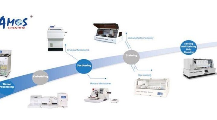
During surgery, once an ovarian mass is resected or biopsied, a small portion of the tissue is rapidly frozen. This frozen tissue is then cut with a microtome, stained, and examined under a microscope by a pathologist. The entire procedure typically takes between 15 to 30 minutes, allowing the surgeon to receive real-time feedback regarding the tumor's histological characteristics. Rapid processing is essential to ensure that the surgical intervention is not unduly prolonged.
Evaluation Criteria
The diagnostic assessment via frozen section considers the cellular architecture, the degree of differentiation, and any features that may indicate malignancy. Pathologists pay close attention to identifying complex areas such as regions suggestive of borderline pathology or mucinous changes, which can sometimes introduce diagnostic challenges. The frozen section findings are then compared with later, more detailed immunohistochemical profiling found in the final histopathological report.
Diagnostic Accuracy and Performance
Overall Performance
Numerous studies have reported high diagnostic accuracy rates ranging from approximately 90% to 97% when using frozen section analysis in ovarian masses. This accuracy rate reflects the technique’s utility in a wide range of ovarian pathologies. The high level of accuracy, particularly for benign tumors, makes it an essential component of intraoperative diagnostic strategies.
An overall accuracy nearing 97% has been documented, and some reports indicate that benign tumors are identified with a sensitivity as high as 99.2%. The ability to rapidly and reliably distinguish between benign lesions and malignant ones enables the surgical team to adjust operative plans promptly and appropriately.
Sensitivity and Specificity
The sensitivity and specificity measures for frozen section analysis vary according to the type of ovarian mass evaluated. For instance:
| Type of Tumor | Sensitivity | Specificity |
|---|---|---|
| Benign Tumors | ~99.2% | High (near 100%) |
| Borderline Tumors | ~88.5% | High specificity but may show variability due to diagnostic challenges |
| Malignant Tumors | ~83% to 89% | Near 100% |
These figures illustrate that while the method is highly reliable, some of the complexities associated with borderline and mucinous tumors can occasionally lead to underdiagnosis. However, despite these challenges, the consensus in clinical practice is that frozen section analysis remains a robust method for immediate intraoperative diagnostics.
Correlation with Final Histopathological Findings
Importance of Concordance
The correlation between frozen section diagnoses and final histopathological findings is of paramount importance, as it validates the accuracy of intraoperative decision-making. Concordance rates are generally high, with studies reporting rates in excess of 90% in many cases. This high concordance ensures that when a frozen section suggests a particular tumor type, the final diagnosis usually confirms these findings, thereby reducing the likelihood of overtreatment or undertreatment.
In instances where discrepancies occur, these often pertain to mucinous and borderline tumors which present with more heterogeneous features. The variability observed with these tumor types can be attributed to sampling errors or the intrinsic complexity of the tumor architecture. When a frozen section analysis does not capture a representative sample, slight deviations from the final diagnosis may occur. Nevertheless, continued refinement in technique and thorough sampling approaches have markedly improved diagnostic reliability.
Clinical Impact on Surgical Decision-Making
The immediate feedback from frozen section analysis plays a critical role in guiding surgical strategy. For example, a diagnosis of a benign ovarian cyst may lead to a conservative approach such as cystectomy, preserving ovarian tissue and, by extension, fertility. Conversely, the identification of malignant features necessitates a more radical approach, with comprehensive surgical staging including procedures like omentectomy, lymph node sampling, and peritoneal washings.
The ability to adjust surgical plans on the fly based on intraoperative findings not only improves patient outcomes but also minimizes the risk of reoperations. For patients with malignant ovarian tumors, timely and appropriate surgical management is essential for reducing metastatic spread and optimizing the prognosis. The use of frozen section analysis ensures that these decisions are based on the best available evidence in real-time.
Technical Considerations and Limitations
Procedure Limitations
While frozen section analysis offers many advantages, it is not without its limitations. For instance, the technique relies on obtaining an adequate sample from the tumor. In cases where the mass is large or heterogeneous, a non-representative sample may lead to diagnostic errors. Furthermore, the rapid freezing process can sometimes introduce artifacts in the tissue, making interpretation more challenging.
Certain types of ovarian tumors, especially those with mucinous components or borderline features, are known to be particularly problematic. The heterogeneity of these tumors means that only portions of the lesion might display distinct histological characteristics, thereby increasing the potential for misdiagnosis. To mitigate these issues, pathologists often rely on their expertise and may request additional sections if the initial sample is inconclusive.
Improving Diagnostic Accuracy
To enhance the overall accuracy of frozen section analysis, several strategies have been employed. Optimizing the sampling technique is vital: ensuring that multiple areas of the tumor are examined can help to capture its full heterogeneity. Moreover, continual quality control programs comparing the intraoperative frozen section with the final histopathological evaluation are essential. These comparative analyses allow for feedback and learning, which in turn helps refine interpretation guidelines and enhance the overall accuracy of the frozen section technique.
Enhanced imaging techniques and improved staining protocols have also contributed to reducing artifacts and improving tissue preservation. As technology advances, the integration of digital pathology and computer-assisted diagnostic tools holds promise for further increasing the reliability and speed of frozen section assessments.
Clinical Implications and Future Directions
Impact on Patient Management
The application of frozen section analysis in the management of ovarian masses represents a significant advancement in surgical pathology. Its high accuracy and rapid turnaround time afford surgeons the confidence to make immediate, informed decisions. This technique is particularly valuable in settings where preserving fertility is a priority. For young patients or those with unilateral ovarian involvement, a benign diagnosis from a frozen section can lead to fertility-sparing surgery.
Moreover, the use of frozen section analysis helps to avoid unnecessary extensive surgery in cases where the mass proves benign on intraoperative examination. This targeted approach minimizes patient morbidity associated with more radical surgical procedures and enhances overall recovery.
Advancements in Diagnostic Tools
Looking ahead, continued research into frozen section analysis is expected to yield even greater diagnostic precision. Future developments may include the integration of artificial intelligence and machine learning algorithms to aid in the rapid interpretation of histopathological slides. The utilization of digital scanners and image processing software may further reduce the occurrence of sampling errors, especially in borderline and heterogeneous tumors.
Additionally, the implementation of more advanced staining techniques and immunohistochemical markers during the frozen section procedure might enable pathologists to better differentiate between tumor subtypes. This evolution in technique not only will improve intraoperative diagnostics but also can provide crucial information regarding tumor biology and prognosis.
Integration of Frozen Section Analysis in Clinical Practice
Interdisciplinary Collaboration
The success of frozen section analysis in the diagnosis of ovarian masses is often a collaborative effort involving surgeons, pathologists, and radiologists. Effective communication among these professionals is crucial for ensuring that tissue samples are handled appropriately, and that the frozen section findings are accurately integrated into the overall clinical picture. This interdisciplinary approach ensures that decisions made in the operating room are comprehensive and based on the best available evidence.
For instance, when a borderline tumor is suspected, radiological imaging prior to surgery may signal irregularities that prompt the pathologist to request multiple samples. The surgeon, aware of these complexities, can then plan for additional intraoperative evaluations if necessary. This collaborative model exemplifies the forward-thinking approaches that modern medicine encourages in the face of diagnostic challenges.
Quality Control and Continuous Improvement
Ongoing quality control measures are integral to maintaining high standards in frozen section diagnosis. Regular audits comparing the frozen section results with final histopathological outcomes help identify areas for improvement. Such audits also contribute to developing standardized protocols, which can be particularly beneficial in training and quality assurance programs.
Training programs for residents and practicing pathologists emphasize the importance of recognizing subtle histological cues in the frozen section environment. Enhanced training not only fosters a deeper understanding of ovarian pathology but also ensures that practitioners are well-equipped to handle challenging cases, ultimately leading to better patient care.
Summary of Findings
Comprehensive Diagnostic Value
Frozen section analysis provides a swift, reliable method for evaluating ovarian masses during surgery. Its high overall accuracy and sensitivity for benign conditions are well-documented in clinical studies. Although limitations exist, particularly in the evaluation of mucinous and borderline tumors, careful sample selection and interdisciplinary collaboration greatly enhance diagnostic precision.
The method not only plays an essential role in guiding the surgical management of ovarian masses but also influences clinical outcomes through its timely feedback. The steady improvement in techniques and the integration of modern technologies promise to further solidify the role of frozen section analysis in oncologic surgery.
References
-
Frozen Section Analysis of Ovarian Masses - PMC
-
Diagnostic Accuracy in Ovarian Tumors - ScienceDirect
- Accuracy of Frozen Section Diagnosis - EJGO
-
Intraoperative Frozen Section Analysis - PMC
- Frozen Section Diagnosis Guide - Diagnostic Histopathology
Recommended Queries for Further Exploration
Last updated March 4, 2025