
Frozen Section Analysis of Ovarian Masses
Insights into Intraoperative Diagnosis and Correlation with Histopathology
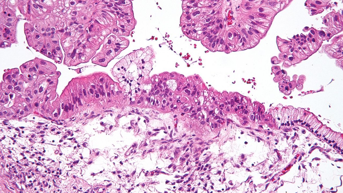
Key Highlights
- High Overall Accuracy: Studies report overall frozen section accuracy between 90% and 97.6%, making it a reliable intraoperative tool.
- Sensitivity Variations: Sensitivity is highest for benign tumors (up to 99.2%), with somewhat lower values for borderline and malignant lesions.
- Diagnostic Challenges: Particular difficulties exist with mucinous and borderline tumors, necessitating careful evaluation and comprehensive sampling.
Introduction to Frozen Section Analysis
Frozen section analysis is a rapid intraoperative diagnostic procedure employed during surgeries involving ovarian masses. Its primary purpose is to provide immediate information regarding the benign, borderline, or malignant nature of a tumor, thereby guiding the surgeon in real time during the operation. The procedure involves exposing a thin slice of the tissue, freezing it rapidly, and then examining it under a microscope so that critical surgical decisions can be made without delay.
This approach is indispensable in situations where the extent of surgical intervention — such as a conservative surgery preserving fertility or a more radical procedure — hinges on the preliminary diagnosis rendered during the operation. The correlation between frozen section findings and the final histopathological diagnosis (performed on more thoroughly processed, permanent tissue samples) has been the focus of numerous studies, underscoring its clinical utility and reliability. The comparison helps not only in validating the initial diagnosis but also in addressing any potential discrepancies that may arise.
Methodology and Diagnostic Process
Procedure for Frozen Section
During an ovarian mass surgery, a representative sample of the tissue is quickly excised and immediately transported to the pathology laboratory. There, the tissue is frozen using a cryostat, allowing it to be thinly sectioned. These sections are then stained and examined microscopically using standard histological techniques. The entire process takes only a few minutes, which is critical in preserving the operative timeline and ensuring that surgical decisions are made with as much current information as possible.
Correlation with Histopathology
After the initial intraoperative diagnosis via the frozen section, the tissue undergoes further processing with fixation, paraffin embedding, and subsequent sectioning and staining. This final histopathological assessment is more detailed and often reveals nuances that the rapid frozen section might not capture. The high concordance rates between the initial frozen section and the final permanent sections underscore the value of this technique.
However, it has been observed that while the overall agreement is high—ranging typically from 85% to 95%—certain tumor types, especially those with mucinous or borderline features, may show discrepancies. These differences often arise from sampling errors, tumor heterogeneity, or technical limitations associated with the rapid freezing process.
Accuracy and Sensitivity Metrics
Overall Accuracy
Multiple studies have established that the overall accuracy of frozen section analysis in evaluating ovarian masses is commendable, typically falling between 90% and 97.6%. This high percentage validates its use as an operative tool for rapid decision-making. In contexts where immediate surgical decisions are critical, this level of reliability provides significant reassurance to both surgeons and patients.
Sensitivity by Tumor Type
The sensitivity of frozen section diagnosis differs among tumor types:
- Benign Tumors: Sensitivity can soar up to 99.2%, meaning that nearly all benign ovarian masses are correctly identified during the intraoperative procedure.
- Borderline Tumors: Sensitivity is slightly lower, with reported values around 88.46%. Borderline tumors pose a diagnostic challenge primarily due to their intermediate pathological features.
- Malignant Tumors: Malignant lesions have a sensitivity of approximately 82.95%, highlighting that while the method is effective, there is still a potential risk for false negatives in more aggressive cases.
Factors Affecting Diagnostic Performance
Several factors contribute to the performance metrics of frozen section analysis:
- The expertise of the pathologist performing the examination.
- The quality and representativeness of the tissue sample taken.
- The inherent complexity of the tumor, especially in cases with mucinous or borderline pathology.
- The utilization of multiple sampling from various regions of the tumor can minimize the risk of misdiagnosis.
Clinical Utility and Implications for Patient Management
Guiding Surgical Decisions
The primary role of frozen section analysis is to facilitate real-time surgical decision-making. For example, if a frozen section indicates a benign tumor, the surgeon might opt for a conservative approach that spares ovarian tissue, particularly in reproductive-age women. Conversely, the identification of borderline or malignant lesions might necessitate more extensive surgical excision, potentially including bilateral salpingo-oophorectomy or even staging procedures.
The immediate results from frozen section analysis thus allow the surgical team to adapt the surgical plan on the fly — an advantage that is difficult to match with any other diagnostic modality.
Impact on Postoperative Management
The correlation between frozen section diagnosis and final histopathological results is not solely of intraoperative importance. It also plays a crucial role in planning postoperative treatment strategies. For instance, if the frozen section suggests a malignant tumor, even if final histopathology indicates a more complex picture, clinicians are better prepared to discuss and plan for adjuvant therapies such as chemotherapy or radiotherapy.
Additionally, when discrepancies occur between the two diagnostic approaches, the case is typically reviewed in a multidisciplinary team meeting to determine the best course of action. The consistent reevaluation of diagnostic procedures contributes significantly to the refinement of both intraoperative and final pathological assessments.
Diagnostic Challenges and Limitations
Borderline and Mucinous Tumors
Despite its high overall accuracy, frozen section analysis faces challenges, particularly in the evaluation of borderline and mucinous ovarian tumors. Such tumors often exhibit complex architectural patterns, where subtle cellular atypia can be easily missed in a frozen state. This may lead to underdiagnosis, where a potentially malignant lesion is initially classified as borderline or even benign.
The heterogeneity of these tumors adds another layer of complexity, as the sampled sections may not truly represent the most aggressive portion of the tumor. Therefore, while frozen section analysis is a powerful tool, it is imperative that pathologists remain aware of its limitations and approach ambiguous cases with a degree of caution.
Technical Considerations
Technical issues such as freezing artifacts or suboptimal quality of the frozen samples can impede accurate interpretation. Pathologists often mitigate these issues by utilizing multiple slides and performing a thorough gross examination of the tumor. Moreover, institutional protocols and standardized procedures for sampling and processing can further enhance diagnostic reliability.
Comparative Analysis: Frozen Section vs. Final Histopathology
Concordance Rates and Discrepancies
The final histopathological examination, which involves more elaborate tissue processing and extensive microscopic evaluation, typically confirms the diagnosis made by the frozen section analysis. Concordance rates between the two approaches generally range from 85% to 95%, indicating that in most cases, the intraoperative assessment is correct.
However, certain discrepancies do occur. They arise mainly due to the limitations inherent in the frozen section technique when dealing with complex tumors. For instance, while the sensitivity for benign tumors is exceptionally high, borderline and malignant lesions occasionally show a lower sensitivity. This is a critical consideration for surgical planning and patient counseling.
Summary Table of Key Diagnostic Metrics
| Parameter | Benign Tumors | Borderline Tumors | Malignant Tumors |
|---|---|---|---|
| Sensitivity | Up to 99.2% | Approximately 88.46% | Approximately 82.95% |
| Overall Accuracy | Between 90% and 97.6% | ||
Implications for Future Practice and Research
Enhancing Diagnostic Protocols
The integration of frozen section analysis with advanced imaging techniques and molecular diagnostics represents a promising path forward in optimizing intraoperative diagnosis. By combining these modalities, healthcare providers can obtain a more comprehensive assessment of the ovarian mass, reducing the likelihood of diagnostic errors.
Ongoing research continues to improve the accuracy of frozen section analysis through refining technical protocols and enhancing pathologist training. Institutions with high surgical volumes and dedicated pathology teams often publish their findings, which contribute to the body of literature vital for standardizing best practices.
Standardized Training and Protocols
It is imperative for surgical and pathology teams to engage in continuing education and training focused specifically on the challenges of frozen section analysis. Standardized protocols help minimize sampling errors, and multidisciplinary case reviews foster an environment of continuous quality improvement.
Clinicians are encouraged to remain updated about new research findings in this field, thereby incorporating emerging techniques and technologies that enhance the overall diagnostic process.
References
-
Role of Frozen Section in Intraoperative Assessment - NCBI PMC
-
Diagnostic Accuracy of Frozen Section in Ovarian Masses - Science Direct
-
Frozen Section Analysis in Ovarian Masses - NCBI PMC
- Intraoperative Frozen Section Findings - Taylor & Francis Online
- Systematic Review on Frozen Section Accuracy - BioMed Central
Recommended Queries
Last updated March 11, 2025