
Unlocking GATA3's Secrets: How Can We Understand Its Role in Young Women's Breast Cancer?
Exploring cutting-edge experiments to decipher GATA3's complex functions and impact on prognosis in younger patients.
Highlights
- Multi-Omics Approach: Integrating genomic, transcriptomic, epigenomic, and proteomic data from young patients is crucial to fully understand GATA3's regulatory networks and functional impact.
- Advanced Model Systems: Utilizing patient-derived cell lines, organoids, and xenografts specific to young breast cancer patients allows for more relevant functional testing of GATA3 mutations and expression changes.
- Clinical Correlation is Key: Linking experimental findings on GATA3 function (e.g., effects on proliferation, metastasis, therapy response) with detailed clinical data from young cohorts is essential for translating research into improved patient outcomes.
Grasping the GATA3 Enigma in Breast Cancer
Why this transcription factor matters, especially for younger women
GATA3 is a pivotal transcription factor essential for normal mammary gland development and maintaining the identity of luminal epithelial cells. In breast cancer, its role is intriguingly complex and context-dependent. Frequently mutated in breast tumors, particularly estrogen receptor (ER)-positive types, GATA3 can act as both a tumor suppressor, often associated with better prognosis and differentiated tumors, and potentially a driver of progression, depending on the specific mutation or cellular environment. Its expression is strongly linked to ER status and luminal subtypes, but alterations can occur across different breast cancer types.
Understanding GATA3's function is particularly important for young breast cancer patients (often defined as under 40 or 50 years old). Breast cancer in younger women can present differently, sometimes exhibiting more aggressive features or distinct molecular subtypes compared to older patients. Investigating how GATA3 expression levels and mutations specifically influence tumor biology, metastasis, and treatment response in this demographic requires tailored experimental strategies. By dissecting its functional effects, we can uncover potential vulnerabilities and therapeutic targets relevant to younger individuals facing this disease.
Key Experimental Strategies for Investigating GATA3 in Young Breast Cancer Patients
A multi-faceted approach from patient samples to molecular mechanisms
To comprehensively investigate the functional effects of GATA3 and its specific role in young breast cancer patients, a combination of advanced experimental techniques is necessary. These approaches span patient sample analysis, cellular and molecular studies, genomic profiling, and in vivo modeling.
Patient Cohort Characterization
Understanding GATA3 starts with the patients themselves, especially focusing on the unique characteristics of breast cancer in younger women.
- Sample Collection and Mutational Profiling: Collect tumor and matched normal tissues from well-characterized cohorts of young breast cancer patients. Perform next-generation sequencing (NGS), including whole-exome or targeted sequencing of the GATA3 gene, to identify the prevalence and types of mutations (e.g., frameshift, missense, intronic) specific to this age group.
- Clinical Data Correlation: Meticulously correlate GATA3 status (expression levels and mutation type) with detailed clinical information: age at diagnosis, tumor subtype (luminal A/B, HER2-positive, triple-negative), stage, grade, hormone receptor status (ER, PR, AR), Ki-67 proliferation index, treatment received, response rates, and long-term outcomes like disease-free and overall survival. This helps stratify risk and identify prognostic significance specific to young patients.
- Immunohistochemistry (IHC) and Spatial Analysis: Use IHC on tumor tissue microarrays (TMAs) or whole sections from young patients to assess GATA3 protein expression levels and subcellular localization. Correlate these findings with histological features and the tumor microenvironment, including immune cell infiltration patterns (e.g., T cells, macrophages). Spatial transcriptomics or proteomics can further map GATA3 activity within the tumor architecture.
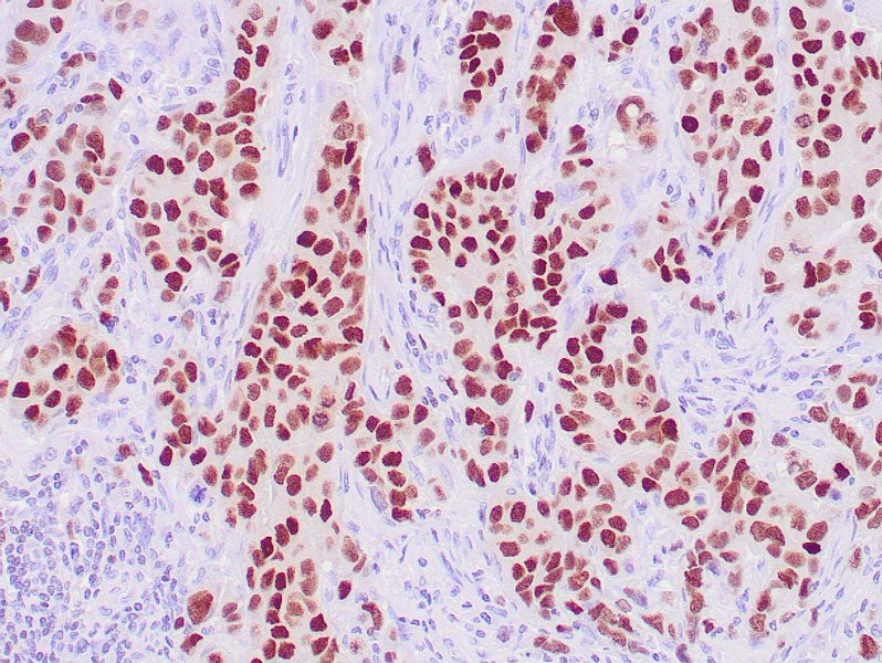
Immunohistochemical staining demonstrating nuclear GATA3 expression in breast carcinoma cells.
Cellular and Molecular Functional Assays
In vitro models allow for direct manipulation of GATA3 to observe its functional consequences.
- Patient-Derived Models: Establish patient-derived cell lines (PDCs) or organoids (PDOs) from fresh tumor tissues of young breast cancer patients harboring specific GATA3 mutations or expression profiles. These models better recapitulate the original tumor's heterogeneity and characteristics.
- Gene Editing and Expression Modulation: Utilize CRISPR/Cas9 technology to precisely introduce specific GATA3 mutations identified in young patients (e.g., R330fs, zinc finger mutations) into relevant breast cancer cell lines (e.g., MCF-7, T47D, MDA-MB-231) or patient-derived models. Alternatively, use RNA interference (siRNA/shRNA) for GATA3 knockdown or lentiviral vectors for stable overexpression of wild-type or mutant GATA3.
- Assessing Phenotypic Effects: Conduct a battery of functional assays on these engineered cells:
- Proliferation assays: (e.g., MTT, Ki67 staining) to measure growth rates.
- Migration and invasion assays: (e.g., wound healing, Transwell assays with Matrigel) to assess metastatic potential.
- Apoptosis assays: (e.g., Annexin V staining, Caspase activity) to evaluate cell death pathways.
- Epithelial-Mesenchymal Transition (EMT) analysis: Assess markers (e.g., E-cadherin, Vimentin, ZEB1/2) via Western blot, qPCR, or immunofluorescence, as GATA3 is known to suppress EMT.
- Investigating Hormone Receptor Interactions: Given GATA3's strong link to ER signaling and potential interactions with the Androgen Receptor (AR), investigate how GATA3 status influences the expression and activity of these receptors, particularly relevant for hormone-sensitive subtypes common in some young patients. Use reporter assays, Western blotting, and study responses to hormonal therapies (e.g., tamoxifen, fulvestrant).

Microscopic view illustrating mammary gland morphogenesis, a process regulated by GATA3.
Genomic and Epigenomic Investigations
Delving into how GATA3 regulates gene expression at a fundamental level.
- Transcriptional Profiling: Perform RNA sequencing (RNA-Seq) on tumor samples from young patients stratified by GATA3 status, or on cell models with manipulated GATA3. Analyze differential gene expression and perform pathway enrichment analysis (e.g., GSEA) to identify biological processes regulated by GATA3 (e.g., differentiation, cell cycle, immune response).
- Mapping GATA3 Binding Sites: Conduct Chromatin Immunoprecipitation sequencing (ChIP-Seq) for GATA3 in relevant cell models or primary tissues. Comparing binding profiles between wild-type and mutant GATA3, or high vs. low GATA3 expression, reveals direct target genes and how mutations might alter DNA binding specificity or occupancy.
- Epigenetic Landscape Analysis: Integrate ChIP-Seq data with assays for epigenetic marks (e.g., H3K27ac for active enhancers, H3K4me3 for active promoters, ATAC-seq for chromatin accessibility) to understand how GATA3 interacts with the chromatin landscape to regulate gene expression.
Protein-Level Investigations
Understanding GATA3 function also involves its interactions with other proteins.
- Identifying Protein Interaction Networks: Use techniques like co-immunoprecipitation followed by mass spectrometry (Co-IP/MS) or proximity ligation assays (PLA) to identify proteins that interact with wild-type and mutant GATA3 in young patient-derived models. This can reveal crucial co-factors or downstream effectors (e.g., interactions with ER, AR, p53, NuRD complex).
- Validating Key Interactions: Confirm significant interactions identified through screening using targeted Co-IP and Western blotting or PLA in multiple model systems.
In Vivo Modeling
Testing GATA3's role in a more complex biological system.
- Xenograft and PDX Models: Inject human breast cancer cells (engineered lines or PDCs) with defined GATA3 status into immunocompromised mice (e.g., NOD/SCID). Patient-derived xenografts (PDXs) established directly from young patients' tumors offer a high-fidelity model.
- Assessing Tumor Growth and Metastasis In Vivo: Monitor primary tumor growth rates using caliper measurements or imaging. Assess metastatic spread to distant organs (e.g., lungs, liver, bone) through bioluminescence imaging (if cells are tagged) or histological analysis post-mortem. Compare outcomes between groups with different GATA3 genotypes/expression levels.
Investigating Therapeutic Relevance
Connecting GATA3 function to treatment strategies.
- Therapy Response Assays: Treat cell lines, organoids, or xenograft models representing young patient tumors (with varying GATA3 status) with standard-of-care therapies (chemotherapy, endocrine therapy, targeted agents like CDK4/6 inhibitors) or novel drugs. Measure sensitivity or resistance (e.g., IC50 values, tumor regression).
- Drug Screening and Synthetic Lethality: Perform high-throughput screening using compound libraries on cells with specific GATA3 mutations or low expression to identify drugs that show selective efficacy. Explore potential synthetic lethal interactions where targeting another pathway becomes effective only when GATA3 function is altered.
Exploring the Immune Context
GATA3 also plays roles in immune regulation, which is critical within the tumor microenvironment.
- GATA3 and the Tumor Immune Microenvironment: Analyze the correlation between GATA3 expression/mutation status in tumor cells and the composition/phenotype of tumor-infiltrating immune cells (TILs) in samples from young patients using multiplex IHC/IF or flow cytometry. Investigate if GATA3 influences the secretion of cytokines or chemokines that modulate immune cell recruitment and function (e.g., its known role in Th2 differentiation).
Integrating Data for a Comprehensive View
Visualizing the multi-dimensional nature of GATA3 research
Understanding GATA3's role requires integrating insights from various experimental dimensions. The radar chart below illustrates a conceptual evaluation of different experimental approaches based on key research parameters. This helps visualize the strengths and potential challenges associated with each method when applied to studying GATA3 in young breast cancer patients. For instance, while in vivo models offer high biological relevance, they can be resource-intensive. Conversely, in vitro assays allow for high-throughput screening but may lack physiological complexity. Combining these approaches provides the most robust understanding.
Mapping the Experimental Landscape
Visualizing the interconnected approaches to GATA3 research
The study of GATA3 in young breast cancer patients involves a network of interconnected experimental strategies. This mindmap visualizes the main research domains and the specific techniques employed within each. It highlights how findings from patient data analysis inform functional studies in vitro and in vivo, which are further deepened by genomic, epigenomic, and proteomic investigations, ultimately aiming to uncover therapeutic vulnerabilities.
(Tumor, Normal, Blood)"] id1b["Clinical Data Correlation
(Age, Subtype, Outcome)"] id1c["GATA3 Mutational Analysis
(NGS, Sequencing)"] id1d["Immunohistochemistry (IHC)
(Protein Expression, Location)"] id1e["Spatial Analysis
(TME Context)"] id2["In Vitro Functional Assays"] id2a["Cell Line Models
(Established, Patient-Derived)"] id2b["Organoid Cultures (PDOs)"] id2c["Genetic Manipulation
(CRISPR, RNAi, Overexpression)"] id2d["Phenotypic Assays
(Proliferation, Invasion, EMT, Apoptosis)"] id2e["Hormone Receptor Studies
(ER/AR Signaling)"] id3["Genomic & Epigenomic Analysis"] id3a["Transcriptomics (RNA-Seq)
(Gene Expression, Pathways)"] id3b["ChIP-Seq
(GATA3 Binding Sites, Target Genes)"] id3c["Epigenetic Profiling
(Histone Marks, Accessibility)"] id4["Protein-Level Studies"] id4a["Interaction Mapping
(Co-IP/MS, PLA)"] id4b["Interaction Validation
(Western Blot)"] id4c["Proteomics
(Expression Levels)"] id5["In Vivo Modeling"] id5a["Xenografts
(Cell Line, PDX)"] id5b["Tumor Growth Monitoring"] id5c["Metastasis Assessment"] id6["Therapeutic Relevance"] id6a["Therapy Response Assays
(In Vitro / In Vivo)"] id6b["Drug Screening"] id6c["Synthetic Lethality"] id7["Immune Microenvironment"] id7a["Immune Cell Profiling
(Flow Cytometry, Multiplex IF)"] id7b["Cytokine/Chemokine Analysis"] id7c["GATA3 Role in Immune Cells"]
Visualizing GATA3 Detection
Understanding immunohistochemistry for GATA3 analysis
Immunohistochemistry (IHC) is a cornerstone technique for assessing GATA3 protein expression in clinical samples. It helps pathologists determine the presence, level, and location of GATA3 within tumor cells, providing valuable diagnostic and prognostic information. Understanding how GATA3 is visualized and interpreted in tissue sections is crucial for correlating its expression with clinical features and experimental findings. The following video provides insights into the significance of GATA3 as a marker in pathology, which is directly relevant to the IHC experiments discussed.
This video discusses GATA3 as an IHC marker, primarily focusing on its utility in distinguishing breast and urothelial carcinomas. While not specific to young patients, it explains the principles of using GATA3 staining in diagnosis, highlighting its sensitivity and the importance of interpretation in the context of other markers. This foundation is essential for designing and interpreting IHC experiments aimed at understanding GATA3's role in specific patient populations like young women with breast cancer.
Comparing Experimental Approaches
A summary of key techniques and their applications
Different experimental techniques provide unique insights into GATA3's function. This table summarizes some of the core methods discussed, outlining their primary focus, strengths, limitations, and specific relevance to investigating GATA3 in the context of young breast cancer patients.
| Technique | Primary Focus | Strengths | Limitations | Relevance to GATA3 in Young Patients |
|---|---|---|---|---|
| NGS / Targeted Sequencing | Identifying GATA3 mutations | Comprehensive mutation detection | Doesn't directly measure function; requires bioinformatics expertise | Identifies specific mutations prevalent in young cohorts for functional studies |
| Immunohistochemistry (IHC) | Protein expression & localization | Clinically established; spatial context | Semi-quantitative; antibody variability | Correlates GATA3 protein levels with clinical features and subtypes in young patients |
| CRISPR/Cas9 Gene Editing | Creating specific GATA3 alterations (KO, KI) | Precise genetic modification | Off-target effects possible; requires cell model validation | Allows direct testing of patient-specific mutation effects on cell behavior |
| RNA Sequencing (RNA-Seq) | Global gene expression profiling | Identifies downstream pathways; unbiased | Measures RNA, not protein/activity; complex data analysis | Reveals transcriptional programs regulated by GATA3 in young patient context |
| ChIP Sequencing (ChIP-Seq) | Mapping genome-wide protein-DNA binding | Identifies direct target genes | Technically demanding; antibody quality crucial | Maps where wild-type vs. mutant GATA3 binds in the genome |
| Patient-Derived Organoids (PDOs) | 3D culture recapitulating tumor features | High patient relevance; drug testing platform | Technically challenging to establish; variable success rates | Models GATA3 function and therapy response in a system closer to young patient tumors |
| Xenografts / PDX Models | In vivo tumor growth & metastasis | Studies tumor behavior in a living system | Expensive; time-consuming; immunocompromised host limitations | Tests the impact of GATA3 status on tumor progression and treatment response in vivo |
| Co-Immunoprecipitation / Mass Spectrometry (Co-IP/MS) | Identifying protein interaction partners | Unbiased discovery of interactome | Can identify non-specific binders; requires validation | Uncovers GATA3's functional networks and how mutations might alter them |
Frequently Asked Questions
Quick answers to common questions about GATA3 research
Why is GATA3 research particularly important for young breast cancer patients? ▼
Breast cancer in young women (often defined as <40 or <50 years) can have distinct characteristics, sometimes being more aggressive or presenting as different subtypes (like triple-negative) compared to cancer in older women. GATA3 plays a key role in differentiation and is linked to hormone receptor status, both factors that can influence tumor behavior and treatment response. Understanding how GATA3 mutations or expression levels specifically impact cancer biology in this younger demographic is crucial for developing age-appropriate diagnostic markers, prognostic tools, and targeted therapies.
Can GATA3 mutations cause breast cancer? ▼
GATA3 mutations are frequently found *in* breast tumors, particularly ER-positive ones, but they are generally considered somatic mutations (acquired during a person's lifetime within the tumor cells) rather than germline mutations that cause inherited cancer susceptibility. While these mutations alter GATA3's function – sometimes disabling its tumor suppressor activity or even conferring tumor-promoting properties – they are typically part of a larger set of genetic changes driving cancer development, rather than the sole initiating cause.
What are the main challenges in studying GATA3 function experimentally? ▼
Several challenges exist. Firstly, GATA3 has a dual role (tumor suppressor vs. oncogene) depending on the mutation type and cellular context, making interpretation complex. Secondly, studying its function requires sophisticated models (like patient-derived organoids or xenografts) that accurately reflect human tumor biology, which can be technically difficult and resource-intensive. Thirdly, dissecting its downstream effects involves integrating multiple data types (genomics, proteomics, etc.). Finally, translating experimental findings into clinical applications requires robust validation in large, well-annotated patient cohorts, particularly for specific subgroups like young patients.
How can patient-derived models help in GATA3 research for young patients? ▼
Patient-derived models, such as cell lines (PDCs), organoids (PDOs), or xenografts (PDXs), are created directly from a patient's tumor tissue. For young breast cancer patients, these models can capture the specific genetic background, mutation profile (including GATA3 status), heterogeneity, and potentially unique biological characteristics of their tumors better than generic, long-established cell lines. This allows researchers to test the functional consequences of GATA3 alterations and screen for drug responses in a system that more closely mimics the actual patient's disease, increasing the translational relevance of the findings.
Recommended Further Exploration
- What are the different types of GATA3 mutations found in breast cancer and how do they affect protein function?
- How does GATA3 expression differ between breast cancer subtypes, particularly in younger versus older patients?
- Investigate the interaction between GATA3 and estrogen receptor signaling pathways in breast cancer therapy response.
- Explore the role of GATA3 in regulating the tumor immune microenvironment in breast cancer.
References
Last updated May 3, 2025