
Innovative 3D Printing Models Transforming Heart Drug Screening
How advanced 3D printed heart models accelerate personalized cardiac drug testing
Key Highlights
- Personalized and Patient-Specific Models: Tailored replicas produce accurate anatomical detail, mimicking patient-specific heart characteristics.
- Integration with Heart-on-a-Chip Platforms: Combines 3D bioprinting with microfluidic technology for real-time drug assessment.
- Revolutionary Drug Screening Efficiency: Enabling high-throughput testing and reducing reliance on animal models, improving overall therapeutic safety.
Introduction to 3D Printing Technology for Heart Drug Screening
In recent years, 3D printing technology has profoundly impacted biomedical research and drug development. Its application in creating heart models for drug screening is one of the most promising advances in personalized medicine. With the ability to fabricate highly detailed and anatomically accurate heart replicas, researchers and clinicians can test candidate drugs on organ-specific models that replicate the complex cardiac physiology. This innovation is not only expediting the drug development process but is also reducing the need for animal testing, thereby providing more reliable predictions of human responses.
Technology Behind 3D Printed Heart Models
The foundation of using 3D printing in cardiac drug screening lies in its capacity to create models that mimic the intricacy of heart tissue and its structures. This technology, especially when combined with bioprinting techniques and heart-on-a-chip platforms, enables the production of controlled environments where researchers can study drug effects in parallel, ensuring both efficacy and safety.
3D Bioprinting and Its Mechanisms
3D bioprinting extends beyond standard 3D printing by integrating living cells, biomaterials, and growth factors in the printing process. Approaches such as the freeform reversible embedding of suspended hydrogels (FRESH) have been instrumental in producing soft, flexible models of cardiac tissue. The FRESH technique allows for the precise deposition of collagen and other biocompatible materials in a supportive hydrogel, ensuring that the printed heart tissues maintain mechanical and functional properties closely resembling native tissues.
FluidForm Bio’s FRESH Bioprinting Technique
Researchers adopting the FRESH method utilize a scaffold-based approach that permits the fabrication of intricate vascular networks as well as flexible heart muscle constructs. Derived from human induced pluripotent stem cell (hiPSC)-cardiomyocytes, these models are instrumental in studying the dynamic responses of heart tissue to drugs. The ability to print heart tissues that closely mimic the native structure provides a robust platform for both testing and understanding cardiac behavior under various physiological and pathophysiological conditions.
Heart-on-a-Chip Systems
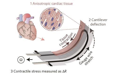
Heart-on-a-chip platforms have emerged as an important adjunct to 3D printing by integrating microfluidic channels and biosensors within the printed constructs. These systems allow continuous monitoring of cardiac tissue function, including contractility, electrophysiological properties, and metabolic activity. The integration of real-time biosensors offers immediate feedback on drug-induced changes, thereby enhancing the precision of toxicity and efficacy studies.
Benefits of 3D Printed Heart Models in Drug Screening
The use of 3D printed heart models in drug screening presents numerous advantages over traditional methods. These benefits not only help in accelerating the drug discovery process but also increase the predictive accuracy of therapeutic safety and effectiveness. Here we detail some of the significant benefits:
Anatomical Accuracy and Customization
3D printed heart models offer unparalleled anatomical precision, capturing detailed structures such as coronary arteries, heart valves, and tissue layers. These models are often patient-specific, created using imaging data such as MRI or CT scans. This customization, or “personalization”, allows researchers to tailor experiments to the specific characteristics of an individual's heart. Such personalized models can reveal unique drug responses and potential adverse reactions, leading to more tailored and effective therapeutic interventions.
Patient-Specific Replicas
Patient-specific models are becoming an essential component of modern drug screening. By incorporating patient-derived cells, scientists generate models that retain the genetic, structural, and functional nuances of individual hearts. This degree of customization makes it possible to predict not only therapeutic success but also potential side effects, thereby narrowing the gap between preclinical trials and clinical outcomes.
High-Throughput Screening and Efficiency
High-throughput screening is a critical advantage of using 3D printed heart models in pharmaceutical research. The ability to rapidly produce multiple models allows researchers to test thousands of compounds in parallel. This high-throughput capability not only speeds up the drug discovery process but also makes it more cost-effective.
Rapid Prototyping and Automation
The mechanization of the 3D printing process coupled with robotic systems has enabled laboratories to continuously generate and assess drug reactions using miniature heart models. This automation is instrumental in rapidly identifying promising drug candidates and discarding compounds with adverse effects before they proceed to more expensive and complex clinical trials.
Enhanced Drug Testing and Toxicity Assessment
One of the most significant impacts of 3D printed heart models is in the realm of drug toxicity assessment. Traditional methods often rely on animal studies, which may not faithfully replicate human physiology, leading to challenges in accurately predicting human responses. In contrast, 3D printed heart models, integrated with heart-on-a-chip platforms, can mimic physiological conditions such as blood flow and tissue interactions.
Real-Time Monitoring and Biosensor Integration
Integrated biosensors within the printed tissues provide continuous monitoring of key cardiac parameters such as electrical activity and contractility. This real-time feedback is critical for understanding the mechanistic effects of drug interactions and promptly identifying signs of cardiotoxicity. Such detailed insights facilitate rapid iterations in drug screening, ultimately leading to safer and more effective cardiovascular therapies.
Challenges and Limitations
Despite significant advances, there remain challenges that researchers must address in 3D printed heart models for drug screening. Some limitations include achieving a sufficient cell density to mimic native tissues fully, replicating the dynamic conditions of blood flow, and maintaining the long-term viability of printed cardiac tissues.
Technological Constraints
One of the primary challenges is the technical complexity involved in printing multi-material and multi-layered heart models. Achieving the detailed structure of myocardial fibers, coronary vasculature, and connective tissues requires high-resolution printing technology. Although current techniques such as FRESH have significantly improved the fidelity of printed tissues, further refinements are needed to replicate the full functionality of the native heart.
Material and Structural Limitations
Advanced biomaterials capable of supporting cellular growth while replicating the mechanical properties of heart tissue are in constant development. Researchers are experimenting with combinations of hydrogels, collagen-based scaffolds, and synthetic polymers to improve the durability and function of printed models. While progress in these areas is promising, continuous advances are required to overcome challenges such as tissue thickness and mass transport limitations, which are essential for long-term tissue viability.
Integration of Complex Physiological Parameters
Integrating complex variables, such as the intricate signaling pathways and biomechanical forces present in a live heart, is a formidable task. Although heart-on-a-chip systems offer great promise, the synchronization of multiple physiological parameters – including pressure dynamics, electrical signaling, and metabolic responses – remains a work in progress. Addressing these challenges is critical for further enhancing the predictive power of 3D printed models.
Comparative Analysis: Traditional Methods vs. 3D Printed Models
To appreciate the transformative potential of 3D printed heart models, it is useful to compare them with traditional methodologies. Conventional drug screening often relies on cell cultures or animal models. These methods, while historically valuable, have several drawbacks including poor replication of human cardiac physiology, variability between specimens, and ethical concerns. In contrast, 3D printed heart models provide a platform that closely matches human anatomy and function, removing many of the uncertainties associated with earlier methods.
Comparative Table of Drug Screening Platforms
| Aspect | Traditional Methods | 3D Printed Models |
|---|---|---|
| Anatomical Accuracy | Limited, often oversimplified models | High, patient-specific and anatomically detailed |
| Customization | Standardized, not patient-specific | Customized to individual patient data |
| High-Throughput Screening | Time-consuming, less scalable | Rapid prototyping and automation support |
| Real-Time Monitoring | Limited integration of biosensors | Integration with heart-on-a-chip platforms |
| Cost and Efficiency | High cost with slower turnaround | Cost-effective with faster drug validation |
| Ethical Considerations | Animal testing raises ethical issues | Reduces or eliminates reliance on animal testing |
Future Directions in 3D Printed Heart Modeling
Continued advancements in 3D printing technology and tissue engineering are expected to further enhance the capabilities of heart models in drug screening. Researchers are focusing on integrating more complex biological systems into printed structures. This includes improvements in vascularization, creating multi-cellular tissue structures that better imitate the in vivo environment, and advancing scaffold materials that support prolonged cell growth and function.
Personalized Medicine and Predictive Analytics
The convergence of 3D printing with personalized medicine aims to produce models that incorporate patient-specific cells and genetic markers. Such models are expected to play a crucial role in predictive analytics by allowing clinicians to simulate patient responses to various drug regimens before clinical administration. This not only optimizes therapeutic strategies but also minimizes the risk of adverse drug reactions.
Advances in Biosensor Technology
Future iterations of heart-on-a-chip platforms are likely to incorporate increasingly sophisticated biosensors that can monitor a wider array of physiological parameters. With real-time data capture and advanced analytics, these systems will enhance our understanding of drug-tissue interactions, making the drug screening process even more robust.
Collaborations and Industry Innovations
Industry collaborations, such as those between academic research institutions, pharmaceutical companies, and biotech startups, are driving the evolution of 3D printed heart models. Joint efforts for technology development and clinical validation are crucial for translating innovative laboratory findings into practical applications that benefit patients. Recent collaborations have focused on eliminating animal testing and establishing regulatory frameworks for 3D printed tissue models, further refining drug discovery pipelines.
References
- Testing Drugs on 3D Printed Heart Tissue - 3DNatives
-
3D Printed Heart Models in Cardiology - NIH
-
Advanced Bioprinting Techniques - NIH PMC
-
Heart-on-a-Chip Systems for Drug Screening - ScienceDirect
- 3D Printed Heart Tissue in Drug Testing - SDSC News
Recommended Related Queries
Last updated March 24, 2025