
Unveiling the Intricate Tapestry: A Comprehensive Exploration of Human Tissues
Delving into the microscopic architecture that forms the foundation of all bodily functions.

- 1.Key Insights into the Body's Building Blocks
- 2.The Core Four: An Overview of Tissue Types
- 3.The Synergy of Tissues: Organ Formation and Clinical Significance
- 4.Quantitative Aspects of Tissue Composition and Specialization
- 5.Deep Dive: Understanding Tissue Function Through Visualization
- 6.Summary of Tissue Types and Their Functions
- 7.FAQ
- 8.Conclusion
- 9.Recommended Further Exploration
- 10.Referenced Search Results
Key Insights into the Body's Building Blocks
- Four Fundamental Types: The human body is meticulously constructed from four primary tissue types: epithelial, connective, muscle, and nervous tissue, each with unique roles.
- Structure Dictates Function: The specific cellular arrangement, extracellular matrix, and specialized components of each tissue type directly determine its physiological capabilities, from protection and support to movement and communication.
- Interconnected Systems: Tissues rarely operate in isolation; their complex interactions and coordinated development are crucial for forming organs and maintaining the body's overall homeostasis.
The human body, an extraordinary feat of biological engineering, owes its remarkable complexity and functionality to the intricate organization of its tissues. These tissues are not merely collections of cells; they are highly specialized ensembles, each designed for particular roles that collectively sustain life. Histology, the microscopic study of these fundamental building blocks, provides a crucial lens through which we can understand health, disease, and the marvel of biological design.
The Core Four: An Overview of Tissue Types
At the highest level of organization beyond individual cells, human tissues are categorized into four primary types. This classification is based on their distinct structures, cellular compositions, and the specific functions they perform within the body. These four types—epithelial, connective, muscle, and nervous tissue—form the bedrock upon which all organs and organ systems are constructed.
This radar chart visually represents the primary functional strengths of each major tissue type. As shown, epithelial tissue excels in protection, absorption, and secretion, while connective tissue provides robust support. Muscle tissue is unparalleled in enabling movement, and nervous tissue is the master of communication. These distinct capabilities highlight the specialization that allows for the complexity of human physiology.
Epithelial Tissue: The Body's Protective Barrier and Selective Gatekeeper
Epithelial tissue, or epithelium, forms expansive sheets that cover external body surfaces and line internal cavities and passageways. It also constitutes the glandular tissue responsible for secretion. Characterized by closely packed cells with minimal extracellular material, epithelial tissues are typically avascular (lacking blood vessels) and rest upon a basement membrane that anchors them to underlying connective tissue. Their primary functions include protection, absorption, filtration, and secretion.
Classification and Key Characteristics
Epithelial tissues are classified based on two main criteria: the shape of the cells and the number of cell layers. Cell shapes include:
- Squamous: Flat, scale-like cells, ideal for diffusion and filtration.
- Cuboidal: Cube-shaped cells, often involved in secretion and absorption.
- Columnar: Tall, column-shaped cells, specialized for absorption and secretion, often featuring cilia or microvilli.
Layers are categorized as:
- Simple: A single layer of cells, found where absorption, secretion, and filtration occur (e.g., lining of the digestive tract, lung alveoli).
- Stratified: Multiple layers of cells, providing protection in areas subject to abrasion (e.g., skin epidermis, lining of the mouth).
Glandular epithelia are a specialized form that produce and release substances like hormones, enzymes, and sweat. Examples range from simple squamous epithelium facilitating gas exchange in the lungs to stratified squamous epithelium forming the robust outer layer of the skin.
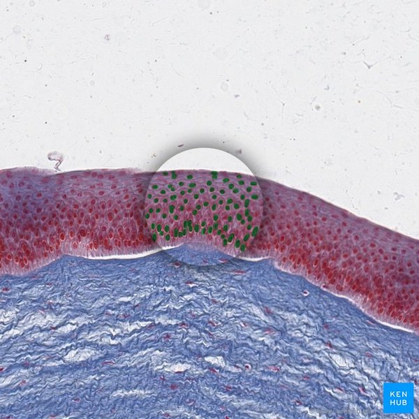
This image displays a microscopic view of epithelial cells, highlighting their densely packed nature and distinct nuclei.
Connective Tissue: The Body's Support System and Integrator
Connective tissue is the most diverse and abundant tissue type in the body, living up to its name by connecting other tissues and providing essential support, protection, and integration. Unlike epithelial tissue, connective tissue is characterized by cells that are often widely spaced and embedded within a rich extracellular matrix. This matrix, secreted by the cells themselves, is composed of protein fibers (collagen, elastic, and reticular) and a ground substance that can range in consistency from fluid to gel-like or solid.
Diverse Subtypes and Their Roles
The vast array of connective tissues includes:
- Connective Tissue Proper:
- Loose Connective Tissue (e.g., areolar tissue): Provides flexible support and acts as a reservoir of water and salts for surrounding tissues.
- Dense Connective Tissue (e.g., tendons, ligaments): Characterized by densely packed collagen fibers, offering high tensile strength and resistance to stretching.
- Specialized Connective Tissues:
- Adipose Tissue (fat): Stores energy, insulates the body, and cushions organs.
- Cartilage: Provides flexible support and reduces friction at joints (e.g., hyaline, elastic, fibrocartilage).
- Bone: Hard, calcified tissue forming the body's skeletal framework, providing rigid support and protection.
- Blood: A fluid connective tissue transporting nutrients, gases, waste products, and immune cells throughout the body.
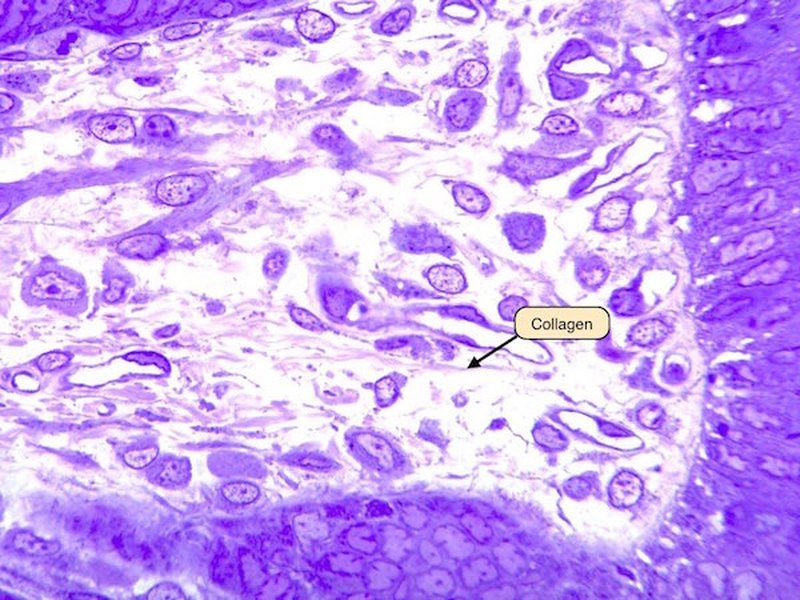
A microscopic image showing the sparse cells and abundant fibers of loose connective tissue, illustrating its supportive role.
Muscle Tissue: The Engine of Movement
Muscle tissue is specialized for contraction, generating the force necessary for movement both externally and internally. Its unique ability to shorten and thicken allows for everything from walking and lifting to the pumping of blood and the movement of food through the digestive tract. There are three distinct types of muscle tissue, each with specific structural features and control mechanisms.
Three Types of Muscular Action
- Skeletal Muscle:
- Control: Voluntary.
- Location: Attached to bones.
- Characteristics: Long, cylindrical cells; multiple nuclei; prominent striations (striped appearance). Responsible for gross body movements.
- Cardiac Muscle:
- Control: Involuntary.
- Location: Exclusively in the heart wall.
- Characteristics: Branching cells; typically single nucleus; striations; interconnected by intercalated discs, which facilitate rapid electrical signal transmission. Responsible for pumping blood.
- Smooth Muscle:
- Control: Involuntary.
- Location: Walls of internal organs (e.g., digestive tract, blood vessels, bladder).
- Characteristics: Spindle-shaped cells; single nucleus; no striations. Responsible for regulating blood flow, moving food, and other involuntary internal processes.
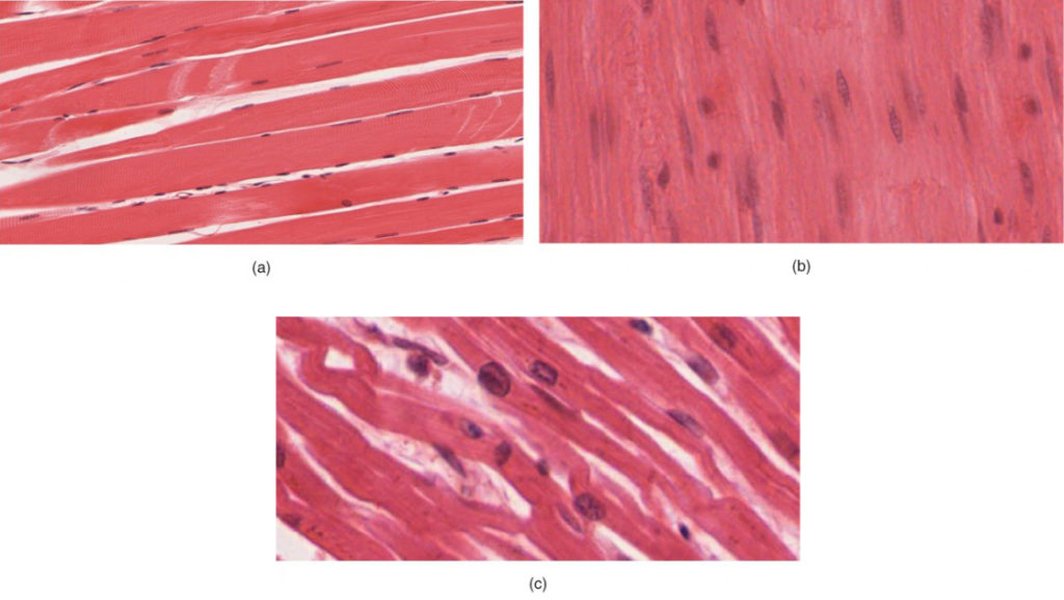
This image illustrates the distinct morphological characteristics of skeletal, smooth, and cardiac muscle tissues, highlighting their different cellular structures and organization.
Nervous Tissue: The Body's Communication Network
Nervous tissue is the master controller of the body, responsible for rapid communication by transmitting electrical signals (nerve impulses) throughout the body. It allows us to perceive sensations, process information, and initiate responses. Found in the brain, spinal cord, and nerves, it is critical for coordinating all bodily functions.
Neurons and Neuroglia
The two main cell types within nervous tissue are:
- Neurons (Nerve Cells): These are the excitable cells specialized for transmitting nerve impulses. Each neuron typically consists of:
- Cell Body (Soma): Contains the nucleus and other organelles.
- Dendrites: Branching extensions that receive signals from other neurons.
- Axon: A long projection that transmits signals away from the cell body to other neurons, muscles, or glands.
- Neuroglia (Glial Cells): These are non-excitable support cells that protect, nourish, and assist neurons. They play crucial roles in maintaining the internal environment of the nervous system and ensuring efficient nerve impulse transmission.
The organization of nervous tissue forms the central nervous system (brain and spinal cord) and the peripheral nervous system (nerves extending throughout the body), facilitating the complex interplay between sensory input, integration, and motor output.
The Synergy of Tissues: Organ Formation and Clinical Significance
The four primary tissue types rarely operate in isolation. Instead, they combine in specific and highly organized arrangements to form organs, each with unique and complex functions. For example, the stomach comprises epithelial tissue for lining and secretion, muscle tissue for churning food, connective tissue for structural support, and nervous tissue to regulate its activities. This intricate interplay allows for sophisticated physiological processes essential for life.
Tissue Interactions and Organogenesis
During embryonic development (organogenesis), these different tissue types are precisely choreographed to create functional organs. Epithelial tissues invariably interface with connective tissues at the basement membrane, muscle tissues are enveloped and supported by connective tissue layers, and nervous tissue relies heavily on neuroglia and a rich vascular supply (a type of connective tissue) for its metabolic needs. This coordinated development ensures that organs are not only structurally sound but also functionally integrated.
This mindmap illustrates the hierarchical organization and interconnectedness of the four primary tissue types in the human body, emphasizing their individual characteristics and collective contributions.
Clinical Correlations and Disease
Pathological changes at the tissue level are fundamental to understanding and diagnosing diseases. For instance, chronic irritation can lead to epithelial metaplasia, a reversible conversion of one mature epithelial type to another. Connective tissue disorders, such as scleroderma, involve excessive collagen deposition (fibrosis). Muscular dystrophies are genetic conditions affecting muscle fibers, leading to progressive weakness. Tumors like gliomas arise from glial cells in nervous tissue.
Histological examination, involving the microscopic study of tissue biopsies, remains a cornerstone of medical diagnosis. Understanding normal tissue architecture is therefore indispensable for recognizing abnormalities and disease states, guiding treatment strategies, and advancing fields like tissue engineering and regenerative medicine.
Quantitative Aspects of Tissue Composition and Specialization
While all four tissue types are essential, their relative abundance and functional specialization vary significantly across different organs and systems. This bar chart offers an opinionated analysis of their contribution to various bodily functions, demonstrating how each tissue type plays a critical, yet distinct, role.
This bar chart illustrates the relative contribution of each tissue type to five critical bodily functions. Connective tissue stands out for structural integrity, while nervous tissue leads in sensory perception. Mechanical force generation is primarily the domain of muscle tissue. Epithelial tissue plays a significant role in metabolic regulation and immune defense, though other tissues also contribute. This further emphasizes the functional specialization and interdependence of tissues within the body.
Deep Dive: Understanding Tissue Function Through Visualization
To further enhance our understanding of human tissues, let's explore a relevant video that visually explains their fundamental characteristics and roles within the body. This particular video provides a clear and engaging overview, suitable for grasping the core concepts discussed.
This embedded YouTube video, "The Four Types of Tissues - Epithelial, Connective, Nervous ...", offers a concise and clear explanation of the four basic tissue types, their structures, and their functions, making complex histological concepts accessible.
Summary of Tissue Types and Their Functions
The table below provides a concise summary of the key features, primary functions, and typical locations for each of the four fundamental tissue types, serving as a quick reference guide to their distinct roles.
| Tissue Type | Key Characteristics | Primary Functions | Typical Locations |
|---|---|---|---|
| Epithelial | Densely packed cells, minimal extracellular matrix, avascular, rests on basement membrane. | Protection, secretion, absorption, filtration, sensation. | Skin surface, lining of digestive tract, glands, kidney tubules. |
| Connective | Cells widely spaced within an extracellular matrix (fibers + ground substance), vascular (mostly). | Support, binding, protection, insulation, transport, energy storage. | Under skin, bones, cartilage, tendons, ligaments, blood, fat. |
| Muscle | Specialized for contraction, excitable cells. | Movement (skeletal), pumping blood (cardiac), internal organ movements (smooth). | Skeletal muscles, heart walls, walls of hollow organs (e.g., stomach, intestines, blood vessels). |
| Nervous | Neurons (transmitting cells) and neuroglia (support cells), excitable. | Rapid communication, sensory perception, integration, motor control. | Brain, spinal cord, peripheral nerves. |
This table highlights the remarkable diversity and specialization among the four tissue types, each contributing uniquely to the complex architecture and physiological capabilities of the human body.
FAQ
What is histology?
Are all tissues in the body identical?
Can tissues repair themselves after injury?
What is the extracellular matrix?
How do different tissues work together?
Conclusion
The human body is an extraordinary example of biological organization, built upon the foundation of four primary tissue types: epithelial, connective, muscle, and nervous tissue. Each type possesses distinct structural characteristics and specialized functions—from forming protective barriers and providing support to enabling movement and facilitating rapid communication. Their intricate interplay and organized integration are what allow for the complex functions of organs and the overall maintenance of life. A thorough understanding of these fundamental tissues is not only essential for comprehending human anatomy and physiology but also forms the bedrock for advancements in medical diagnostics, pathology, and the burgeoning fields of tissue engineering and regenerative medicine.
Recommended Further Exploration
- Discover the nuances of epithelial tissue classification and their specific locations in the body.
- Explore the complex composition and vital roles of the extracellular matrix in various connective tissues.
- Investigate the molecular mechanisms that drive muscle contraction and relaxation in different muscle types.
- Delve into the diverse types of neuroglia and their essential functions in supporting the nervous system.