
How to Perform the Khoury Dental Technique
A Comprehensive Guide to Advanced Autogenous Bone Grafting for Dental Implants
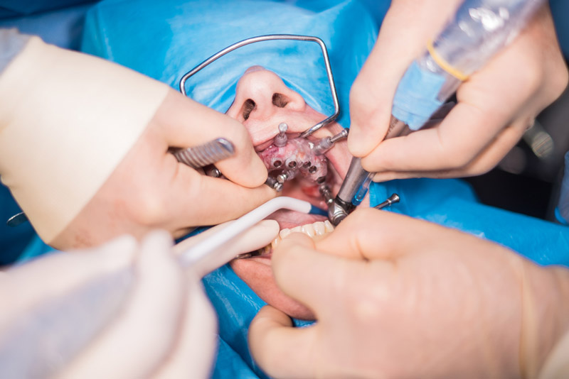
Key Takeaways
- Autogenous Bone Grafting: Utilizes the patient's own bone for optimal biocompatibility and integration, ensuring high success rates.
- Comprehensive Procedure: Encompasses detailed steps from patient evaluation to implant placement, ensuring thorough preparation and execution.
- Advantages and Considerations: Offers benefits such as reduced rejection and healing time, while requiring skilled surgical expertise for optimal outcomes.
Introduction to the Khoury Dental Technique
The Khoury Dental Technique, also known as the "Split Bone Block Technique" or "Biological Bone Augmentation," is an advanced method of autogenous bone grafting developed by Prof. Fouad Khoury. This technique is widely recognized in the field of oral implantology for its effectiveness in reconstructing significant bone deficiencies, thereby facilitating successful dental implant placements. By utilizing the patient's own bone, the Khoury technique ensures superior biocompatibility, reduces the risk of graft rejection, and promotes natural bone integration.
Patient Evaluation and Planning
Clinical and Radiographic Assessment
The first crucial step in the Khoury Dental Technique involves a comprehensive evaluation of the patient's oral and overall health. This assessment includes:
- Clinical Examination: Evaluates the extent of bone deficiency, oral hygiene status, and the presence of any underlying conditions that may affect grafting.
- Radiographic Imaging: Utilizes advanced imaging techniques, primarily Cone Beam Computed Tomography (CBCT) scans, to assess bone volume, density, and anatomical structures. Accurate imaging is essential for precise planning and execution of the grafting procedure.
- Digital Planning: Incorporates digital tools to create a detailed surgical plan, ensuring accurate placement and shaping of the bone graft.
Harvesting Autogenous Bone
Donor Sites and Techniques
Autogenous bone harvesting is the cornerstone of the Khoury technique, leveraging the patient's own bone to enhance graft success. Key aspects include:
- Donor Sites: Common intraoral donor sites include the mandibular ramus and symphysis (chin area). These sites are preferred due to their accessibility and the quality of bone available.
- Harvesting Procedure: Specialized instruments are employed to obtain thin bone plates from the donor site. The process involves meticulous surgical techniques to minimize trauma and ensure adequate bone quantity and quality.
- Biocompatibility: Using the patient's bone eliminates the risk of immune rejection and enhances the integration of the graft with existing bone structures.
Preparation of the Recipient Site
Creating the Graft Site
Preparing the recipient site is critical for the successful integration of the bone graft. This involves:
- Flap Design: A well-designed flap is raised to expose the deficient bone area. The design ensures adequate access while preserving blood supply to the surrounding tissues.
- Creating a Tunnel or Space: A precise tunnel or cavity is created within the deficient area to accommodate the bone graft. This space must be adequately vascularized to support graft integration.
- Debridement: The recipient site is thoroughly debrided to remove any infected or necrotic tissues, ensuring a clean environment for graft placement.
Graft Placement
Shaping and Securing the Bone Plates
Proper placement of the bone graft is essential for achieving the desired bone augmentation. The process includes:
- Shaping the Bone Plates: The harvested bone plates are meticulously shaped to fit the contours of the recipient site. Custom shaping ensures that the graft fills the deficient area seamlessly.
- Securing the Graft: The shaped bone plates are fixed in place using biocompatible screws or other fixation devices. Stable placement prevents movement during the healing process, promoting effective osseointegration.
- Perimeter Formation: In some cases, a perimeter or "box-like" structure is created around the graft site to provide additional support and stability.
Soft Tissue Management
Ensuring Graft Protection
Managing the soft tissues surrounding the graft site is vital to protect the graft and promote healing. This involves:
- Flap Closure: The surgical flap is carefully sutured to achieve primary closure, ensuring that the graft remains covered and protected from the oral environment.
- Tension-Free Suturing: Suturing techniques are employed to minimize tension on the flap edges, reducing the risk of dehiscence or graft exposure.
- Soft Tissue Healing: Adequate soft tissue coverage facilitates proper healing and reduces the likelihood of complications such as infection or graft failure.
Healing and Integration
Monitoring and Osseointegration
The healing period is a critical phase where the bone graft integrates with the existing bone structure. Key considerations include:
- Healing Timeline: Typically, 4-6 months are required for the graft to fully integrate and for sufficient bone volume to be achieved. This duration can vary based on the extent of augmentation and individual patient factors.
- Follow-Up Appointments: Regular monitoring through clinical evaluations and imaging (such as CBCT scans) ensures that the graft is integrating properly and identifies any potential issues early.
- Osseointegration: The biological process where the grafted bone fuses with the existing bone, creating a stable foundation for dental implants.
Implant Placement
Finalizing the Procedure
Once the graft has successfully integrated and adequate bone volume is established, the final step involves the placement of dental implants:
- Assessment of Bone Quality: Prior to implant placement, the quality and quantity of the augmented bone are reassessed to ensure readiness for implants.
- Surgical Implant Placement: Dental implants are then surgically placed into the augmented bone using precise techniques to ensure optimal positioning and stability.
- Prosthetic Integration: After implant placement, prosthetic components (such as crowns or bridges) are attached, completing the dental restoration process.
Advantages of the Khoury Technique
- High Success Rate: Utilizes autogenous bone, ensuring excellent biocompatibility and integration, leading to predictable and successful outcomes.
- Reduced Healing Time: Compared to other bone grafting methods, the Khoury technique often requires a shorter healing period (approximately 4 months) for effective bone regeneration.
- Versatility: Suitable for complex cases involving both vertical and horizontal bone augmentation, making it adaptable to various clinical scenarios.
- Durable Results: Achieves long-term stability and natural bone volume, providing a robust foundation for dental implants.
- Minimized Rejection Risks: Since the graft is derived from the patient's own bone, there is minimal risk of graft rejection or immune response.
Key Considerations and Risks
-
Advanced Surgical Skills Required: The Khoury technique demands a high level of surgical expertise and experience to ensure precise execution and minimize complications.
-
Potential Complications: Risks include infection, non-integration of the graft, soft tissue exposure, and donor site morbidity. Proper surgical technique and post-operative care are essential to mitigate these risks.
-
Patient Selection: Ideal candidates are those with sufficient overall health, adequate soft tissue, and specific bone deficiencies suitable for autogenous grafting. Comprehensive evaluation is necessary to determine eligibility.
-
Post-Operative Care: Adherence to post-operative instructions, including oral hygiene practices and activity restrictions, is crucial for optimal healing and graft success.
Comparative Table of Bone Grafting Techniques
| Technique | Graft Source | Healing Time | Advantages | Disadvantages |
|---|---|---|---|---|
| Khoury Technique | Autogenous bone (patient's own bone) | 4-6 months | High biocompatibility, reduced rejection, versatile for complex augmentations | Requires advanced surgical skills, potential donor site morbidity |
| Allograft | Donor bone from a different individual of the same species | 6-9 months | No need for a second surgical site, widely available | Risk of disease transmission, slower integration |
| Xenograft | Bone from a different species (e.g., bovine) | 6-12 months | Extensive availability, structural support | Higher risk of rejection, longer healing time |
| Synthetic Grafts | Biocompatible materials (e.g., hydroxyapatite) | 6-9 months | No risk of disease transmission, customizable shapes | Lower integration rates, may require additional stabilization |
Conclusion
The Khoury Dental Technique represents a significant advancement in the field of oral implantology, offering a reliable and effective solution for patients with substantial bone deficiencies. By utilizing autogenous bone, this method ensures superior biocompatibility and integration, resulting in high success rates and durable outcomes. While the technique demands a high level of surgical expertise and careful patient selection, its advantages in reducing rejection risks and healing time make it a preferred choice among dental professionals. Comprehensive planning, meticulous execution, and rigorous post-operative care are essential components that contribute to the success of the Khoury technique. As dental implantology continues to evolve, the Khoury Dental Technique stands out as a benchmark for effective bone augmentation and implant integration.
References
Last updated January 20, 2025