
Histopathological Analysis of Treated Liver Specimens
Understanding Extensive Hepatocyte Degeneration and Cytoplasmic Vacuolization
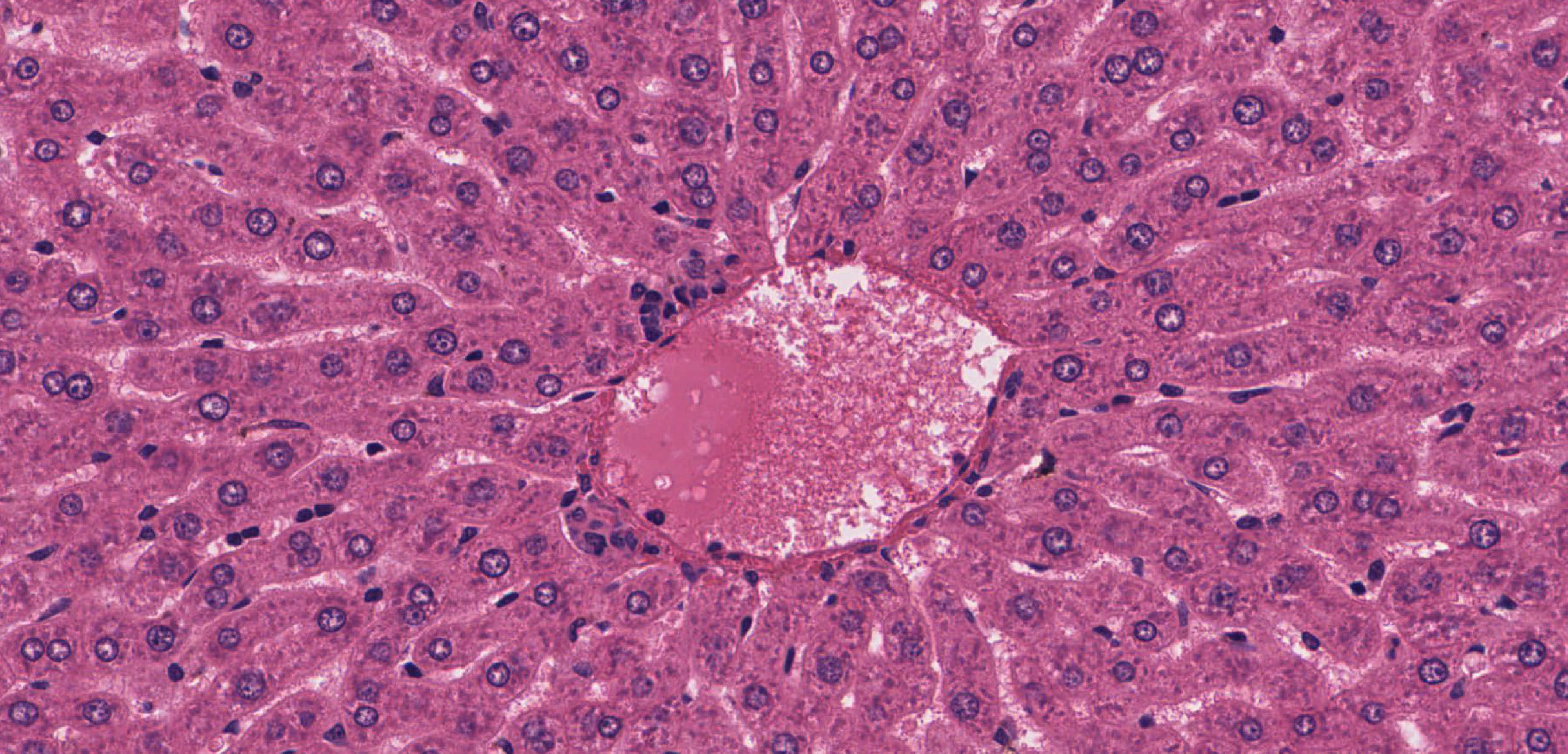
Highlights
- Extensive degeneration indicates severe hepatocyte injury potentially due to toxins, metabolic disturbances, or viral infections.
- Cytoplasmic vacuolization represents a cellular reaction that may involve adaptive responses or degenerative alterations.
- Pathological implications require thorough diagnostic work-up to distinguish between reversible adaptive changes and irreversible liver damage.
Introduction
The histopathological investigation of liver tissues in treated groups has revealed extensive degeneration of hepatocytes, with a notable presence of cytoplasmic vacuolization. This observation is critical as it may signify a spectrum of underlying pathological processes ranging from adaptive responses and metabolic alterations to severe toxic injuries or infectious insults. Given the multifaceted nature of liver diseases, understanding the cellular and molecular underpinnings of these findings is pivotal for accurate diagnosis and further research.
Pathological Description and Cellular Mechanisms
Hepatocyte Degeneration
Extensive hepatocyte degeneration refers to a comprehensive breakdown of liver cell structure and function. In a treated group, such changes typically suggest significant cellular injury which may be associated with necrosis, ballooning degeneration, and eventual cell death. Ballooning degeneration is characterized by enlarged hepatocytes with a pale or wispy cytoplasm, indicating a loss of cellular integrity that may precede apoptosis or necrosis. The degeneration can be observed as a continuum, ranging from reversible cellular stress responses to irreversible cell death, depending on the nature of the insult.
Mechanisms of Injury
Several mechanisms contribute to hepatocyte degeneration:
- Toxin Exposure: Chemicals or drugs can directly harm hepatocytes, triggering cellular stress responses, oxidative damage, and subsequent degeneration.
- Metabolic Disturbances: Conditions such as non-alcoholic fatty liver disease (NAFLD) or steatohepatitis may instigate lipid accumulation and oxidative stress, culminating in cellular injury.
- Viral Infections: Hepatitis viruses can induce inflammatory responses and direct cellular damage, exacerbating degeneration.
Cytoplasmic Vacuolization
Cytoplasmic vacuolization occurs when vacuoles, which are small, clear spaces, form within the cytoplasm of a cell. In the context of liver pathology, vacuolization can have various interpretations:
Adaptive or Degenerative Phenomena?
It is important to note that vacuolization is not solely indicative of degeneration. In some instances, it represents an adaptive response:
- Adaptive Response: Under conditions of mild to moderate cellular stress, hepatocytes may accumulate glycogen or other metabolites in vacuoles as a protective mechanism to counteract injurious stimuli.
- Degenerative Process: In cases where the injury is more severe, such as extensive toxic or viral insult, vacuolization may reflect the cell's inability to adequately manage metabolic disturbances. The presence of vacuoles may then be a prelude to lytic cell death or apoptosis.
Cytoplasmic vacuoles may contain a variety of substances including fat, water (edema), glycogen, or metabolic byproducts. The specific composition of these vacuoles can provide clues as to the underlying cause of the change. For example, vacuoles resulting from lipid accumulation are often observed in steatohepatitis, whereas those filled with water might indicate cellular edema.
Clinical Implications
The presence of both extensive degeneration and cytoplasmic vacuolization in hepatocytes suggests a significant disruption in liver architecture and function. Clinically, these histological findings can be associated with several conditions, each requiring specific diagnostic and therapeutic considerations.
Differential Diagnoses
When such features are observed in liver tissue samples, several diagnostic possibilities should be considered:
Toxic Hepatitis
Exposure to hepatotoxic compounds, whether through drug toxicity or chemical exposures, may yield extensive hepatocyte degeneration and vacuolization. The liver, being the primary organ for detoxification, is especially vulnerable to such insults.
Non-Alcoholic and Alcoholic Steatohepatitis
Both non-alcoholic steatohepatitis (NASH) and alcoholic steatohepatitis (ASH) are characterized by hepatocyte injury, inflammation, and fat accumulation. In these conditions, vacuolization can indicate both the presence of fat droplets and cellular stress responses. The progression to severe degeneration often reflects ongoing metabolic derangements and inflammatory damage.
Viral Hepatitis
Viral hepatitis (such as hepatitis C) can also trigger these histopathological changes. The immune-mediated response to viral infection, coupled with direct cytopathic effects, results in hepatocyte degeneration and potential vacuolization as part of the cellular stress response.
Diagnostic Approaches
Accurate diagnosis of the underlying cause of hepatocyte degeneration and vacuolization necessitates a multi-faceted approach:
Histological Examination
Liver biopsies remain the gold standard for evaluating these conditions. Histological analysis can delineate the extent of cellular degeneration, identify vacuolization patterns, and assess the presence of additional features such as inflammation, fibrosis, or steatosis. Specific staining techniques may be employed to differentiate between types of inclusions present in vacuoles.
Biochemical and Molecular Testing
Complementary biochemical tests (such as liver enzyme assays) and molecular markers can aid in interpreting liver injury. For instance, elevated transaminases might confirm hepatocyte damage while molecular assays can help detect underlying viral infections or genetic predispositions contributing to metabolic liver diseases.
Table Summary of Key Histopathological Features
| Feature | Description | Clinical Correlate |
|---|---|---|
| Degeneration | Disruption of hepatocyte architecture often with ballooning degeneration. | Toxin exposure, viral hepatitis, metabolic disorders |
| Cytoplasmic Vacuolization | Formation of vacuoles in the cytoplasm that may contain fat, water, or glycogen. | Adaptive stress response, fatty liver disease, hepatotoxicity |
| Infiltration and Inflammation | Accompanying inflammatory response often detected in liver biopsies. | Indicates ongoing liver injury and immune response |
| Fibrosis | Variable degree of fibrosis may accompany chronic injury. | Progression to cirrhosis |
Molecular and Biochemical Insights
Beyond histological examination, understanding the molecular basis of hepatocyte damage is essential. Oxidative stress, a principal factor in many forms of liver injury, leads to the production of reactive oxygen species (ROS), which can damage cellular membranes, proteins, and DNA. This type of injury might be particularly evident in toxic insults or metabolic derangements, where the balance of detoxification mechanisms is overwhelmed.
Emerging research underscores the role of signaling pathways and stress response proteins that may be activated in response to hepatocellular injury. For example, the activation of stress kinases and subsequent initiation of apoptotic pathways can lead to cellular degeneration. Conversely, sub-lethal vacuolization may sometimes reflect an upregulation of cellular repair mechanisms, where hepatocytes accumulate specific metabolites in vacuoles as a provisional means of counteracting damage.
Interpreting the Findings in Clinical Settings
Integrating the observed histopathological changes with clinical data allows for a nuanced interpretation of liver injury. A case with extensive hepatocyte degeneration combined with widespread cytoplasmic vacuolization warrants an in-depth evaluation:
Correlation with Clinical History
Detailed patient history and exposure analysis are critical in discerning the etiology of liver damage. Recent use of hepatotoxic drugs, alcohol consumption, exposure to industrial toxins, or a history of metabolic syndrome can guide the clinician toward a specific diagnosis. Additionally, certain viral infections that target the liver would also be strongly considered.
Imaging and Laboratory Evaluation
Radiological imaging (such as ultrasound, CT scans, or MRI) can help ascertain the extent of liver damage and reveal structural changes consistent with steatosis or fibrosis. Typically, elevated liver enzymes (AST, ALT) in serum biochemical tests would support the histological findings of hepatocyte degeneration. When results are ambiguous, liver biopsy remains the definitive method for diagnosis.
Therapeutic Implications
Recognizing the specific histopathological patterns facilitates targeted therapy:
- Toxic Injuries: If a toxic agent is identified, cessation of exposure is paramount. Supportive treatments including antioxidants and agents that facilitate hepatic regeneration may be employed.
- Metabolic Liver Diseases: Management may involve lifestyle interventions, insulin-sensitizing agents, and lipid-lowering drugs, in order to mitigate underlying metabolic dysregulation.
- Viral Hepatitis: In cases associated with viral infections, antiviral therapies aim to control the infection and attenuate further hepatocyte damage.
Integrative Discussion and Future Perspectives
The dual finding of extensive hepatocyte degeneration and cytoplasmic vacuolization in treated liver samples presents a complex challenge, where both adaptive and deleterious cellular responses interplay. While vacuolization might be protective in isolated instances, its extensive presence combined with overt degeneration clearly signifies a tipping point where cellular reparative mechanisms are overwhelmed.
Research Directions
Future research should focus on elucidating the temporal sequence of histological changes post-injury. Advanced imaging techniques, such as multiphoton microscopy and confocal laser scanning, along with molecular profiling of stress response pathways, could help distinguish between early adaptive vacuolization and irreversible cell damage.
Molecular Markers and Therapeutic Targets
Identification of specific molecular markers could be pivotal in early detection of cell degeneration. For example, monitoring the expression levels of anti-apoptotic and pro-apoptotic proteins, as well as markers of oxidative stress, could assist in determining the viability of hepatocytes under injurious conditions. Understanding these pathways would not only deepen our knowledge of liver pathology but also offer novel therapeutic targets to arrest or reverse progression.
In conclusion, effective management of liver injuries with such histopathological characteristics requires a collaborative approach integrating clinical, histological, and molecular diagnostics. Careful correlation of these data will help tailor interventions that may minimize further hepatocellular damage and promote hepatic recovery.
Conclusion
The extensive degeneration of hepatocytes accompanied by cytoplasmic vacuolization in the examined treated liver group reflects a critical state of liver injury. This pathological picture points to a variety of potential underlying causes ranging from toxic exposures and metabolic disorders to viral hepatitis. Importantly, while vacuolization can sometimes denote a reversible adaptive response, when observed alongside severe degeneration it suggests that liver cells are overwhelmed by injury processes. Diagnostic work-up should therefore include comprehensive histopathological evaluation, supportive laboratory tests, and appropriate imaging studies to fully elucidate the cause and extent of liver damage.
Clinically, early detection and subsequent targeted therapeutic interventions are vital. Whether the injury stems from a toxin, metabolic syndrome, or viral infection, recognizing the pattern of hepatocellular changes can guide the treatment strategy, potentially harnessing opportunities to reverse or mitigate the damage before it advances to cirrhosis or irreversible liver failure.
References
-
PubMed Abstract - PubMed
- Springer PDF Article - Springer
- PubMed Abstract - PubMed
-
PMC Article - NCBI PMC
-
PMC Article - NCBI PMC
Recommended Further Queries
Last updated February 19, 2025