
Unlocking Precision in Lower Limb Anesthesia: A Comprehensive Guide to Nerve Stimulator-Guided Lumbar Plexus Blocks
Master the meticulous technique of lumbar plexus blockade using only nerve stimulation for effective anesthesia and analgesia.
Highlights of the Technique
- Precision through Physiology: The nerve stimulator-only technique relies on eliciting specific muscle twitches, primarily of the quadriceps, to confirm needle proximity to the lumbar plexus, offering a functional endpoint for accurate local anesthetic placement.
- Anatomical Landmark Cruciality: Accurate identification of surface anatomical landmarks, such as lumbar spinous processes and the iliac crest, is foundational to guiding the needle towards the deeply situated psoas compartment where the lumbar plexus resides.
- Systematic Current Reduction: A key safety and efficacy feature involves starting nerve stimulation at a higher current (e.g., 1.5 mA) and gradually reducing it to a low threshold (0.2-0.5 mA) once the target motor response is observed, ensuring the needle tip is close to the nerve before injection.
Understanding the Lumbar Plexus Block
A lumbar plexus block (LPB), often referred to as a psoas compartment block, is a regional anesthesia technique targeting the lumbar plexus. This intricate network of nerves is formed by the anterior rami of the L1 to L4 spinal nerves, with occasional contributions from T12 and L5. It is situated deep within the psoas major muscle, anterior to the transverse processes of the lumbar vertebrae. The primary nerves arising from the lumbar plexus that are targeted by this block include:
- Femoral nerve (L2-L4): Innervates the anterior thigh, knee, and medial leg/foot.
- Lateral femoral cutaneous nerve (L2-L3): Provides sensation to the anterolateral thigh.
- Obturator nerve (L2-L4): Innervates the adductor muscles of the medial thigh and provides sensory input to the medial knee and thigh.
This block is highly effective for providing anesthesia and/or analgesia for surgical procedures involving the hip (e.g., hip replacement, femur fracture), anterior and medial thigh, and knee. When performed using only a nerve stimulator, the technique relies on precise anatomical knowledge and the interpretation of elicited motor responses to guide needle placement, as direct visualization of structures (like with ultrasound) is absent.
Anatomy of the Lumbar Plexus, situated within the Psoas Major muscle.
Essential Preparations for the Procedure
Thorough preparation is paramount for the safe and successful execution of a lumbar plexus block using a nerve stimulator.
Patient Positioning
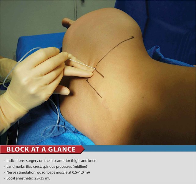
The patient is typically positioned in the lateral decubitus position, with the side to be blocked uppermost. Key aspects include:
- The back should be straight and parallel to the edge of the operating table to aid in landmark identification.
- The hip and knee of the operative side may be slightly flexed, and the foot on the side to be blocked can be positioned over the dependent leg. This positioning helps to stabilize the patient, relax the psoas muscle, and allow for clear observation of quadriceps muscle twitches (patellar movement).
- Ensure the patient is comfortable and adequately supported.
Necessary Equipment
A standardized setup ensures efficiency and safety. The required equipment includes:
- Sterile gloves, gown, mask, and drapes.
- Antiseptic solution (e.g., chlorhexidine) for skin preparation.
- Local anesthetic for skin infiltration (e.g., 1% lidocaine).
- An insulated nerve block needle: Typically 10-15 cm in length and 18-22 gauge, designed for deep blocks and effective current delivery.
- A peripheral nerve stimulator with a connecting cable. The cathode (negative terminal) is connected to the block needle, and the anode (positive terminal, grounding electrode) is placed on the patient's skin, often on the ipsilateral thigh.
- Syringes and needles for drawing up and injecting local anesthetic.
- The chosen local anesthetic solution for the block (e.g., 0.25-0.5% ropivacaine or bupivacaine).
- Standard monitoring equipment: ECG, non-invasive blood pressure cuff, pulse oximeter.
- Emergency resuscitation equipment and medications (including lipid emulsion for local anesthetic systemic toxicity - LAST).
The Step-by-Step Procedure
The lumbar plexus block using only a nerve stimulator is a technique that demands meticulous attention to detail at each stage.
Identifying Anatomical Landmarks
Accurate landmark identification is the cornerstone of this technique:
- Locate the Lumbar Spinous Processes: Palpate the spinous processes of the lumbar vertebrae. The L4 spinous process is often identified by drawing an imaginary line connecting the highest points of the iliac crests (intercristal line or Tuffier's line), which typically crosses the L4 spinous process or the L4-L5 interspace.
- Determine the Insertion Level: The block is commonly performed at the L3, L4, or sometimes L5 level.
- Mark the Needle Insertion Point: The insertion point is marked approximately 3-5 cm lateral to the midline (spinous process) at the chosen vertebral level. Some practitioners use a point two to three patient's fingertip widths lateral to the spinous process at the level of the anterior superior iliac crest (which often corresponds to L4).
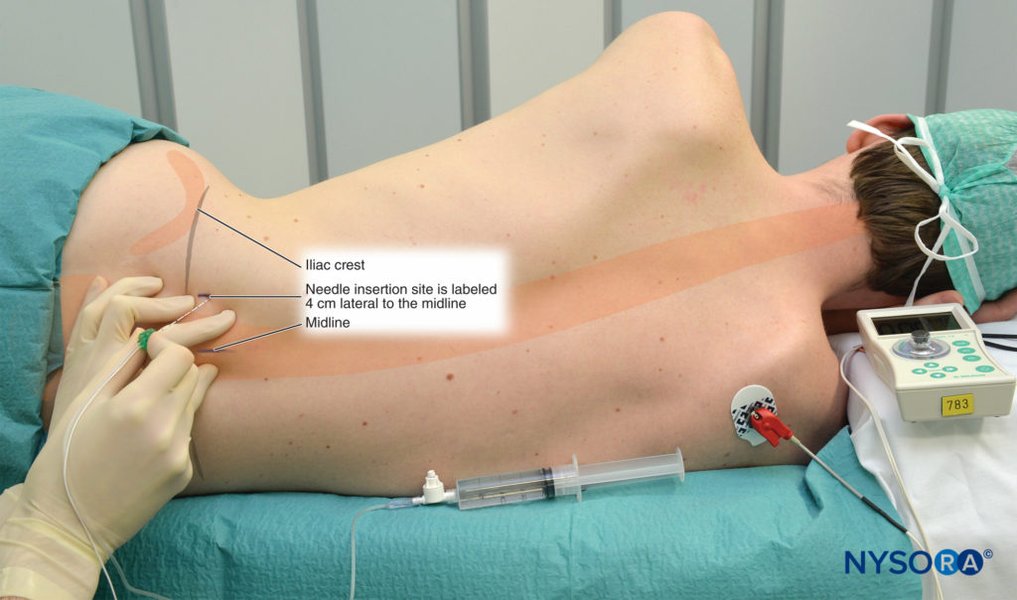
Key anatomical landmarks for identifying the needle insertion site for a lumbar plexus block.
Initial Setup and Skin Preparation
After confirming patient consent and establishing intravenous access and standard monitoring, the skin over the marked area is prepared with an antiseptic solution and draped in a sterile manner. Local anesthetic is infiltrated into the skin and subcutaneous tissues at the needle insertion site to minimize discomfort during needle passage.
Needle Insertion and Nerve Stimulation
The nerve stimulator is set up and tested before needle insertion:
- Initial Nerve Stimulator Settings: The stimulator is typically set to deliver a current of 1.0 to 1.5 mA, a pulse duration of 0.1 milliseconds (ms), and a frequency of 1 to 2 Hertz (Hz).
- Needle Insertion: The insulated block needle, connected to the nerve stimulator, is inserted perpendicular to the skin at the marked entry point.
- Advancing the Needle: The needle is advanced slowly and cautiously. As it passes through the paraspinous muscles, local muscle twitches may be observed. These are not the target response.
- Contacting the Transverse Process: The needle is advanced until it contacts the transverse process of the target lumbar vertebra. This usually occurs at a depth of 3-6 cm, depending on the patient's body habitus. Contacting bone provides a crucial depth reference.
- "Walking Off" the Transverse Process: Once bone is contacted, the needle is slightly withdrawn and then redirected ("walked off") either cephalad (superiorly) or caudad (inferiorly) to pass beyond the transverse process and enter the psoas compartment where the lumbar plexus lies. The needle should be advanced an additional 1-2 cm past the depth at which the transverse process was encountered. The depth to the plexus is typically 6-8 cm, but can be more in larger patients.
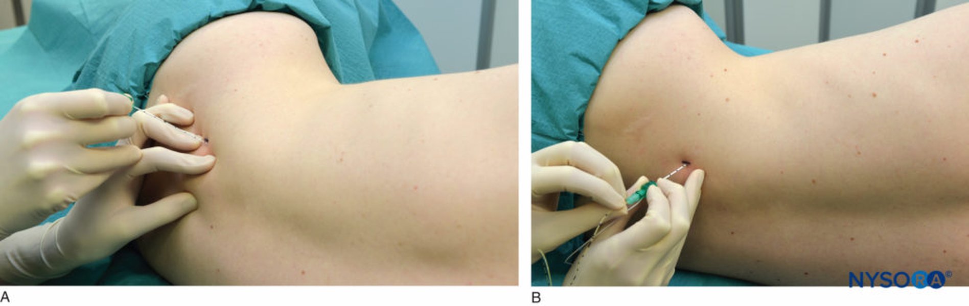
Illustration of needle trajectory for lumbar plexus block, aiming for the psoas compartment.
Achieving the Target Motor Response
The primary goal is to elicit a motor response from muscles innervated by the lumbar plexus:
- Desired Response: The most sought-after and reliable motor response is a visible or palpable contraction of the quadriceps femoris muscle (patellar twitch or "dancing patella"). This indicates proximity to the femoral nerve component of the lumbar plexus. Twitches of the adductor muscles (obturator nerve) or lateral thigh muscles (lateral femoral cutaneous nerve) can also occur.
- Current Reduction: Once a satisfactory quadriceps twitch is obtained, the stimulating current is gradually decreased. The aim is to maintain the quadriceps twitch at a current between 0.2 mA and 0.5 mA.
- A response at a current <0.2 mA may suggest intraneural needle placement, increasing the risk of nerve injury. In such cases, the needle should be slightly withdrawn until the twitch is lost and then repositioned until it reappears at a current of 0.2-0.5 mA.
- If a twitch is only present at a current >0.5 mA, the needle is likely not close enough to the plexus, and gentle repositioning is required.
Local Anesthetic Administration
Once the needle is deemed to be in the correct position (quadriceps twitch at 0.2-0.5 mA):
- Aspiration: Careful aspiration is performed in multiple planes (after gentle needle rotation) to rule out intravascular or intrathecal needle placement. This must be repeated after every 3-5 mL of injectate.
- Test Dose (Optional but Recommended): Some practitioners administer a small test dose (e.g., 3 mL of local anesthetic with epinephrine) to further check for intravascular injection.
- Incremental Injection: The main volume of local anesthetic is injected slowly, in 5 mL increments, with intermittent aspiration between each increment. This minimizes the risk of rapid systemic absorption and allows early detection of adverse reactions.
- Monitoring During Injection: The patient should be closely monitored for any signs of local anesthetic systemic toxicity (LAST), such as tinnitus, metallic taste, circumoral numbness, dizziness, or seizures. Injection should be stopped immediately if any such signs occur. Resistance to injection should also prompt reassessment of needle position.
Following injection, the motor response to nerve stimulation should diminish or disappear, indicating successful nerve blockade.
Pharmacological Considerations
Local Anesthetics Used
Long-acting local anesthetics are typically preferred for lumbar plexus blocks to provide extended postoperative analgesia. Common choices include:
- Ropivacaine: Often used in concentrations of 0.2% to 0.5%.
- Bupivacaine: Commonly used in concentrations of 0.25% to 0.5%. Levobupivacaine, the S-enantiomer of bupivacaine, may offer a slightly better safety profile.
Dosage Guidelines
The volume of local anesthetic required for a lumbar plexus block is relatively large due to the extensive nature of the plexus within the psoas compartment.
- Volume: Typically, 20 to 40 mL of local anesthetic solution is administered. Some dosing guidelines suggest 0.3 to 0.5 mL/kg of body weight.
- Maximum Dose: It is crucial to adhere to maximum recommended doses for the chosen local anesthetic to prevent systemic toxicity. For example, the maximum dose of bupivacaine is often cited as 2 mg/kg (not exceeding a total dose like 150-175 mg). Patient factors such as age, comorbidities, and lean body mass should be considered.
Visualizing Key Success Factors
The success and safety of a nerve stimulator-guided lumbar plexus block depend on several interacting factors. The radar chart below illustrates the relative importance and considerations for these key elements. While operator skill is paramount, understanding these components helps optimize the technique.
This chart highlights that precise landmarking, meticulous needle handling, and appropriate use of nerve stimulator settings (especially current thresholding) are of paramount importance and significantly influenced by operator experience.
Mindmap of the Lumbar Plexus Block Procedure
The following mindmap provides a visual summary of the key stages and considerations involved in performing a lumbar plexus block using a nerve stimulator.
This mindmap provides a quick overview of the procedural flow, from initial patient setup through to post-procedure care, highlighting the critical decision points and actions.
Advantages and Limitations of the Nerve Stimulator-Only Technique
Benefits
- Functional Confirmation: Provides direct physiological evidence (motor twitch) of nerve proximity, which can be reassuring for the operator.
- Widely Accessible: Does not require specialized imaging equipment like ultrasound, making it feasible in settings with limited resources.
- Established Technique: It has a long history of use with well-documented outcomes and procedural steps.
Challenges and Potential Risks
Despite its utility, the nerve stimulator-only technique has inherent challenges and potential complications:
- Deep and Variable Anatomy: The lumbar plexus is situated deep within the psoas muscle, and its exact location can vary between individuals. Anatomical variations or obesity can make landmark identification and needle placement challenging.
- "Blind" Technique: Unlike ultrasound-guided blocks, this method does not allow real-time visualization of the needle, surrounding anatomical structures (vessels, kidneys, peritoneum), or local anesthetic spread.
- Potential Complications:
- Epidural or Subarachnoid Spread: If the needle is advanced too medially, local anesthetic can spread into the epidural or subarachnoid space, leading to a high neuraxial block or total spinal anesthesia.
- Local Anesthetic Systemic Toxicity (LAST): Due to the large volume of local anesthetic used and the vascularity of the psoas muscle.
- Hematoma: Retroperitoneal or psoas hematoma can occur, especially in anticoagulated patients or if a blood vessel is inadvertently punctured.
- Nerve Injury: Direct needle trauma to nerve roots or intraneural injection can cause neurological deficits.
- Kidney Puncture: The kidneys are located anterolateral to the psoas muscle, and there's a risk of renal injury if the needle is directed too anteriorly or laterally, particularly on the right side.
- Block Failure or Incomplete Block: May occur due to inaccurate needle placement or insufficient local anesthetic spread.
- Operator Dependent: Success and safety are highly dependent on the operator's anatomical knowledge, experience, and meticulous technique.
Key Parameters Summary
The following table summarizes critical parameters for the nerve stimulator-guided lumbar plexus block:
| Parameter | Setting/Value | Notes |
|---|---|---|
| Initial Stimulating Current | 1.0 - 1.5 mA | For initial localization of the plexus; elicits twitches when further from the nerve. |
| Pulse Duration | 0.1 ms | A short pulse duration is typically used for motor nerve localization. |
| Stimulation Frequency | 1 - 2 Hz | Allows for clear observation of muscle twitches without causing tetany. |
| Target Motor Response | Quadriceps femoris muscle contraction (patellar twitch) | Most reliable indicator of femoral nerve (and thus lumbar plexus) proximity. |
| Final Stimulating Current (Confirmation) | 0.2 - 0.5 mA | Confirms close proximity of the needle tip to the nerve before injection. Responses below 0.2 mA may indicate intraneural placement. |
| Insulated Needle | 10-15 cm length, 18-22 gauge | Length necessary to reach the deep plexus; insulation directs current to the tip. |
| Local Anesthetic Volume | 20 - 40 mL | Large volume needed for adequate spread within the psoas compartment. |
Post-Procedure Care and Monitoring
After the block is performed:
- The patient should be monitored in a recovery area with continuous ECG, blood pressure, and pulse oximetry.
- Assess the onset and extent of sensory and motor blockade in the distribution of the femoral, lateral femoral cutaneous, and obturator nerves. This typically involves checking for warmth, loss of sensation to cold or pinprick, and ability to move the relevant muscle groups.
- Monitor for signs of complications, including LAST (which can have a delayed onset), hematoma formation (e.g., flank pain, hypotension), or neurological deficits.
- Provide clear instructions to the patient regarding limb numbness and weakness, and precautions to prevent injury.
Visual Demonstration: PNS Guided Lumbar Plexus Block
The following video provides a visual demonstration and discussion of performing a lumbar plexus block guided by a peripheral nerve stimulator (PNS). It covers anatomical landmarks, the technique of using the nerve stimulator, and eliciting the appropriate motor responses. Understanding the visual cues and procedural flow can be very helpful for clinicians learning or refining this technique.
This video demonstrates the PNS guided lumbar plexus block, highlighting anatomy, landmarks, and nerve stimulation technique.
Observing the procedure, even in a demonstration format, can reinforce the importance of careful landmarking, needle handling, and interpretation of nerve stimulator feedback. The video complements the textual description by providing a dynamic perspective on achieving a successful and safe lumbar plexus block.
Frequently Asked Questions (FAQ)
Which nerves are targeted by a lumbar plexus block?
A lumbar plexus block primarily targets the major nerves originating from the lumbar plexus (L1-L4, with contributions from T12 and L5). These include the femoral nerve, the lateral femoral cutaneous nerve, and the obturator nerve. Successful blockade of these nerves provides anesthesia and analgesia to the anterior and medial thigh, the knee, and parts of the leg and foot.
What is the most reliable motor response to look for during a lumbar plexus block with a nerve stimulator?
The most reliable and sought-after motor response is a contraction of the quadriceps femoris muscle, which manifests as a visible or palpable twitch of the patella (often called the "patellar tap" or "dancing patella"). This indicates that the needle tip is in close proximity to the femoral nerve, which is a major component of the lumbar plexus.
Why is the electrical current reduced once a motor response is elicited?
The electrical current is gradually reduced to confirm that the needle tip is very close to the nerve. Eliciting the target motor response (e.g., quadriceps twitch) at a low current (typically 0.2 mA to 0.5 mA) indicates optimal needle placement for local anesthetic injection. If the response is lost at a higher current, the needle is likely too far away. If a response is obtained at a current less than 0.2 mA, it may suggest intraneural placement, and the needle should be slightly withdrawn before injecting.
What are the primary risks associated with a lumbar plexus block using only a nerve stimulator?
Primary risks include epidural or spinal anesthesia (if the needle is too medial), local anesthetic systemic toxicity (LAST) due to the large volume of anesthetic, retroperitoneal hematoma, nerve injury (from direct trauma or intraneural injection), kidney puncture, and block failure. The "blind" nature of the technique (without ultrasound) contributes to these risks, emphasizing the need for meticulous technique and anatomical knowledge.
How is the correct needle depth determined when using only landmarks and a nerve stimulator?
The correct needle depth is estimated based on patient anatomy and by first contacting the transverse process of the target lumbar vertebra (typically 3-6 cm deep). After contacting bone, the needle is "walked off" the transverse process and advanced slightly further (usually 1-2 cm deeper) into the psoas compartment where the plexus lies. The final confirmation of depth and position relies on eliciting the appropriate motor response (quadriceps twitch) at the target low current (0.2-0.5 mA). The overall depth to the plexus is often in the range of 6-8 cm but can vary significantly.
Recommended Further Exploration
- What are the comparative advantages and disadvantages of performing a lumbar plexus block with a nerve stimulator versus ultrasound guidance?
- How can potential complications associated with lumbar plexus blocks, such as local anesthetic systemic toxicity or hematoma, be effectively managed?
- In what ways do anatomical variations in the lumbar plexus or psoas muscle impact the success rate of nerve stimulator-guided blocks?
- What is the specific role and efficacy of lumbar plexus blocks as part of a multimodal analgesic regimen for major hip surgeries?
References
Last updated May 6, 2025