
Lymphoma and Tumor Volume
Understanding the Role of Tumor Volume Measurement in Lymphoma Management
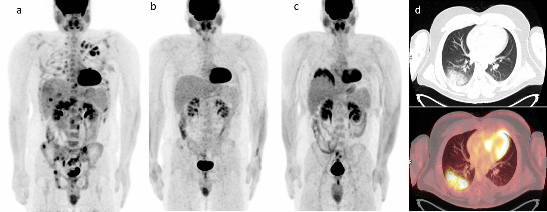
Essential Insights
- Total Metabolic Tumor Volume (TMTV) is a critical prognostic marker in lymphoma.
- TMTV measurement via FDG-PET/CT scans aids in risk stratification and treatment planning.
- Standardization of TMTV measurement continues to evolve, enhancing personalized care.
Overview of Lymphoma and the Importance of Tumor Volume
Lymphoma, a type of cancer originating in the lymphatic system, affects key components such as the lymph nodes, spleen, thymus, and bone marrow, which are crucial for the immune response. A significant challenge in managing lymphoma is assessing the extent of the disease, for which tumor volume measurement has become an essential clinical tool. In this context, Total Metabolic Tumor Volume (TMTV) plays a central role, as it provides a quantifiable measure of the metabolically active tumor burden across the body.
Traditional imaging methods, such as CT and MRI, have been used primarily to evaluate the structural aspects of the disease. However, advancements in imaging technology have shifted the focus toward functional imaging—specifically, [18F-fluorodeoxyglucose positron emission tomography/computed tomography (FDG-PET/CT)]. This technique not only assesses anatomical details but also provides pivotal metabolic information that is integral to evaluating aggressive disease behavior.
Detailed Analysis of Tumor Volume in Lymphoma
Defining Total Metabolic Tumor Volume (TMTV)
Total Metabolic Tumor Volume (TMTV) is defined as the summation of metabolically active tumor regions identified during FDG-PET/CT imaging. Through computational methods that involve segmenting PET-avid lesions, TMTV quantifies the overall energetic and volumetric burden of lymphoma. This metric considers both the size and the metabolic intensity of lesions, making it highly relevant for assessing the active components of the disease.
The process of measuring TMTV involves using specialized software that delineates tumor boundaries based on thresholds usually set as a percentage of the maximum standardized uptake value (SUVmax) within a lesion (commonly around 40-41%). Once these regions are identified, their individual metabolic volumes are summed up to yield the TMTV, expressed in cubic centimeters (cm³) or milliliters (mL).
Prognostic Implications of TMTV in Lymphoma
Numerous studies have consistently shown that a higher TMTV at baseline is associated with poorer clinical outcomes across various subtypes of lymphoma. For instance, patients with Diffuse Large B-Cell Lymphoma (DLBCL), Hodgkin lymphoma (HL), and follicular lymphoma (FL) who display elevated TMTV values typically demonstrate reduced progression-free survival (PFS) and overall survival (OS) rates. The prognostic value of TMTV is well-documented:
- In DLBCL, models integrating TMTV with other clinical parameters such as performance status have been powerful in identifying patients at greater risk of refractory disease or relapse before treatment initiation.
- In follicular lymphoma, a baseline TMTV exceeding a certain threshold (e.g., >510 cm³) has been associated with a shorter duration of remission and overall survival.
- In Hodgkin lymphoma, the variability in optimal cutoffs (ranging between 89 to 268 cm³) highlights the need for tailored evaluation depending on patient and disease characteristics.
These insights underscore that TMTV is not only a reflection of tumor bulk but also of the biologically active fraction of the disease that may influence treatment responsiveness.
Clinical Applications and Risk Stratification
The incorporation of TMTV into clinical practice is proving transformative in the management of lymphoma. Its use extends beyond mere diagnostic assessment, influencing both prognostic evaluations and therapeutic decisions:
Risk Stratification
By integrating TMTV values into risk stratification models, clinicians can effectively identify patients who may benefit from more aggressive treatment regimens. For instance:
- Patients with lower TMTV might be considered for standard treatment protocols, potentially minimizing exposure to overtreatment and its accompanying side effects.
- Conversely, individuals with high TMTV values can be earmarked for intensified therapy or enrollment in clinical trials evaluating novel treatments.
The data derived from TMTV assessments can also be used in conjunction with other metabolic and clinical parameters, such as beta-2-microglobulin levels and patient performance scores, to create multidimensional prognostic models, further refining the personalization of care.
Therapeutic Decision-Making
Beyond risk stratification, TMTV measurements actively contribute to the formulation of treatment plans. For example, in cases where imaging reveals bulky disease (often defined by a tumor mass exceeding 10 cm in diameter), the intensity and combination of chemotherapeutic agents may be adjusted accordingly. Moreover, the dynamic changes in TMTV observed during interim FDG-PET/CT scans can serve as early indicators of treatment efficacy, leading to timely modifications in therapeutic strategies.
As emerging research continues to validate the importance of TMTV, there is growing enthusiasm for its integration into routine clinical protocols, thus potentially improving overall survival and quality of life for lymphoma patients.
Challenges and Future Directions
Standardization of Measurement Techniques
Despite the significant clinical potential of TMTV, standardization remains one of the foremost challenges. Variability in imaging protocols, segmentation software, and threshold criteria can lead to disparities in TMTV measurements across different institutions. This inconsistency impacts the reproducibility of results and may complicate treatment recommendations. Ongoing research is aimed at achieving consensus on standardized measurement techniques, including:
- Developing uniform threshold settings for FDG uptake segmentation.
- Enhancing software algorithms to minimize inter-operator variability.
- Conducting multicenter trials to harmonize TMTV-based prognostic assessments.
The realization of standardized TMTV measurements has the potential to facilitate broader clinical adoption, support more robust comparative studies, and ultimately improve prognostic accuracy.
Integrating TMTV with Other Prognostic Indicators
In addition to TMTV, other biomarkers and clinical parameters continue to be explored for comprehensive prognostic modeling in lymphoma. For example, factors such as patient age, performance status, lactate dehydrogenase (LDH) levels, and genetic mutations are frequently considered alongside TMTV. The future lies in developing predictive models that combine these multidimensional data inputs, providing a more accurate and personalized risk assessment. Such models could:
- Refine prognostic stratification by incorporating both metabolic imaging and molecular diagnostics.
- Facilitate early identification of high-risk patients who may benefit from innovative therapies.
- Allow dynamic monitoring of treatment efficacy through serial measurements of TMTV and related biomarkers.
Advancements in Imaging Technology
The evolution of imaging technology continues to enrich the assessment of tumor volume in lymphoma. Enhancements in FDG-PET/CT imaging, including improved spatial resolution and quantitative analysis capabilities, are expected to augment the accuracy of TMTV measurements. Researchers are also investigating alternative radiotracers that may offer superior specificity for lymphoma compared to 18F-FDG. The progression of these innovations holds promise for:
- Providing more precise delineation of metabolically active tumor regions.
- Allowing earlier detection of treatment response and disease progression.
- Enhancing the overall predictive power of imaging-based assessments.
A Comprehensive Comparison of Traditional and Novel Metrics
Traditional metrics such as the sum of the product of diameters (SPD) or maximum standardized uptake value (SUVmax) have long been used to assess tumor burden in lymphoma. While these parameters provide valuable data, they are limited in that they offer a unidimensional assessment of the disease. TMTV, on the other hand, provides a three-dimensional, volumetric perspective of metabolically active tumor tissue. The following table summarizes the key differences between these approaches:
| Metric | Description | Advantages | Limitations |
|---|---|---|---|
| TMTV | Sum of metabolically active tumor volume from FDG-PET/CT scans | Comprehensive assessment of active tumor burden; strong prognostic indicator | Requires specialized software; variability in segmentation methods |
| SUVmax | Maximum standardized uptake value within a tumor lesion | Simple to measure; widely available | Does not account for overall tumor volume; may miss heterogeneity |
| SPD | Product of the longest diameters of tumor lesions | Traditional method; easy clinical application | Limited to two-dimensional assessment; less predictive of metabolic activity |
This table highlights that while traditional metrics still hold value, TMTV offers a more nuanced evaluation of tumor dynamics, particularly when assessing aggressive or widespread disease.
Future Research and Clinical Integration
Ongoing Studies
Research into TMTV and its clinical implications is rapidly evolving. Ongoing studies are focused on:
- Validating optimal TMTV cutoff values across different lymphoma subtypes.
- Integrating TMTV with emerging biomarkers to create comprehensive prognostic models.
- Assessing the impact of TMTV-guided treatment modifications on long-term patient outcomes.
As these studies yield data, the incorporation of TMTV into clinical guidelines is expected to become more widespread, promoting personalized and nuanced treatment strategies.
Clinical Implementation
The clinical implementation of TMTV measurement is expected to transform how oncologists approach lymphoma treatment by providing:
- Early identification of high-risk patients, enabling proactive treatment adjustments.
- More accurate prediction of treatment responses and potential relapses.
- A framework for integrating imaging data with other clinical and molecular parameters to ensure individualized treatment plans.
With continued technological advancements and increased collaboration among research institutions, TMTV is poised to become an integral aspect of lymphoma management protocols.
References
-
PMC Article - NCBI
- PubMed Article - PubMed
-
PMC Article - NCBI
- Blood Journal - ASH Publications
-
ScienceDirect Article - ScienceDirect
Recommended Queries for Further Insights
Last updated March 17, 2025