
Focusing Specimens: Low Power vs. High Power
Understanding the Critical Steps in Microscope Observation
Highlights
- Enhanced Navigational View: Using the low power objective provides a wide field that makes it easier to locate and center the specimen.
- Increased Depth and Safety: The greater depth of field and longer working distance in LPO reduce the risk of damaging your slides or high power lenses.
- Smooth Transitioning: Proper initial focus with the LPO enables efficient and precise shifts to high magnification with minimal adjustments.
Introduction
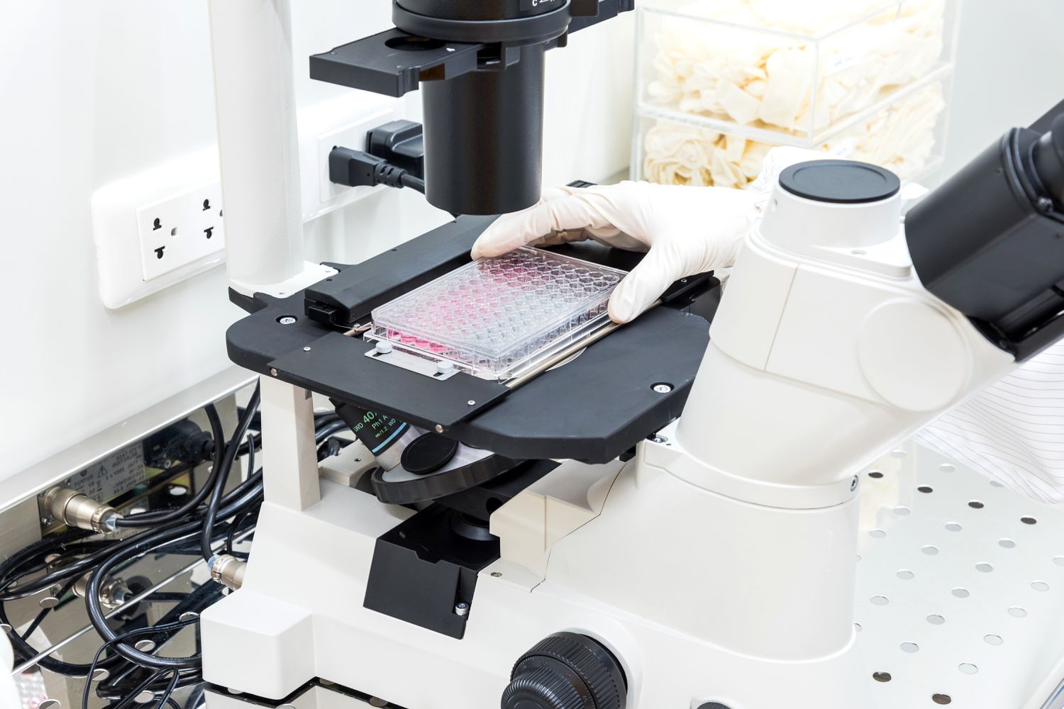
In the realm of microscopy, proper focus is paramount to obtaining both a clear overall view and highly detailed insights into the specimen under observation. One of the fundamental practices in achieving high-quality imagery is beginning the focusing process with the Low Power Objective (LPO) before transitioning to the High Power Objective (HPO). This procedure is not arbitrary; rather, it is underpinned by several scientific and practical reasons that contribute collectively to what is known as an optimal focusing workflow.
The Role of the Low Power Objective (LPO)
The LPO is the starting point for most microscopic examinations because it offers a wide field of view, excellent depth of field, and an overall easier focusing experience. This objective is designed to capture a large area of the specimen, which is crucial when initially surveying the sample. By utilizing lower magnification, the LPO allows one to rapidly identify the specimen's overall structure, locate points of interest, and position the slide so that the area of concern is centered in the field of view.
Wider Field of View
A major advantage of using the LPO is its ability to deliver a significantly wider field of view compared to the HPO. When a specimen is observed under the LPO, the larger viewing area ensures that even if the sample is not perfectly aligned, the user can still locate and roughly center it on the slide. This makes it far easier to begin focusing without getting lost in details that are irrelevant at this stage of examination.
Greater Depth of Field
The concept of depth of field refers to the vertical range within which the specimen remains in focus. The LPO, by virtue of its lower magnification, possesses a greater depth of field, meaning that a larger portion of the sample remains sharply defined simultaneously. This is particularly beneficial when first adjusting the focus because it allows the user to see a sufficiently extensive area of the specimen clearly. A broad depth of field minimizes the risk of missing any part of the specimen that might be of interest.
Ease and Efficiency with Coarse Adjustment
Given the broader field of view and improved depth of field, the LPO allows for more effective and forgiving use of the coarse adjustment knob. Coarse adjustments are essential for making large focus shifts quickly, and the LPO’s characteristics prevent the user from overshooting the correct focal plane. This makes the initial focusing process not only simpler but also more efficient, as it establishes a clear baseline focus that can be later refined.
The Transition to High Power Objective (HPO)
Once a specimen has been roughly focused under the LPO, the next stage is the transition to the HPO. The HPO is engineered to provide much higher magnification, enabling the user to observe fine details and minute structures within the specimen. However, the HPO comes with certain limitations and risks that justify starting with the LPO.
Narrow Field of View and Precision
The HPO, while excellent for magnifying small details, offers a much narrower field of view. This can be challenging if the specimen is not already centered and in focus. By first using the LPO, the user ensures that the area of interest is in the center of the field. This greatly reduces the likelihood of having to search again for the correct region once the magnification is increased.
Reduced Depth of Field
As magnification increases, the effective depth of field dramatically diminishes. In practical terms, this means that only a very thin layer of the specimen is in focus at any one time when using the HPO. Starting with the LPO, which grants a greater depth of field, provides the user with a clear initial focus, reducing the task of re-focusing or potentially missing important details when transitioning upwards in magnification.
Protection Against Damage
Another significant concern when shifting to high magnification is the potential risk of damaging the microscope or the specimen itself. The HPO has a shorter working distance, meaning that the physical gap between the objective lens and the specimen is minimal. An error in focusing could result in the lens colliding with the slide. By commencing with the LPO, the operator sets up a safe, controlled focus environment that minimizes the risk of accidental contact or physical damage.
Efficiency in Focusing and Transitioning
Starting with the LPO helps establish the overall orientation of the specimen. This initial setup includes pinpointing the exact area of interest and ensuring that the primary focus is achieved with ease. Once these elements are successfully administered, the transition to the HPO involves only fine adjustments using the fine adjustment knob. This methodical progression from a broad overview to detailed examination contributes significantly to a smoother and more efficient focusing journey.
Comparative Analysis: LPO vs. HPO Attributes
To further elucidate the reasons behind the prescribed focusing sequence, it is beneficial to compare the characteristics and capabilities of the LPO and HPO across several dimensions. The following table summarizes some key differences:
| Characteristic | Low Power Objective (LPO) | High Power Objective (HPO) |
|---|---|---|
| Field of View | Wide and expansive | Narrow and limited |
| Depth of Field | Greater depth with more elements in focus | Shallow depth of field requiring fine adjustments |
| Working Distance | Longer, reducing the risk of collision | Shorter, increasing potential contact risks |
| Ease of Focusing | More forgiving with coarse adjustments | Requires precise fine adjustments |
| Specimen Overview | Provides a comprehensive overview | Facilitates detailed inspection |
The tabulated comparison clearly illustrates that the inherent design features of the LPO make it uniquely suited for establishing the initial focus and layout of the specimen. In contrast, the HPO is optimized for detailed inspection once the preliminary focus and positioning have been efficiently managed.
Detailed Technical Principles Behind the Focusing Sequence
Beyond the observable practical benefits, several underlying optical principles justify the recommended practice of beginning with the LPO. These principles are rooted in the optics of light behavior, lens mechanics, and the physical constraints imposed by the equipment.
Light Path and Optical Alignment
In any optical system, the path of light is critical to the clarity and quality of the final image. When using a microscope, light enters the specimen, is focused through various lenses, and ultimately forms an image for the viewer. The wide-angle capture via the LPO ensures that the light passing through the specimen is collected over a broader area, thereby reducing potential aberrations. With a more comprehensive capture of the light path, the overall image remains balanced and minimizes optical errors.
Role of Parfocality
Many modern microscopes are designed with parfocal lenses. This means that once the specimen is in focus with one objective (typically the LPO), it remains approximately in focus when switching to the HPO, requiring only minimal fine adjustments. Initiating the focusing process with the LPO leverages this design trait effectively. It ensures that the user’s time is not wasted in re-focusing completely after changing magnification, thereby streamlining the diagnostic or observational process.
Adjustability and Specimen Safety
The mechanical design of microscopes, particularly the spacing and alignment of the objective lenses, necessitates careful manipulation. The coarse adjustment knob, used predominantly with the LPO, provides large-scale focus changes without risking abrupt or dangerous movements of the specimen stage. Conversely, the HPO's short working distance means that even slight misadjustments can lead to collisions with the specimen. By ensuring safe focus under LPO conditions, the chance of accidental damage is dramatically reduced.
Fine Detail Visualization Versus General Observation
When starting with the LPO, the user is afforded the ability to gain a holistic view of the sample, allowing for the identification of regions that merit more detailed examination. This "overview first" approach contrasts sharply with jumping directly to high magnification, where the narrow focus might lead you to miss context or important larger-scale structures. In practice, the LPO establishes a contextual map of the specimen, ensuring that any subsequent high-powered observations are both targeted and informed.
Practical Implications and Step-by-Step Workflow
The practical workflow of using a microscope typically involves a sequence of deliberate steps that maximize both accuracy and safety. This workflow helps ensure that observers can maintain focus on areas of interest without unnecessary delays or disruptions.
Step 1: Initial Setup with the LPO
Begin by placing the specimen on the microscope slide and positioning it onto the stage. Using the LPO, adjust the focus using the coarse adjustment knob. The wider view of the LPO aids in quickly locating the specimen, ensuring that the entirety of the sample is visible. At this point, the user should take care to:
- Ensure the specimen is properly centered and evenly illuminated.
- Adjust the lighting and condenser settings to achieve the ideal contrast and clarity.
- Utilize the larger depth of field to find the correct focal plane.
Step 2: Transitioning the Focus
After establishing a clear and broad view under the LPO, the next step involves carefully switching to the HPO. Since modern microscopes are generally parfocal, the specimen remains largely in focus, requiring only adjustments via the fine adjustment knob. It is critical during this transition to make small increments in focus loss, thereby avoiding any abrupt changes that could potentially move the slide or damage the objective lens.
Step 3: Detailed Observation with the HPO
With the specimen now centered and properly focused under the high magnification provided by the HPO, the observer can begin the detailed examination. The narrow field of view at this stage highlights fine structures and details that were not resolvable under the LPO. The user can now perform a more in-depth analysis, carefully tweaking the focus with precision to ensure the clearest possible image.
Benefits in Academic and Research Settings
In educational and research laboratories, the practice of using the LPO before the HPO is not just a matter of tradition–it is a safety and efficiency protocol. Academic settings often include multiple users of varying expertise, and starting with the LPO ensures that even novice users can observe samples without causing erroneous adjustments or incurring damage to the equipment. Research laboratories benefit similarly, as time and precision are critical in ensuring that the data collected is both accurate and reproducible.
Safety Considerations
One of the paramount concerns in any laboratory setup is the safety of both the specimen and the microscope. Focusing under the LPO first creates a buffer zone that safeguards against the potential of damaging the slides or the lenses. Because the distance between the specimen and the objective is larger with the LPO, there is considerably less risk of the lens making contact with the sample during the initial focusing phase. This not only prolongs the life of the microscope but also ensures that specimens remain intact and undistorted for accurate analysis.
Training and Best Practices
Training protocols for new microscope users consistently emphasize the significance of starting with the LPO. Best practices include:
- Familiarization with the coarse adjustment mechanism and recognizing the broad patterns of the specimen.
- Recognizing the differences in depth of field and field of view between the LPO and HPO.
- Systematic transitioning to high power magnification, ensuring that the specimen remains centered and in focus.
This structured approach not only enhances the accuracy of the observations but also instills a disciplined methodology that is critical in scientific research.
Additional Considerations and Technical Nuances
While the fundamental reasons for using the LPO first are well-established, advanced users may take into account a few additional considerations to further refine their workflow.
Optical Aberrations and Corrections
Optical aberrations can distort the image if the focus is not properly established. Using the LPO reduces the impact of minor optical defects since the inherent design of low power objectives minimizes such aberrations over a wider field. This initial broad focus helps in quickly identifying and mitigating any distortions before proceeding to finer adjustments.
Specimen Movement and Stability
Stability is a critical factor in microscopy. Slight movements of the specimen stage can lead to misalignment or loss of the desired focus area, particularly when switching to a high-magnification objective. The stabilization provided by the LPO’s wider focus allows the user to confirm that the specimen is securely and optimally positioned, minimizing disruptions during the delicate phase of high-power observation.
Fine-Tuning with Digital Enhancements
In modern microscopy, digital enhancements and image processing tools may be used following the initial focusing procedure. By starting with a well-focused image obtained under the LPO, users are better able to apply digital filters and adjustments accurately. Any digital magnification or enhancement applied post-LPO focusing benefits from the stable and clear baseline image, ensuring that subsequent analyses are both reliable and reproducible.
Summarizing the Advantages
To summarize, beginning your microscopic analysis with the LPO is justified by a combination of technical, practical, and safety considerations. The advantages encapsulated in this approach include:
- Improved overview and orientation: A wide field of view facilitates initial specimen location, ensuring that areas of interest are accurately identified.
- Enhanced depth of field: The increased depth of field under low magnification keeps more of the specimen in focus, reducing the trial-and-error needed during focusing.
- Risk reduction: With a greater working distance, the risk of damaging the specimen or high power lens is minimized, making focusing safer.
- Smooth and efficient transitioning: Establishing focus on the specimen with the LPO allows for minimal fine adjustments when shifting to the HPO, ensuring a more systematic examination process.
Conclusion and Final Thoughts
In conclusion, the practice of focusing specimens under the Low Power Objective (LPO) before transitioning to the High Power Objective (HPO) is grounded in a robust framework of optical physics and practical laboratory safety protocols. The LPO's wide field of view, enhanced depth of field, and forgiving nature during coarse adjustments make it the ideal first step in microscope focusing. This initial phase not only facilitates the efficient location and centering of the specimen but also minimizes the risk of collision and optical errors when shifting to high magnification.
As the user increases the magnification, the switch to the HPO allows for more detailed observation, leveraging the precise fine-focus capabilities while relying on the robust initial setup achieved via the LPO. This systematic approach enhances overall efficiency, accuracy, and safety in microscopic work, making it indispensable for educational, research, and clinical applications. Ultimately, the sequence of beginning with the LPO before using the HPO provides a logical, effective, and safe method for detailed specimen analysis.
References
- Socratic Q&A - Socratic
- Quora - Science&Technology
- Brainly - Educational Q&A
- University of Hawaii - Microscopy Tutorial
- NAU - Microscope Notes
- Sciencing - Science Explanation
Recommended
Last updated February 20, 2025