
Why Microscope Images Appear Inverted
Understanding Image Inversion Through Simple Lens Mechanics
Key Highlights
- Lens Interaction: The objective and eyepiece lenses both play a role in flipping the image.
- Light Refraction: The bending of light via curved (convex) lenses causes a natural inversion.
- Fundamental Optics Principle: Image inversion is an inherent property of the physics behind light and lenses.
Introduction
When observing an object under a microscope, you might notice that the image appears inverted – that is, it looks upside down and sometimes even reversed from left to right. Understanding why this happens does not require advanced knowledge of optics; instead, it is all based on the simple physics of light refraction through curved lenses. This comprehensive explanation will break down the topic step by step, offering an in-depth yet simplified reasoning that combines fundamental principles of optics with the practical workings of a microscope.
How a Microscope Works
Basic Components of a Microscope
A typical compound microscope is composed of two primary sets of lenses: the objective lens and the eyepiece (or ocular) lens. The objective lens is positioned near the specimen and is responsible for creating an initial magnified and inverted image. The eyepiece lens then further magnifies this image so that it can be observed by the viewer. Although there may be additional components, such as the condenser and light source, the inversion of the image is primarily due to the way the lenses handle the incoming light.
Objective Lens
The objective lens is the first lens that interacts with light from the specimen. It is a convex lens, which means it is curved on both sides. When light passes through this lens, it bends (or refracts) due to the change in medium from air to glass. The curvature of the lens causes the light rays to cross over, flipping the image vertically (upside down) and sometimes horizontally.
Eyepiece Lens
The eyepiece lens, located at the top of the microscope, takes the magnified, already inverted image produced by the objective lens and amplifies it further. It does not re-invert the image. Instead, it simply increases the size so that the details become more apparent to the observer.
The Role of Light Refraction in Image Inversion
The principle at work here is known as refraction. Refraction is the bending of light as it passes from one medium to another – in this case, from air into glass. The curved surfaces of the lenses mean that light is not traveling in straight lines; instead, it bends in a way that causes the top part of the light beam to be directed as if it were coming from the bottom, and vice versa. This bending mechanism is what ultimately results in the image being flipped.
Understanding Refraction
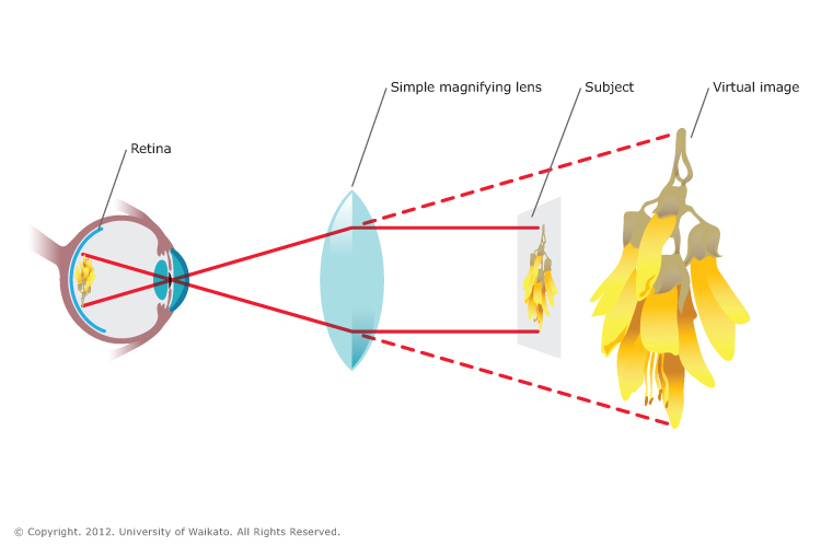
Imagine holding a regular magnifying glass over an object. If you move the glass around, you might notice that the orientation of the object seems altered. This is because, as light goes through the curved glass, it bends. In a microscope, this same concept is applied on a much more sophisticated level, with two successive lenses working together:
| Component | Function | Effect on Image |
|---|---|---|
| Objective Lens | Collects light from the specimen and performs initial magnification | Inverts (flips) the image |
| Eyepiece Lens | Further magnifies the image for easier observation | Maintains the inversion created by the objective lens |
The table above outlines how each component of the microscope contributes to the overall process, emphasizing that the image inversion occurs as an intrinsic part of how light is manipulated. It's not a flaw but a predictable outcome based on the laws of physics.
Breaking Down the Science in Simple Terms
Step-by-Step Explanation
Step 1: Light Leaves the Specimen
Every object emits or reflects light. In the case of a specimen under a microscope, light passes through or reflects off the tiny details within it. This light is what ultimately carries the image information.
Step 2: Interaction with the Objective Lens
Once the light leaves the specimen, it enters the objective lens. The objective lens gathers the light rays and bends them. Due to its convex shape, the light rays crossing inside the lens lead to the formation of an image that is inverted vertically. This means that what was originally on the top now appears on the bottom, and vice versa.
Step 3: The Eyepiece Lens Adds Magnification
After the objective lens forms an inverted image, this intermediate image is relayed to the eyepiece. The eyepiece further magnifies this image without flipping it back. Thus, the viewer sees an enlarged but still inverted image.
Why It Is Natural for Microscopes to Invert Images
In most simple optical systems that use lenses, such as magnifying glasses and telescopes, inversion of images is a common result of the way light flows through curved glass. The inversion happens because lenses do not “understand” the concept of “up” or “down”; they simply refract light based on the curvature and index of refraction of the material. In a microscope, since there are two sets of lenses, the effect is compounded if nothing is done to re-invert the image.
Correcting Our Perception
Although the inverted image might seem unusual at first, our brains are remarkably adaptable. When you set up a microscope or look at an inverted image, your brain often compensates by "flipping" the image in your mind so that you can make sense of the observation. However, for many scientific applications, the inversion is left uncorrected because the relative positions of structures are still accurate, and the scientist is already aware of the inversion.
Additional Considerations
Historical Perspective and Practical Implications
The phenomenon of image inversion in optical devices has been known since the early days of lens use. Early microscopes and telescopes all exhibited inverted images because the technology simply relied on the basic properties of optics. Over time, scientists and engineers have designed corrective devices such as erecting lenses or mirror systems for applications where a correctly oriented image is necessary, like in photography or certain types of telescopic observations.
Practical Applications in Modern Microscopy
In biological and material sciences, the inversion of the image does not interfere with the analysis since the relative positioning of structures remains consistent. Researchers know that the image is flipped and can adjust their interpretations accordingly. In some advanced instruments, electronic imaging systems can reorient the image for display purposes, though the optical path itself still inverts the light.
Alternative Optical Systems
It is not only microscopes that produce inverted images. Many optical systems that employ lenses, including cameras and projectors, also exhibit some form of image transformation due to the way light is refracted. However, in these systems, additional components are often included to correct or compensate for the inversion. For example, a digital camera might use software to flip the image automatically before it is displayed.
Understanding It Through Everyday Examples
Consider everyday objects like a magnifying glass or even swimming goggles. When you look into a curved surface, you might notice the reversal of left and right or top and bottom under certain conditions. This everyday experience is directly related to what happens in a microscope. The difference is simply one of scale and precision. In a microscope, the same bending of light that slightly alters the image with a magnifying glass is taken to a much higher degree, making the inversion very apparent.
Common Questions and Clarifications
Do All Microscopes Invert Images?
While the vast majority of compound microscopes invert images as a natural outcome of using convex lenses, there are some specialized microscopes designed for different purposes that may incorporate additional optical elements to correct the inversion. However, for standard biological and laboratory microscopes, an inverted image is normal.
Can the Inversion Be Fixed?
In principle, it is possible to design an optical system that re-inverts the image so that it appears right-side up. This would usually involve adding another lens or mirror system to reverse the inversion process. However, such modifications are not commonly used in standard microscopes because the inversion does not hinder the analysis of the specimen. Instead, the consistent inversion is factored into the way scientists learn to interpret microscopic images.
Does Magnification Affect the Inversion?
Magnification itself does not affect whether an image is inverted; it only enlarges the already inverted image. The inversion is determined by the optical path and the way the lenses are arranged, rather than the degree of magnification. Whether you are looking at a slightly magnified or highly magnified image under a microscope, the inversion is a result of the light bending through the lens systems.
In-Depth Analysis of the Optical Process
Mathematical Insight into Refraction
Although a full dive into the mathematical derivation is beyond the scope of this simplified explanation, the basic idea can be captured with Snell's law, which relates the angles of incidence and refraction for a light ray passing between two media. The equation is given by:
$$n_1 \sin(\theta_1) = n_2 \sin(\theta_2)$$
Here, $$n_1$$ and $$n_2$$ are the refractive indices of the two different media (for example, air and glass), and $$\theta_1$$ and $$\theta_2$$ represent the angles of incidence and refraction, respectively. The curvature of the lens changes these angles in such a way that crossing of light rays occurs, leading directly to image inversion.
Understanding Through Diagrams and Visualizations
Visual aids are often very useful for comprehending optical phenomena. While we cannot display a drawn diagram here, imagine the following sequence:
- Light rays emanate upward from the top of a specimen.
- The objective lens collects these rays, bending them so that they cross over, making the top rays appear as if they are coming from the bottom of the specimen.
- After the crossover, the eyepiece magnifies this new inverted image and presents it to your eye.
This step-by-step visual process emphasizes why the image inversion is not an error, but simply the natural outcome of the geometric arrangement of the lenses.
Practical Implications for Microscope Users
Learning to Interpret Inverted Images
For students and researchers using microscopes, getting accustomed to reading inverted images is part of the training. Initially, the inversion might seem disorienting, but with practice, it becomes second nature. The regular inversion means that if you know the layout of the specimen, you will consistently see it flipped, which allows you to develop strategies for interpretation that leverage the predictable nature of the optics.
Tips for Beginners
1. Assume Consistency: Since the inversion is systematic, once you familiarize yourself with the orientation, you can mentally flip the image while analyzing details.
2. Use Markers: Some laboratory slides have orientation markers that help you determine which part is which, thereby reducing confusion.
3. Practice with Simple Specimens: Begin by examining specimens with clear, distinct features so that you can quickly learn to interpret the inverted image.
Enhanced Technologies and Digital Corrections
While traditional optical microscopes provide an inverted image by design, modern digital microscopy offers the possibility to automatically correct the image orientation. Cameras attached to microscopes can process the captured image in real time, flipping it right-side up for display purposes. This feature is particularly useful in educational settings or in presentations where an intuitive, correctly oriented image is desirable.
Summarizing the Core Reasons for Image Inversion
A Simplified Recap
In summary, the reason why images appear inverted when you examine them under a microscope is straightforward:
- The objective lens first takes the light from the specimen and bends it to form an inverted image.
- The eyepiece lens then magnifies this already inverted image, resulting in the final view as seen through the microscope.
- This inversion is a natural outcome of light refraction through convex lenses and is consistent with the basic principles of optics.
By understanding these steps, it becomes clear that the culprit is not a flaw in the microscope or a malfunction in the lenses, but rather an inherent aspect of how light interacts with curved glass surfaces. This predictable inversion allows users to adjust their observations without compromising the utility of the microscopic details.
Conclusion and Final Thoughts
To conclude, the inversion of images seen under a microscope is a direct consequence of how convex lenses bend light. The process begins when light from a specimen enters the objective lens, where it is refracted and crossed over, thereby creating an inverted image. This image is then magnified by the eyepiece lens without any corrective measures to revert the flip. The phenomenon is not an error but a natural refraction effect intrinsic to optical instruments. Understanding this optical behavior is crucial for students, researchers, and anyone interested in microscopy, as it can enhance one’s ability to interpret and work with microscopic images effectively. Moreover, modern technological advancements in digital imaging can mitigate the confusion caused by inversion by automatically adjusting the image orientation.
This explanation not only simplifies the underlying science but also emphasizes that image inversion is a predictable and manageable aspect of using microscopes. By embracing the principles of refraction and the fundamental workings of thin lenses, users can better appreciate the marvels of optical instruments and the intricate details they reveal about the micro-world.
References
- Do Microscopes Invert Images and Why? - Science Selector
- Do Microscopes Invert Images? Why Does it Happen? - 3D Biology
-
3.2A: Microscopy - Biology LibreTexts
- Do Microscopes Invert Images? - Microscope Clarity
Recommended Further Readings and Queries
Last updated February 20, 2025