
Introduction to Microscopes
Explore the fascinating world of magnified imagery and scientific discovery
Highlights
- Essential Components: Learn how lenses, light sources, and precision mechanics transform tiny specimens into visible images.
- Diverse Types: Discover the differences between light, electron, compound, and stereo microscopes and their unique applications.
- Historical Impact: Understand how microscopes revolutionized science from early discoveries to modern technological breakthroughs.
Fundamentals of Microscopy
What is a Microscope?
A microscope is an optical instrument that allows us to visualize objects which are too small to be seen with the naked eye. It significantly enlarges the image of microscopic specimens, revealing intricate details and structures that otherwise remain hidden. The principal function of the microscope is to produce a magnified image, separate fine details within a specimen, and make these details visible to observers. This fundamental capability has expanded human knowledge in fields ranging from biology and medicine to materials science and engineering.
Historical Context
The invention of the microscope marked a monumental leap in scientific progress. Emerging in the late 16th and 17th centuries, early microscopes were rudimentary devices that laid the groundwork for subsequent scientific discoveries. Pioneers like Antonie van Leeuwenhoek and Robert Hooke utilized these early instruments to observe microscopic organisms and plant structures. Hooke’s illustrations of cork cells and van Leeuwenhoek’s observations of bacteria and protozoa demonstrated the latent potential hidden in everyday materials, igniting developments in cell biology and microbiology. Over the centuries, technological advancements have refined microscope design, introducing precise optics, enhanced illumination systems, and novel imaging modalities.
Basic Operating Principle
The core principle behind microscope operation involves the manipulation of light or electron beams through a series of lenses to magnify and delineate images of small objects. In the simplest terms, a light microscope employs visible light that passes through or reflects off a specimen. The system of lenses then bends (refracts) this light, magnifying the image. Key to this process are the objective lens, which captures the specimen's details, and the eyepiece lens, which further enlarges the image for viewing. In contrast, electron microscopes replace visible light with electrons that have much shorter wavelengths, permitting significantly higher resolution and magnification. The enhanced detail provided by such microscopes has been critical in exploring structures at the molecular and atomic levels.
Types of Microscopes
Light Microscopes
Light microscopes are the most commonly used instruments in both educational settings and research laboratories. They use visible light combined with multiple lenses to magnify specimens. One of the central types of light microscopes is the compound microscope, which is characterized by having at least two sets of lenses: the objective lenses located near the specimen and the eyepiece lenses. Magnifications typically range from 40x to 1000x, although they can sometimes exceed these limits when using special techniques.
Components of Light Microscopes
Light microscopes comprise several key components:
- Eyepiece: The lens through which the observer views the magnified image.
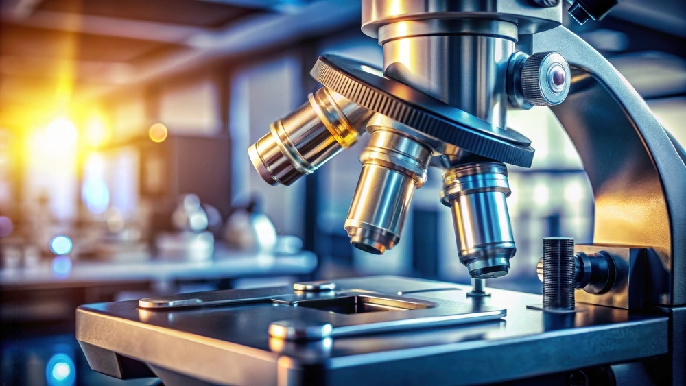
- Objective Lenses: A set of lenses of varying magnification powers mounted on a rotating nosepiece.
- Stage: The platform where the specimen is secured for observation.
- Focus Knobs: These adjust the distance between the lens and the specimen, ensuring a clear image is obtained.
- Condenser: Gathers light from the source and focuses it on the specimen for improved illumination.
- Light Source: Provides the necessary illumination to view the specimen. In modern microscopes, this can be a built-in LED or halogen lamp.
Electron Microscopes
Electron microscopes have transformed scientific study by enabling magnification levels far beyond those achievable with light microscopes. Instead of using photons, these devices accelerate electrons to produce images with sub-nanometer resolution. There are two principal types of electron microscopes:
Transmission Electron Microscopes (TEM)
TEMs work by passing a beam of electrons through an ultrathin specimen. As the electrons interact with the specimen, they are scattered and transmitted, forming an image on a detector. This method yields highly detailed images that reveal the internal structure of cells, tissues, and materials. TEM is especially useful in virology, cellular biology, and nanotechnology.
Scanning Electron Microscopes (SEM)
In contrast to TEMs, SEMs scan the surface of a specimen with a focused electron beam. The electrons interact with the atoms on the surface of the specimen, emitting secondary electrons that are detected to produce detailed three-dimensional images. SEM is particularly advantageous for examining surface morphology and texture in materials science, engineering, and biological applications.
Other Types of Microscopes
Beyond the conventional light and electron microscopes, several specialized instruments are designed to meet specific research needs:
- Stereo or Dissecting Microscopes: Provide lower magnification with a three-dimensional view, ideal for studying larger, opaque objects such as insects or plant parts.
- Fluorescence Microscopes: Use fluorescent dyes to tag specific components of a specimen, which emit light of different wavelengths when excited by a particular light source. This allows for the tracking of various cellular components in biomedical research.
- Confocal Microscopes: Utilize laser light to collect data in layers, offering high-resolution and three-dimensional reconstructions of specimens, which is particularly useful in biological imaging.
- Phase Contrast Microscopes: Enhance contrast in transparent samples, such as living cells, by converting phase shifts in light into variations in brightness.
Core Concepts in Microscopy
Magnification and Resolution
Two fundamental concepts in microscopy are magnification and resolution. Magnification refers to the process of enlarging the appearance of the specimen, making its features more visible. However, simply increasing magnification does not inherently provide clearer details.
Resolution is the ability of the microscope to distinguish between two closely spaced points. Higher resolution implies that minute details of the specimen can be observed distinctly. In light microscopy, resolution is limited by the wavelength of visible light, while electron microscopes, using electrons with substantially shorter wavelengths, overcome many of these limitations to achieve much higher resolution.
Both features are interdependent: a microscope must balance amplification with the ability to resolve fine details. Even when an image is highly magnified, insufficient resolution can result in a blurry or indistinct view.
The Role of Contrast
Contrast enhancement is another significant aspect in achieving clear microscopic images. Contrast determines how well the features of a specimen can be distinguished from each other or from the background. Various techniques, such as staining in biological specimens or using different illumination techniques, are implemented to boost contrast. In fluorescence microscopy, for example, contrast is increased by tagging cellular components with fluorescent labels, thus isolating specific parts of the specimen for better visualization.
Practical Considerations in Microscopy
Operating a microscope effectively requires some practical knowledge. Correct positioning of the specimen on the stage, proper focusing, and adjustment of the light source are all crucial to obtaining clear images. For instance, when using a compound microscope, the depth of field—the vertical range in which the specimen remains sharply in focus—is very shallow; hence, manual or automated fine focusing is often necessary to acquire the best possible view.
Additionally, the quality of the lenses and the design of the microscope play pivotal roles. Advanced microscopes are built with high-quality optical components that reduce aberrations and improve image clarity. Moreover, some of the latest improvements include digital imaging systems that integrate with microscopes, allowing real-time viewing and even automated analysis of the viewed images.
Microscopy: In-Depth Look through a Comparative Table
The following table summarizes the main types of microscopes, highlighting their key features, advantages, and common applications:
| Type | Magnification Range | Key Components | Typical Applications |
|---|---|---|---|
| Compound Light Microscope | 40x to 1000x (and beyond with techniques) | Eyepiece, multiple objective lenses, stage, condenser, focus knobs | Biology, medical diagnostics, educational labs |
| Electron Microscope (TEM) | Up to 2,000,000x | Electron beam generator, electromagnetic lenses, ultra-thin specimen holders | Cellular ultrastructure, virology, nanotechnology |
| Electron Microscope (SEM) | Up to 1,000,000x | Electron scanning system, detectors for secondary electrons | Surface morphology studies in materials science, forensics |
| Stereo (Dissecting) Microscope | 10x to 50x | Two separate optical paths for 3D imaging, low power objectives | Dissection, examining large organisms and artifacts |
| Fluorescence Microscope | Similar to light microscopes but with specialized illumination | Filters, dichroic mirrors, fluorescent dye excitation sources | Molecular biology, immunology, tracking cellular processes |
Applications of Microscopy
Biological and Medical Research
In the realm of biology and medicine, microscopes have opened a previously hidden world of cellular and subcellular structures. Light microscopes allow researchers and medical professionals to study tissue samples, diagnose diseases, and detect cellular abnormalities. Pathologists often rely on these instruments to examine biopsy specimens, while in molecular biology, fluorescence and confocal microscopes enable the visualization of proteins, nucleic acids, and other cellular constituents with high specificity.
Materials Science and Engineering
Materials science leverages high-resolution electron microscopes to analyze the microstructure of metals, polymers, and composite materials. The detailed images provided by SEMs and TEMs are invaluable for understanding the properties of materials at the nano and microscale, which is crucial in fields such as nanotechnology, metallurgy, and electronic device fabrication.
Environmental and Forensic Applications
Besides traditional biological and material investigations, microscopy also plays an essential role in environmental studies and forensic science. Microscopes assist in identifying pollutants, analyzing soil composition, and examining trace evidence in criminal investigations. Their ability to enhance minuscule details makes them indispensable across diverse investigative fields.
Advancements and Future Directions
Technological Innovations
The field of microscopy continues to evolve with advancements that push the boundaries of what can be observed. Innovations such as super-resolution microscopy, which circumvents traditional limits imposed by diffraction, have revolutionized the visualization of cellular components at unprecedented scales. Additionally, integration with digital imaging and automated analysis is streamlining research processes and enabling quantitative studies of microscopic specimens.
Interdisciplinary Impact
Modern microscopy is not confined to a single discipline; its impact spans across biology, medicine, engineering, and even art conservation. By providing insights into the microstructure of artworks, specialists can perform restoration and preservation with great precision, ensuring that historical artifacts can be maintained for future generations. Similarly, in the field of pharmacology, microscopes aid in drug discovery by allowing detailed examination of cellular responses to new therapeutic agents.
Educational Importance
Microscopes are fundamental in education, serving not only as teaching tools in biology and chemistry labs but also as gateways to instilling scientific curiosity in students. Hands-on experience with microscopes nurtures observational skills and provides an experiential understanding of how scientific inquiry is conducted.
Advanced Concepts and Techniques
Digital Microscopy and Image Analysis
With the advent of digital technology, contemporary microscopes have been augmented with digital cameras and sophisticated image processing software. This fusion of optics and digital technologies allows for enhanced image capture, real-time analysis, and data sharing among researchers around the globe. Techniques such as time-lapse imaging further enhance the study of dynamic biological processes, enabling the observation of cellular mechanisms over time.
Multimodal Imaging
Multimodal imaging techniques are emerging as powerful tools that integrate various imaging methodologies into one platform. Combining modalities such as fluorescence, phase contrast, and electron microscopy, researchers can achieve a more comprehensive understanding of the specimen under study. This holistic approach facilitates a deeper analysis of both structural and functional aspects of cells and materials.
Quantitative Microscopy
Quantitative microscopy transforms images into data that can be analysed statistically, thereby turning qualitative observations into measurable parameters. This is particularly important in high-throughput research environments where precise measurements of changes in cell morphology, fluorescence intensity, or material grain sizes provide key insights into scientific experiments and industrial processes.
Challenges and Considerations
Despite these technological progressions, microscopy is not without challenges. The preparation of specimens can be a complex process that may introduce artifacts if not handled meticulously. Additionally, the high cost of advanced microscopes, maintenance requirements, and the need for specialized training remain hurdles in some research and clinical settings. However, ongoing technological improvements and increasing accessibility continue to mitigate these issues, ensuring that microscopy remains at the forefront of scientific discovery.
Practical Examples and Case Studies
Cellular Structure Analysis
One of the most instructive applications of microscopy is in cellular structure analysis. Researchers utilize various types of microscopes to study cells from different tissues and organisms. For example, fluorescence microscopy has been used to highlight specific proteins within cancer cells, thereby aiding in the understanding of tumor progression and aiding targeted therapy development. By staining cells with unique dyes, scientists can differentiate between cellular organelles, observe the dynamics of cytoskeletal structures, and monitor signal transduction pathways.
Material Durability and Defect Analysis
In the field of materials science, microscopes are crucial in assessing the durability and integrity of components. SEM and TEM are used extensively to examine the microstructural properties of metals and semiconductors. Detailed imaging of grain boundaries, phase distributions, and the presence of micro-cracks or other defects can provide insight into the causes of material failure. These findings not only guide quality control in manufacturing processes but also help in the development of more resilient materials.
Environmental Microscopy
Environmental studies also benefit significantly from microscopy. Analyzing water samples, soil particles, or atmospheric particulates under a microscope allows researchers to monitor pollution, study the biodiversity of microbial life, and assess the impacts of environmental changes. These microscopic evaluations are essential in forming environmental policies and in the management of natural resources.
Conclusion and Final Thoughts
In summary, the microscope is far more than a mere tool; it is an indispensable window into the microcosm that exists beyond the limits of human vision. From its early development during the Renaissance to the sophisticated digital and electron microscopes utilized in modern research, its evolution has continually enhanced our ability to explore the natural world. The diverse types of microscopes, each tailored to specific observations—from the broad, three-dimensional views provided by stereo microscopes to the ultra-high resolution offered by electron microscopes—illustrate the instrument's versatility across scientific disciplines.
Understanding microscopy involves more than just recognizing the basic operational principles. It requires an appreciation of the intricate balance between magnification, resolution, contrast, and the role of advanced technologies in augmenting these parameters. As science pushes toward ever more refined imaging capabilities, innovations such as super-resolution techniques, digital integration, and multimodal imaging are reshaping our approach to research and discovery. These advancements not only bring greater clarity to existing questions but also continuously open new avenues of inquiry that impact fields as varied as cellular biology, environmental science, materials engineering, and forensic analysis.
Ultimately, the microscope stands as a testament to human ingenuity, bridging the gap between the visible and the unseen. As an educational tool, it cultivates a spirit of curiosity and analytical thinking, thereby fostering the next generation of scientists. In research, it reveals hidden complexities and guides experimental design, ensuring that our understanding of both the living and inanimate worlds keeps pace with technological progress. Microscopy, by magnifying the minute details of the world around us, has fundamentally transformed our approach to science and continues to challenge the boundaries of what we can observe.
References
- Microscope Basics - Get Started with Microscopes! - Microscope Genius
- Microscope Basics - BioNetwork
- Microscope - Wikipedia
- Introduction to Microscope Basics - Zeiss Campus
Recommended Searches
Last updated February 20, 2025