
Widefield vs. Fluorescence Microscope
Exploring the differences and applications of widefield and fluorescence microscopy
Key Highlights
- Illumination Methods: Widefield microscopy illuminates the entire sample while fluorescence microscopy uses selective illumination with specialized probes.
- Image Formation: Widefield offers rapid imaging of overall structures; fluorescence provides high contrast images of specific molecules.
- Application Focus: Widefield is ideal for dynamic live-cell imaging and high-throughput applications, whereas fluorescence microscopy excels in targeted molecular studies and high-resolution imaging.
Understanding the Fundamentals
Microscopy is an essential tool in biological and biomedical sciences, enabling researchers to visualize and study specimens at various scales. Among the different techniques available, widefield and fluorescence microscopy are two widely used methods, each with distinct operational principles, advantages, and limitations. While these two techniques may appear similar at first glance, they differ significantly in the way they illuminate samples, form images, and are applied to research questions.
Widefield Microscopy
Principle and Illumination
Widefield microscopy employs a straightforward approach by illuminating the entire sample uniformly with a broad light source—this can include halogen, LED, or mercury lamps. Since the entire field of view is exposed to light at once, this technique is exceptionally well suited for capturing fast dynamic changes in live cells. However, one drawback is that because all planes are illuminated, out-of-focus light from other planes can contribute to a decrease in image clarity.
Image Formation and Technical Aspects
In widefield microscopes, the light passes through the specimen and is captured by the optical system (lenses and a detector or eyepiece), forming a complete image. This method benefits from a simpler optical design which allows for rapid image acquisition—a feature particularly valuable in high-throughput studies and routine imaging. Despite its simplicity, this technique may encounter challenges such as background noise and reduced contrast when dealing with thicker specimens.
Applications and Limitations
Widefield microscopy is effectively used in:
- Brightfield imaging for observing overall sample morphology.
- Phase contrast and differential interference contrast (DIC) imaging to enhance the visualization of transparent specimens.
- Live-cell imaging where fast temporal resolution is required.
Limitations include its reduced ability to reject out-of-focus light, which can cause blurring and limit resolution—especially in thicker specimens.
Fluorescence Microscopy
Principle and Illumination
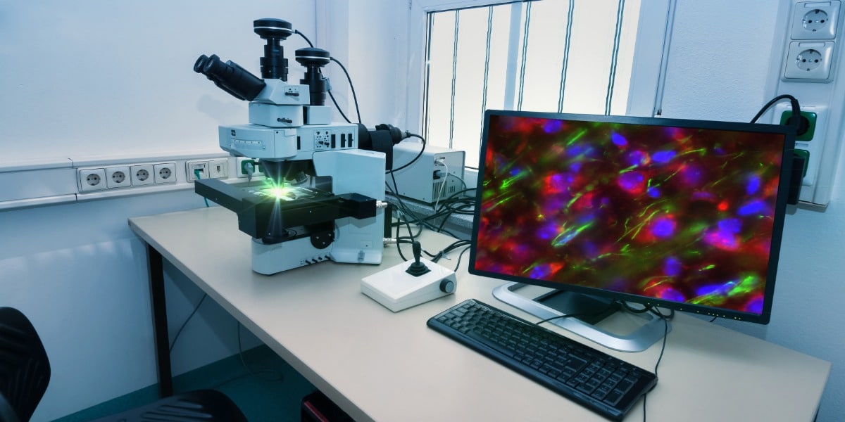
Fluorescence microscopy harnesses the ability of specific organic molecules or proteins (fluorophores) to absorb light at one wavelength and emit it at another, longer wavelength. Unlike widefield microscopy, this technique does not simply illuminate the sample indiscriminately; rather, it selectively excites fluorophores that have been introduced into the sample through labeling. This selective illumination significantly improves image contrast and specificity, enabling the detailed visualization of particular proteins, organelles, or other cellular structures.
Image Formation and Technical Aspects
In fluorescence microscopy, the image is formed by detecting light specifically emitted from excited fluorophores. To achieve this, the microscope is equipped with specialized filters such as excitation, emission, and dichroic mirrors which are critical in separating the excitation light from the emitted fluorescence signal. This precise separation not only improves contrast but also allows for high-resolution imaging of targeted areas. The technical constraints include a more complex optical setup and the potential need for advanced methods like confocal microscopy to further improve the rejection of out-of-focus light.
Applications and Limitations
Fluorescence microscopy is extensively used in the fields of cell biology, molecular biology, and biochemistry due to its ability to achieve:
- High sensitivity in detecting specific molecules or structures.
- Detailed mapping of subcellular structures using fluorescent dyes, proteins, or probes.
- Dynamic studies to monitor cellular processes and interactions in real time.
Its limitations, however, include potential photobleaching (where the fluorophores fade with prolonged exposure) and the necessity to use specialized fluorophores which might not always be compatible with all specimens.
Comparative Analysis Through a Detailed Table
| Feature | Widefield Microscopy | Fluorescence Microscopy |
|---|---|---|
| Illumination | Uniform, whole-sample illumination using broad-spectrum sources. | Selective excitation of fluorophores, using specific wavelengths. |
| Image Formation | Direct capturing of light passing through the sample; may include out-of-focus light. | Formation of images based on emitted fluorescence, significantly improving contrast. |
| Speed | Fast imaging, ideal for live-cell applications and high-throughput screening. | Relatively slower due to the need for precise excitation and filtering; can be enhanced with modern techniques. |
| Cost and Complexity | Simpler, less complex, and generally less expensive. | More expensive, with a complex setup including filters and sensitive detectors. |
| Resolution | Sufficient for many applications but compromised by background noise from out-of-focus light. | Provides higher resolution of specific structures, especially when utilized in confocal setups. |
| Applications | Brightfield, DIC, phase contrast imaging; overall morphology and dynamics of the sample. | Targeted imaging of proteins, organelles, and cellular interactions; tracing molecular pathways. |
| Limitations | Blurred images in thicker specimens; limited molecular specificity. | Potential photobleaching; requires specialized fluorophores; complexity in setup and maintenance. |
Integrating Applications, Strengths, and Weaknesses
Broad Application Spectrum
The decision to use either widefield or fluorescence microscopy largely depends on the specific research goal. For projects that require simple, rapid identification of structural features and dynamic behavior in live cells, widefield microscopy stands out as a reliable technique. In contrast, when the aim is to study particular proteins, localize organelles, or monitor specific molecular interactions within cells, fluorescence microscopy is indispensable.
Often in modern laboratories, these techniques are not mutually exclusive. Widefield fluorescence microscopes combine the advantage of broad illumination with the benefits of fluorescence markers, providing a compromise that capitalizes on rapid imaging with high specificity.
Enhancement Through Advanced Systems
Technology Evolution: Confocal Microscopy and Beyond
An evolution within fluorescence microscopy is the development of confocal microscope systems. Confocal microscopy further refines the image by using a spatial pinhole to eliminate out-of-focus light, yielding images with improved resolution and depth discrimination. While these advanced systems retain the benefits of fluorescence techniques, they also share the inherent complexities and higher costs associated with fluorescence microscopy setups.
Recent advances have led to high-speed confocal and spinning disk confocal microscopes that minimize the time lag traditionally associated with fluorescence imaging. These systems cater to live-cell imaging requirements while still providing detailed, high-contrast visualizations.
Strategic Considerations for Microscope Selection
Sample Characteristics
When selecting the appropriate microscope, consideration of the specimen's properties is key. For thin samples with minimal background autofluorescence, widefield microscopy, particularly in its fluorescence variant, is efficient. However, thicker specimens often benefit from the enhanced depth resolution of confocal systems, which mitigate the influence of out-of-focus signals.
Experimental Objectives
Researchers must weigh the trade-offs between imaging speed, resolution, and specificity. The rapid data acquisition of widefield microscopy is a tremendous asset for kinetic studies and high-throughput analyses. In contrast, experiments that require precise localization and identification of cellular components rely heavily on fluorescence microscopy’s high-contrast imaging capabilities.
Instrumentation and Economic Trade-offs
Instrumentation cost and operational complexity have a significant influence on method selection in both academic and industrial settings. Widefield microscopes offer a more economical solution for routine imaging, making them widely accessible. On the other hand, the specialized components required for fluorescence systems – such as high-performance cameras, specific filters, and high-intensity light sources – typically result in higher investment costs.
Furthermore, widefield systems are simpler to operate due to their minimal need for calibration and fewer moving parts. This simplicity enhances their utility in environments where ease of use and fast turnaround times are critical. In contrast, the enhanced specificity and resolution offered by fluorescence microscopy justify the additional complexity and cost in advanced molecular and cellular research.
Comparative Perspective Through a Case Study
Case Study: Live-Cell Imaging Versus Targeted Molecular Mapping
Consider a scenario in which a research team aims to monitor dynamic cellular responses to a pharmaceutical agent. For live-cell imaging, a widefield microscope can capture rapid changes across entire cell populations, providing critical contextual information about the overall cellular morphology. However, if the objective shifts to identifying the precise location of a specific receptor or protein within these cells, the team might adopt widefield fluorescence microscopy. By employing fluorescent markers that target the protein of interest, the researchers can observe its distribution with high contrast and specificity – a task that would be less discernible with traditional widefield imaging alone.
This dual approach illustrates how the inherent strengths of both methodologies can be synergistically employed to achieve comprehensive insights. The initial broad overview yields necessary context while targeted fluorescence imaging hones in on the specific molecular details.
Key Takeaways for Researchers
- Select based on goal: Choose widefield for rapid, general imaging and fluorescence for detailed, molecule-specific studies.
- Weigh cost vs. performance: Consider the complexities, costs, and technical support required when deciding on fluorescence microscopy setups, especially for high-resolution applications.
- Integrate techniques: In many state-of-the-art laboratories, combining widefield fluorescence methods can offer the best of both worlds—balancing speed, resolution, and specificity.
References
- IF Imaging: Widefield vs. Confocal Microscopy - PTG Lab
- Comparing Confocal and Widefield Fluorescence Microscopy - Evident Scientific
- Focal Wars: Widefield vs. Confocal - BioCompare
- Differences Between Widefield and Confocal Microscopy - Visikol
- Differences Between Confocal and Widefield Microscopy - Axiom Optics
Recommended Searches
Last updated March 20, 2025