
Unlocking the Bladder's Secrets: How Nerves and Reflexes Control Urination
Explore the intricate neural pathways governing the essential process of micturition, from automatic reflexes to conscious control.
Essential Insights
- The Micturition Reflex: This is an automatic process triggered by bladder stretching, involving signals between the bladder, spinal cord, and brain to initiate urination.
- Dual Nerve Control: The bladder is controlled by both the autonomic (parasympathetic for emptying, sympathetic for storage) and somatic (voluntary sphincter control) nervous systems.
- Coordinated Action: Normal urination requires precise coordination between bladder muscle (detrusor) contraction and urethral sphincter relaxation, managed by complex neural circuits.
Understanding Micturition: More Than Just Letting Go
Micturition, commonly known as urination or voiding, is the physiological process of expelling urine from the urinary bladder through the urethra. It might seem simple, but it's orchestrated by a sophisticated interplay between the bladder muscles, the nervous system, and conscious control. Central to this is the micturition reflex, a complex neural pathway that governs when and how we urinate.
The Two Phases of Bladder Function
The bladder cycle primarily consists of two distinct phases:
1. The Storage Phase (Filling)
During this phase, the bladder functions as a compliant reservoir, gradually filling with urine produced by the kidneys. To maintain continence (prevent leakage), several coordinated actions occur:
- Detrusor Muscle Relaxation: The smooth muscle wall of the bladder (the detrusor muscle) remains relaxed. This allows the bladder to expand and accommodate increasing volumes of urine without a significant rise in internal pressure. Sympathetic nerve activity helps maintain this relaxation.
- Sphincter Contraction: Both the internal urethral sphincter (smooth muscle, involuntary control) and the external urethral sphincter (skeletal muscle, voluntary control) remain tightly closed. Sympathetic nerves contribute to keeping the internal sphincter closed, while the somatic nervous system maintains tone in the external sphincter.
2. The Voiding Phase (Emptying)
This phase involves the active expulsion of urine from the bladder. It is initiated by the micturition reflex, which is triggered when the bladder reaches a certain level of fullness.
The Micturition Reflex: Step-by-Step
The micturition reflex is a fundamental spinal reflex that can be modulated (inhibited or facilitated) by higher brain centers. Here's how it unfolds:
- Bladder Distension & Sensory Signals: As the bladder fills (typically reaching 300-400 mL in adults), stretch receptors embedded within the bladder wall become activated.
- Afferent Pathway: These receptors send nerve impulses (afferent signals) via the pelvic nerves to the sacral region of the spinal cord (specifically segments S2-S4).
- Spinal Cord Processing: Within the sacral spinal cord, these signals initiate a reflex arc. The information is also relayed upwards to higher brain centers, including the pons and cerebrum, creating the conscious sensation of needing to urinate.
- Efferent Parasympathetic Response: The spinal cord sends signals back to the bladder via parasympathetic efferent fibers (also within the pelvic nerves).
- Detrusor Contraction: Parasympathetic stimulation causes the detrusor muscle to contract, increasing pressure inside the bladder.
- Internal Sphincter Relaxation: Simultaneously, parasympathetic activity (and inhibition of sympathetic activity) leads to the relaxation of the internal urethral sphincter.
- External Sphincter Relaxation: For urination to occur, the external urethral sphincter must also relax. This involves inhibition of the somatic motor neurons travelling via the pudendal nerve. In infants, this relaxation is purely reflexive. In adults, it's under voluntary control.
- Role of Higher Brain Centers: The pontine micturition center (PMC) in the brainstem acts as a crucial coordination center. It receives input about bladder fullness and signals from the cerebral cortex (regarding the appropriateness of urinating). The PMC can facilitate the reflex by coordinating detrusor contraction and sphincter relaxation, or inhibit it via descending pathways if the time isn't right. Voluntary initiation involves consciously relaxing the external sphincter and allowing the pontine center to facilitate the reflex.
This reflex is described as "self-regenerative" because the initial detrusor contraction further stimulates the stretch receptors, reinforcing the signals to the spinal cord and strengthening the contraction until the bladder is sufficiently emptied.
The Neural Highway: Bladder Innervation
Effective bladder control relies on a complex network of nerves connecting the bladder and sphincters to the spinal cord and brain. This network includes components from the autonomic (involuntary) and somatic (voluntary) nervous systems.
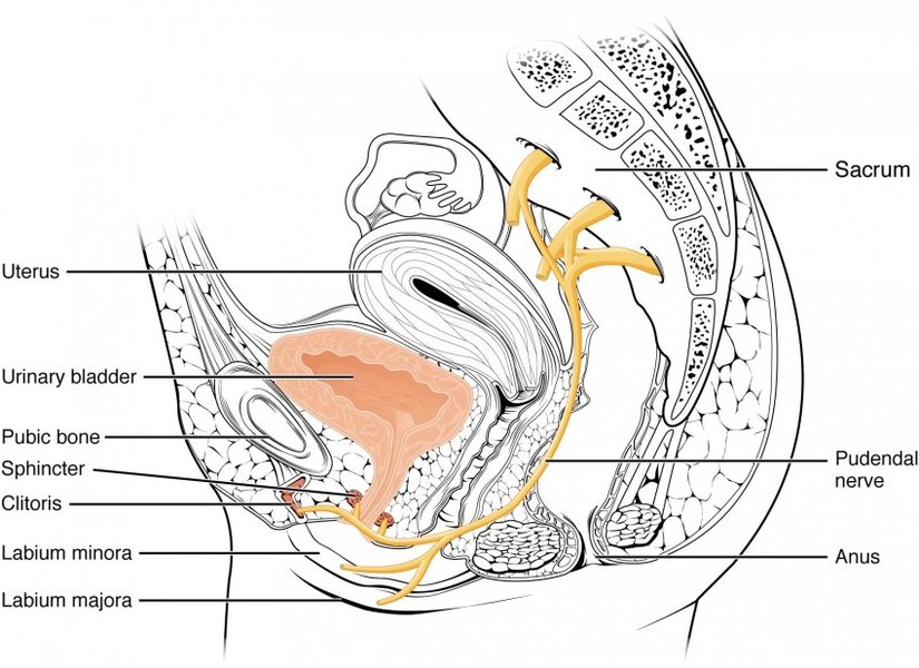
Diagram showing the sympathetic, parasympathetic, and somatic nerves innervating the bladder and urethral sphincters.
Autonomic Nervous System Control
The autonomic system manages the involuntary aspects of bladder function – storage and the reflexive part of emptying.
Parasympathetic Innervation (The "Emptying" Nerves)
- Origin: Arises from the sacral spinal cord segments (S2, S3, S4).
- Nerves: Travels via the pelvic splanchnic nerves to the bladder wall and internal sphincter.
- Function: Primarily responsible for the voiding phase. Stimulation causes:
- Contraction of the detrusor muscle.
- Relaxation of the internal urethral sphincter.
- Sensory Role: Also carries afferent sensory information (like stretch/fullness and pain) from the bladder back to the spinal cord.
Sympathetic Innervation (The "Storage" Nerves)
- Origin: Arises from the thoracolumbar spinal cord segments (T10/T11 to L2).
- Nerves: Travels via the hypogastric nerves to the bladder wall and internal sphincter.
- Function: Primarily responsible for the storage phase. Stimulation causes:
- Relaxation of the detrusor muscle (allowing filling).
- Contraction of the internal urethral sphincter (preventing leakage).
Somatic Nervous System Control
The somatic system provides voluntary control over urination.
Somatic Innervation (Voluntary Control)
- Origin: Arises from the sacral spinal cord segments (S2, S3, S4).
- Nerves: Travels via the pudendal nerve.
- Function: Innervates the external urethral sphincter (a skeletal muscle). Provides tonic contraction to maintain continence and allows for conscious relaxation to initiate or permit urination. It also allows for voluntary interruption of urine flow.
Visualizing the Micturition Pathway
This mindmap provides a simplified overview of the key components and processes involved in micturition control, illustrating the relationship between the bladder, nervous system components, muscles, and functional phases.
(Pelvic Nerves S2-S4)"] id2a1a["Contracts Detrusor"] id2a1b["Relaxes Internal Sphincter"] id2a2["Sympathetic
(Hypogastric Nerves T10-L2)"] id2a2a["Relaxes Detrusor"] id2a2b["Contracts Internal Sphincter"] id2b["Somatic"] id2b1["Pudendal Nerve (S2-S4)"] id2b1a["Controls External Sphincter
(Voluntary)"] id2c["Control Centers"] id2c1["Sacral Spinal Cord (S2-S4)
Reflex Center"] id2c2["Pontine Micturition Center (PMC)
Coordination"] id2c3["Cerebral Cortex
Voluntary Control"] id3["Micturition Reflex"] id3a["Trigger: Bladder Stretch"] id3b["Pathway: Afferent -> Spinal Cord -> Efferent"] id3c["Outcome: Detrusor Contraction & Sphincter Relaxation"] id4["Phases"] id4a["Storage Phase
(Sympathetic Dominant)"] id4b["Voiding Phase
(Parasympathetic Dominant)"]
Coordination: Putting It All Together
Normal urination is a marvel of neural coordination. During storage, sympathetic and somatic pathways dominate, keeping the bladder relaxed and the outlet sealed. As the bladder fills, sensory signals increase. When the time and place are appropriate, conscious control centers in the brain signal the PMC. The PMC then orchestrates a switch: sympathetic and somatic outflow are inhibited, while parasympathetic outflow is activated. This leads to detrusor contraction and coordinated relaxation of both sphincters, resulting in efficient bladder emptying.
Summary of Nerve Actions in Micturition
The following table summarizes the primary roles of the different nerve types during the storage and voiding phases of micturition:
| Nerve Type | Origin | Nerve Pathway | Target Muscle(s) | Primary Action in Storage Phase | Primary Action in Voiding Phase |
|---|---|---|---|---|---|
| Sympathetic | T10-L2 | Hypogastric Nerves | Detrusor Muscle, Internal Sphincter | Relaxes Detrusor, Contracts Internal Sphincter | Inhibited |
| Parasympathetic | S2-S4 | Pelvic Splanchnic Nerves | Detrusor Muscle, Internal Sphincter | Minimal Activity | Contracts Detrusor, Relaxes Internal Sphincter |
| Somatic | S2-S4 | Pudendal Nerve | External Sphincter | Contracts External Sphincter (Tonic/Voluntary) | Relaxes External Sphincter (Voluntary Inhibition) |
Neural Activity During Micturition Phases
This radar chart illustrates the relative activity levels of key neural components during the bladder's storage and voiding phases. Note how sympathetic and somatic activity dominate during storage to maintain continence, while parasympathetic activity surges during voiding to facilitate emptying. Sensory input increases significantly as the bladder fills, triggering the voiding reflex.
Visualizing the Process: Micturition Animation
This animation provides a clear visual explanation of the neural control involved in the micturition reflex, showing how signals travel and coordinate the actions of the bladder and sphincters during both filling and emptying phases. It helps illustrate the complex interactions described above.
Clinical Relevance: When Control Goes Wrong
Understanding the micturition reflex and bladder innervation is crucial for diagnosing and managing urinary problems. Damage to the nerves controlling the bladder, often due to spinal cord injury, multiple sclerosis, stroke, diabetes, or pelvic surgery, can lead to **neurogenic bladder**. This condition encompasses various dysfunctions, including:
- Urinary Incontinence: Difficulty storing urine due to an overactive detrusor muscle or weak sphincters.
- Urinary Retention: Difficulty emptying the bladder due to an underactive detrusor muscle or sphincters that don't relax properly.
Treatment often involves medications, behavioral therapies, or other interventions aimed at restoring more normal bladder function by targeting specific aspects of the neural control pathway.
Frequently Asked Questions (FAQ)
What exactly triggers the urge to urinate?
The urge to urinate is primarily triggered by the stretching of the bladder wall as it fills with urine. Specialized stretch receptors in the detrusor muscle detect this distension. When the bladder volume reaches a certain threshold (variable, but often around 200-400 mL), these receptors send increasingly frequent signals via afferent nerves (pelvic nerves) to the sacral spinal cord and then up to the brain, creating the conscious sensation of bladder fullness and the need to void.
How does voluntary control override the reflex?
Voluntary control, primarily learned during toilet training, involves higher brain centers (like the cerebral cortex) modulating the basic micturition reflex. These centers can send inhibitory signals down the spinal cord to:
- Suppress the pontine micturition center, preventing it from facilitating the voiding reflex.
- Maintain or increase the contraction of the external urethral sphincter via the somatic (pudendal) nerve.
This allows a person to delay urination even when the bladder is full and the reflex is initiated. Conversely, voluntary urination involves consciously relaxing the external sphincter and allowing the brain centers to facilitate the pontine micturition center's action.
What roles do the internal and external sphincters play?
Both sphincters act as valves to control urine outflow:
- Internal Urethral Sphincter: Located at the junction of the bladder and urethra, this is made of smooth muscle and is under involuntary (autonomic) control. It remains contracted during the storage phase (aided by sympathetic nerves) and relaxes during the voiding phase (due to parasympathetic activity and sympathetic inhibition).
- External Urethral Sphincter: Located further down the urethra, this is made of skeletal muscle and is under voluntary (somatic) control via the pudendal nerve. It provides the primary mechanism for consciously holding urine and initiating or stopping the stream.
Can the micturition reflex be affected by emotions?
Yes, emotional states like anxiety or stress can influence bladder function. The brain centers involved in processing emotions have connections to the areas controlling micturition (like the PMC and cortical areas). Strong emotions can sometimes trigger an urgent need to urinate or, conversely, make it difficult to relax enough to void, demonstrating the link between the brain's emotional and autonomic control centers.
Recommended Reading
- Explore the detailed anatomy of the urinary bladder and urethra.
- Investigate the causes and available treatments for neurogenic bladder dysfunction.
- Learn how toilet training helps children develop voluntary control over the micturition reflex.
- Understand the key differences in bladder control mechanisms between infants and adults.
References
Last updated May 2, 2025