
Unveiling the Neural Mysteries of Mood Disorders
Exploring which brain activity claim does not align with neuroimaging findings
Key Takeaways
- Anterior cingulate misconception: Contrary to common assertions, activity in the anterior cingulate is often increased (especially in the subgenual region) in depression rather than diminished.
- Validated correlations: Elevated striatal activity correlates with mania, and amygdala overactivity is well-documented in depression.
- Complex hippocampal dynamics: Bipolar disorder features hippocampal changes, but evidence suggests that the simplified view of diminished hippocampal activity does not capture the full nuance.
Comprehensive Analysis of Brain Function in Mood Disorders
The human brain is a sophisticated organ with multiple circuits orchestrating behavior, emotions, and cognitive functions. In mood disorders such as depression and bipolar disorder, neuroimaging studies have revealed variances in the functioning of several brain regions. Researchers have utilized advanced techniques like functional magnetic resonance imaging (fMRI) and positron emission tomography (PET) to explore these differences in brain activation.
Dissecting the Four Statements
We are tasked with identifying which of the four statements about brain function in people with mood disorders is not correct. Let’s evaluate each option individually:
Statement A: Mania and Elevated Striatum Activity
Mania, a hallmark of bipolar disorder, is associated with an abnormal surge in emotional arousal, impulsivity, and goal-directed behavior. Many neuroimaging studies have reported that during manic episodes, the brain's reward centers—most notably the striatum—display significantly elevated activity. The striatum is instrumental in processing rewards, motivation, and decision-making. Elevated striatal activity corresponds with the heightened emotional and motivational states seen in manic individuals. Therefore, this statement aligns with current neuroscientific findings and is accepted as accurate.
Statement B: Anterior Cingulate Activity and Depression
The anterior cingulate cortex (ACC) plays a central role in emotion regulation, integration of cognitive inputs, and error detection. In many cases of depression, particularly when focusing on the subgenual anterior cingulate cortex (sgACC), neuroimaging studies have demonstrated hyperactivity rather than diminished activity. This hypermetabolism in the ACC appears linked to the persistent negative mood states and difficulties in modulating emotional responses. Although some early literature might have suggested reduced activation under specific conditions, the prevailing consensus is that the ACC, particularly the sgACC, tends to have increased activity in depressed individuals. Thus, the statement asserting that it is "diminished" does not accurately reflect these robust findings.
Statement C: Amygdala Overactivity in Depression
The amygdala is a critical hub in the brain for processing emotions, notably fear, anxiety, and emotional memory. In patients with depression, there is substantial evidence from neuroimaging studies showing hyperactivity in the amygdala during tasks that involve emotional processing. This overactivity is believed to contribute to the heightened sensitivity to negative emotional stimuli and the emotional dysregulation that is characteristic of depression. Accordingly, the statement regarding amygdala overactivity is in line with current research.
Statement D: Bipolar 1 Disorder and Hippocampal Activity
Bipolar 1 disorder involves dramatic mood swings between manic and depressive episodes, and the hippocampus plays a significant role in memory and emotion regulation. Many studies have identified structural and functional alterations in the hippocampus in individuals with bipolar disorder, including reductions in volume. While reduced hippocampal volume is a consistent finding, the complexity of functional activation in the hippocampus during various mood states means that a blanket statement asserting “diminished hippocampal activity” may oversimplify the reality. However, current evidence does tend to support some aspects of reduced hippocampal engagement, particularly associated with the chronicity of the illness. Nevertheless, compared to the more definitive evidence related to striatum and amygdala, the simplified description of hippocampal activity in bipolar disorder remains a topic open to nuance.
In synthesizing the above evaluations, the majority of research indicates that:
- The assertion that manic episodes correlate with elevated striatum activity is well-supported.
- The observation that there is overactivity in the amygdala in depression is also corroborated by extensive studies.
- The complexities linked to hippocampal activity in bipolar disorder are recognized, but the consensus often points toward alterations that may include diminished volume and functional changes.
- The description of anterior cingulate activity as being diminished in depressed individuals stands in contradiction to the preponderance of evidence indicating that this region is commonly hyperactive, particularly in its subgenual area.
Therefore, the statement that is not correct is the one suggesting:
"Anterior cingulate activity is diminished in people with depression."
This incorrect portrayal highlights the need to understand that in many cases of depression, it is the dysregulated increase in activity within regions like the subgenual anterior cingulate cortex that contributes to the pathology rather than a decrease.
Brain Function in Mood Disorders: A Visual Synthesis
To provide a comprehensive visualization of the relationships between different brain regions discussed in relation to mood disorders, the following radar chart summarizes our comparative evaluation. The chart includes multiple datasets that reflect the varied opinions on activity levels in key brain regions during different mood disorder states.
Supplementary Visuals and Resources
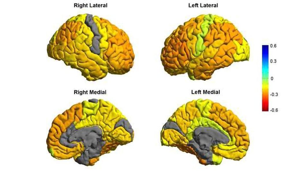
In addition to the radar chart summarizing brain function insights, the following images help illustrate neuroimaging findings and brain scans associated with mood disorders:


The first image depicts brain imaging research related to mood disorders, highlighting regions involved in emotional regulation. The second image provides an overview of various brain scan types, including those that detect structural and functional changes in conditions such as depression and bipolar disorder.
Detailed Comparative Table
The table below organizes our analysis of the statements regarding brain function in mood disorders. It delineates the brain region involved, the typical activity state in relevant conditions, and a brief note on research consensus.
| Brain Region | Mood Disorder Association | Activity State | Research Note |
|---|---|---|---|
| Striatum | Mania | Elevated | Increased reward and goal-directed behavior observed in manic episodes. |
| Anterior Cingulate Cortex (ACC) | Depression | Increased (Hyperactive, especially in the subgenual region) | Current evidence demonstrates hyperactivity rather than diminished function. |
| Amygdala | Depression | Overactive | Heightened emotional processing, particularly negative emotions, is evident. |
| Hippocampus | Bipolar 1 Disorder | Altered (Often reduced volume) | Structural changes noted; functional activity can be complex and state-dependent. |
Multimedia Insight
Below is a relevant video that discusses the neural underpinnings of mood disorders, including the role of various brain regions in mediating emotional states. This video by Yale Medicine explains how depression affects brain function, offering insights that complement the discussion provided here.
This video is particularly relevant because it visually demonstrates the neuroimaging findings pertinent to depression and highlights the alterations in activity within the anterior cingulate cortex, amygdala, and other key regions.
Expandable FAQ Section
References
-
Mood Disorders: What They Are, Symptoms & Treatment - Cleveland Clinic
-
Brain structural and functional abnormalities in mood disorders - PMC
- Mood Disorder - StatPearls - NCBI Bookshelf
- Mayo Clinic - Mood Disorders
- Mood Disorders | Johns Hopkins Medicine
Recommended Further Queries
Last updated April 3, 2025