
Pioneering Multicellular 3D-Printed Heart Models for Enhanced Drug Screening
Innovative strategies in 3D bioprinting for realistic human heart models
Key Insights
- Multicellular Integration: Incorporate diverse cardiac cell types to mimic native heart tissue complexity.
- Advanced Biomaterials & Bioinks: Utilize materials that simulate the extracellular matrix and support cell functionality.
- Vascularization & Dynamic Stimulation: Integrate vascular networks and apply mechanical/electrical stimuli to mature tissue architecture.
Overview of Multicellular 3D-Printed Heart Model Advancements
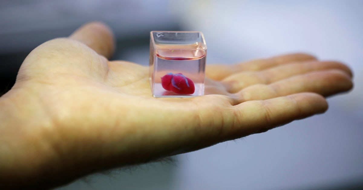
The journey towards enhancing drug screening capabilities using 3D-printed human heart models is multifaceted. By leveraging multicellular approaches, researchers aim to develop constructs that closely imitate the intrinsic architecture and physiology of native cardiac tissue. This advancement not only improves the predictive accuracy of drug testing but also facilitates the study of complex cellular interactions under both normal and pathophysiological conditions. Several key areas of innovation include the integration of diverse cell types, development of sophisticated bioinks, incorporation of vascular networks, and implementation of biofabrication technologies that enable precise cellular patterning.
Strategic Areas for Advancing Multicellular 3D-Printed Heart Models
1. Multicellular Integration and Cellular Heterogeneity
A critical strategy involves incorporating multiple cardiac cell types into a single 3D-printed construct to mirror the complexity of the human heart:
Diverse Cell Types
Native cardiac tissue comprises not only cardiomyocytes but also fibroblasts, endothelial cells, and smooth muscle cells. The inclusion of these cells in 3D prints ensures that the model recapitulates the cellular heterogeneity of the heart, thereby enabling better simulation of physiological responses to pharmacological agents.
Stem Cell-Derived Cardiac Cells
Utilizing induced pluripotent stem cells (iPSCs) to derive cardiomyocytes and other cardiac cell types further personalizes and enhances the accuracy of the models. These patient-specific cells can be used to study drug responses in a manner that reflects individual variability in cardiac function and disease states.
2. Advanced Biomaterials and Bioink Innovation
The creation of realistic heart models demands the development of bioinks and biomaterials that emulate the extracellular matrix (ECM) found in native cardiac tissue. These materials must foster cell adhesion, viability, proliferation, and differentiation.
Designing Bioactive Materials
Advanced bioinks, particularly those loaded with growth factors and nanoengineered to mimic the ECM, are pivotal. They enable a supportive microenvironment that drives the functional assembly of cells. Hydrogels, specifically engineered to provide the right balance of mechanical strength and biodegradability, enhance the structural fidelity of printed tissues.
Biomimicry in Bioink Formulation
Engineering bioinks that accurately reflect the mechanical properties of native heart tissue ensures that the 3D-printed models are not only structurally robust but also physiologically relevant. This approach has a direct impact on the contractile behavior and functional maturation of the cardiomyocytes.
3. Vascularization and Perfusion Strategies
In native heart tissue, dense vascular networks are essential for oxygen and nutrient delivery, as well as waste removal. Replicating these networks in 3D-printed heart models is crucial for maintaining cellular viability and function.
Developing Vascular Networks
Techniques such as sacrificial templating and co-printing with endothelial cells can be utilized to produce intricate vascular structures. These strategies ensure that the printed tissues remain viable during long-term experiments and accurately represent the flow dynamics present in human hearts.
Integration of Microfluidic Systems
Incorporating microfluidic channels into the heart model recapitulates blood flow conditions. This dynamic environment facilitates real-time monitoring of drug diffusion, tissue perfusion, and metabolic waste clearance, leading to more precise drug screening outcomes.
4. Enhanced Bioprinting Techniques and Precision Printing
The evolution of 3D printing methodologies has dramatically improved the resolution and complexity of tissue constructs. Enhanced printing techniques enable the spatial arrangement of multiple cell types in defined architectures.
Multi-material and High-resolution Printing
Techniques such as extrusion-based and laser-assisted bioprinting allow for the controlled deposition of cells and bioinks. This precision is critical for establishing complex tissue geometries that closely mimic in vivo heart structures.
Freeform Reversible Embedding of Suspended Hydrogels (FRESH)
FRESH bioprinting offers a promising approach to construct delicate and intricate cardiac structures with high fidelity. This method supports the formation of micro-scale features that are essential for replicating the native heart microenvironment.
5. Functionalization and Real-Time Assessment
Beyond structural mimicry, functional accuracy is imperative for drug screening applications. The implementation of advanced sensing and stimulation techniques plays a key role in this regard.
Electrical and Mechanical Stimulation
Applying electrical pulses or mechanical conditioning promotes cellular alignment, contractility, and tissue maturation. Such stimulation is crucial for mimicking the electrophysiological properties of the heart, thereby enhancing the model’s responsiveness to drug treatment.
Integration of Biosensors
Embedding biosensors within the 3D-printed heart models enables real-time monitoring of cellular activities such as calcium handling and contraction kinetics. This integrated approach aids in evaluating the pharmacodynamics of drugs under study.
6. Standardization, Validation, and Ethical Considerations
To ensure that 3D-printed heart models become widely accepted tools in drug screening, the establishment of standardized protocols and rigorous validation is essential.
Developing Standard Protocols
Consistency in fabrication processes, culture conditions, and data recording must be maintained across laboratories. Standardizing these methods facilitates reproducibility and enables valid cross-study comparisons.
Conducting Comprehensive Validation Studies
Comparative studies that correlate drug responses in 3D-printed models with clinical outcomes are fundamental to validate the predictive power of these systems. Such data-driven validation supports the integration of these models into routine drug development pipelines.
Ethical and Regulatory Frameworks
As advancements progress, engaging with regulatory bodies to establish guidelines for 3D printed models is vital. Addressing ethical concerns related to the use of stem cells and patient-derived materials secures public trust and promotes responsible research practices.
Comparative Table of Key Strategies
| Strategy Area | Description | Benefits |
|---|---|---|
| Multicellular Integration | Incorporates diverse cell types including cardiomyocytes, fibroblasts, and endothelial cells. | Accurately mimics native tissue complexity and improves drug response prediction. |
| Advanced Biomaterials | Development of bioinks that simulate the extracellular matrix with proper mechanical properties. | Enhances cell adhesion, growth, and differentiation, resulting in robust tissue formation. |
| Vascularization | Creating networks using sacrificial templating and microfluidic integration. | Ensures nutrient delivery and waste removal, thereby supporting long-term tissue viability. |
| High-Resolution Bioprinting | Utilization of techniques such as FRESH, extrusion, and laser-assisted printing. | Enables precise spatial placement of multiple cell types, fostering complex tissue architectures. |
| Functionalization | Embedding biosensors and applying electrical/mechanical stimuli. | Promotes tissue maturation and real-time monitoring of drug effects, enhancing screening accuracy. |
| Standardization & Validation | Developing protocols and comparative studies to ensure reproducibility. | Strengthens the reliability of the models and supports regulatory acceptance. |
Integrating Advanced Technologies for Future Innovations
The future of 3D-printed human heart models lies at the intersection of advanced biofabrication, material science, and integrated sensor technologies. Researchers are exploring the use of machine learning to optimize printing parameters. This can result in improvements in tissue morphology and enhanced predictive accuracy of drug responses. Automation in bioprinting can also streamline the fabrication process and reduce variability.
Additionally, combining high-resolution imaging techniques with 3D bioprinting opens new avenues for real-time monitoring of tissue development. Optical coherence tomography (OCT) and fluorescence microscopy might be integrated within culture systems to provide continuous feedback during tissue maturation. These advancements ensure that the heart models not only look anatomically accurate but also perform functionally similar to their biological counterparts.
Another emerging dimension is the integration of computational modeling with printed constructs. Simulation techniques, based on finite element analysis (FEA), can assist in predicting the mechanical behavior of 3D printed tissues under various physiological conditions. When combined with real-world experimental data, these models provide deeper insights into tissue engineering and drug screening processes.
Opportunities and Future Directions
The continuous evolution of biofabrication techniques points to significant opportunities in personalized medicine. By using patient-specific cells and designing customizable tissue architectures, researchers can create heart models tailored for individual drug screening. This personalized approach not only minimizes adverse reactions but also contributes to the development of targeted therapies.
Ongoing improvements in bioprinting resolution and multi-material applications are expected to lead to even more sophisticated models that combine cellular, structural, and functional fidelity. Integration with automated monitoring systems and real-time analytics will further enhance the ability of these models to serve as reliable platforms for preclinical drug testing.
References
- MDPI - Advances in Cardiac 3D Bioprinting
- Nature - 3D Bioprinted Cardiac Tissue
- PubMed - Multicellular Vascular Models
- Oxford Academic - Stem Cell-Derived Cardiac Tissue
- Frontiersin - Bioengineering and Biotech Advances
Recommended Related Queries
Last updated March 23, 2025