
Unveiling Brain Networks: How Prepulse Inhibition and Functional Neurological Disorders Reshape Our Understanding
Exploring the intricate neural circuits underlying sensorimotor gating and FND, focusing on key brain regions and connectivity.
Understanding the brain's complex mechanisms requires looking closely at specific processes and disorders. Prepulse Inhibition (PPI) offers a window into sensorimotor gating – the brain's ability to filter sensory information – while Functional Neurological Disorder (FND) highlights disruptions in brain communication pathways. Neuroimaging techniques like functional Magnetic Resonance Imaging (fMRI) and functional connectivity analyses are crucial tools in mapping the neural correlates of both.
Key Insights into PPI and FND Neural Correlates
- Shared Hubs: Regions like the insula and temporoparietal junction (TPJ) emerge as critical nodes involved in modulating both PPI and the altered neural processing seen in FND.
- Network Disruptions: FND is characterized by changes in how brain networks function and communicate, rather than structural damage, impacting sensorimotor control, emotional regulation, and predictive processing.
- Sensorimotor Gating: PPI relies on a complex circuit including the striatum, thalamus, insula, and temporal/parietal areas to effectively filter sensory inputs and prevent motor system overload. Deficits are linked to various neuropsychiatric conditions.
Decoding Prepulse Inhibition (PPI): The Brain's Sensory Filter
What is PPI and Why Does it Matter?
Prepulse Inhibition (PPI) is a fundamental neuropsychological process. It describes the phenomenon where a weak sensory stimulus (the "prepulse") presented shortly before a strong, startle-inducing stimulus (the "pulse") significantly reduces the magnitude of the startle reaction. Think of it as the brain's automatic volume control for sudden, loud noises or unexpected touches, preventing an exaggerated response when a preceding cue signals something is coming. This mechanism, known as sensorimotor gating, is crucial for filtering irrelevant sensory information and focusing attention.
Deficits in PPI are not just a laboratory curiosity; they serve as an important biomarker or endophenotype for several neuropsychiatric conditions, including schizophrenia and Tourette Syndrome, suggesting underlying problems in how the brain processes sensory information and inhibits motor responses.

Illustration of the Prepulse Inhibition Paradigm
This image depicts the typical setup for measuring PPI, showing how a prepulse stimulus presented before a startling pulse stimulus reduces the resulting startle response (measured, for example, by eye blink). Studies often use acoustic or tactile stimuli.
Neural Circuits Orchestrating PPI
Functional neuroimaging, particularly fMRI, has been pivotal in mapping the brain networks responsible for PPI. Research points to a core circuitry involving several interconnected regions:
The Cortico-Striato-Thalamo-Pontine Pathway
This pathway is considered central to PPI. Studies consistently show activation and modulation within these areas:
- Striatum (including the Putamen): Plays a critical role in gating sensory information and modulating motor output. fMRI studies show activation here during successful PPI, and dysfunction is linked to PPI deficits in conditions like schizophrenia. The putamen specifically contributes to motor control aspects of the startle response and its inhibition.
- Thalamus: Acts as a major sensory relay station, filtering information before it reaches the cortex. Its interaction with the striatum is vital for gating.
- Pons (Pontine Reticular Formation): Contains the primary startle pathway nuclei, where the inhibition ultimately occurs.
Modulatory Roles of Cortical and Limbic Regions
Beyond the core pathway, other brain areas modulate PPI, integrating sensory, emotional, and cognitive information:
- Insula: Increasingly recognized for its role in sensory integration, interoception (sensing the body's internal state), and emotional awareness. fMRI studies using tactile and acoustic PPI paradigms show insula activation, suggesting it helps evaluate the significance of the prepulse and modulate the gating process. Its connectivity with other regions like the TPJ and prefrontal cortex is crucial.
- Temporoparietal Junction (TPJ): A hub for integrating sensory information, attention, and self-awareness. Functional connectivity studies highlight its interaction with the insula during PPI, potentially facilitating the coordination of sensory predictions and attentional allocation required for effective gating. Rightward asymmetry in TPJ-insula connectivity has been noted.
- Superior Temporal Gyrus (STG), Planum Temporale, Supramarginal Gyrus: These temporal and parietal areas are heavily involved in auditory and multimodal sensory processing. They contribute to the initial processing and filtering of the prepulse stimulus, especially in acoustic PPI paradigms. Their functional connectivity with the insula and TPJ helps form the network that attenuates the startle response.
- Prefrontal Cortex (PFC): Involved in top-down control and attention. Studies suggest areas like the dorsolateral PFC (dlPFC) exert inhibitory control over startle circuitry, potentially via connections with the insula, especially when attention modulates PPI.

Brain Activation during Tactile PPI
This fMRI image highlights brain regions, including temporal and parietal areas, showing differential activation during a tactile prepulse inhibition task, illustrating the complex network involved.
Functional Connectivity Insights
Functional connectivity studies, often using fMRI, examine how these brain regions interact dynamically during PPI. They reveal distinct networks for startle mediation and PPI modulation. For instance, inverse connectivity patterns (where activity in one area decreases as activity in another increases) between regions like the anterior insula and dlPFC suggest active inhibitory control mechanisms are engaged during successful PPI.
Understanding Functional Neurological Disorder (FND)
When Brain Communication Goes Awry
Functional Neurological Disorder (FND), sometimes previously termed conversion disorder, is a condition characterized by neurological symptoms (e.g., weakness, paralysis, tremor, sensory loss, seizures, gait problems) that are real and distressing but are not caused by traditional, structurally defined neurological diseases. Instead, FND is understood as a disorder of brain function – a problem with how the brain's hardware is operating, leading to disruptions in the communication pathways between the brain and the body.
The core issue lies in altered brain network activity and connectivity, affecting how signals for movement, sensation, and even awareness are generated, processed, and transmitted.
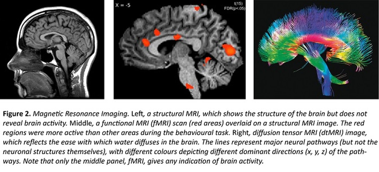
Neuroimaging Techniques like fMRI
Functional MRI (fMRI) is a key tool used to study brain activity changes in conditions like FND and during tasks like PPI by detecting changes associated with blood flow.
Neural Mechanisms Implicated in FND
Neuroimaging research in FND is rapidly evolving, moving away from searching for a single "lesion" towards understanding distributed network dysfunction. Key findings include:
- Altered Sensorimotor Control Networks: Studies often show abnormal activity and connectivity in brain regions responsible for planning, executing, and monitoring movement, even when structural scans are normal.
- Disrupted Predictive Processing: Some models suggest FND involves faulty "predictive coding," where the brain's expectations or predictions about sensory input or movement outcomes don't match reality, leading to symptoms. Areas involved in generating these predictions and processing prediction errors show altered function.
- Emotional Regulation and Limbic System Involvement: Regions involved in processing emotions (like the amygdala and insula) and regulating responses often show altered activity and connectivity in FND, potentially interacting with sensorimotor networks. Stress and past trauma are known risk factors.
- Role of Attention and Agency: Aberrant attentional processes and disruptions in the sense of agency (the feeling of controlling one's own actions) are also implicated, often linked to altered function in parietal regions like the TPJ and prefrontal areas.
Involvement of Specific Brain Regions
Many of the brain regions highlighted in PPI research also show alterations in FND:
- Insula: Dysfunction in the insula is frequently reported in FND, potentially related to altered interoception, emotional processing, and integration of bodily signals.
- Temporoparietal Junction (TPJ): Altered activity and connectivity involving the TPJ are linked to FND symptoms, possibly due to its role in multisensory integration, body representation, and agency.
- Putamen and Basal Ganglia: While primarily functional, some studies suggest subtle structural or functional changes in basal ganglia circuits, including the putamen, which could contribute to motor symptoms like tremor or weakness.
- Supplementary Motor Area (SMA) and Prefrontal Cortex: These areas involved in motor planning and executive control often show hypoactivity during FND motor symptoms.
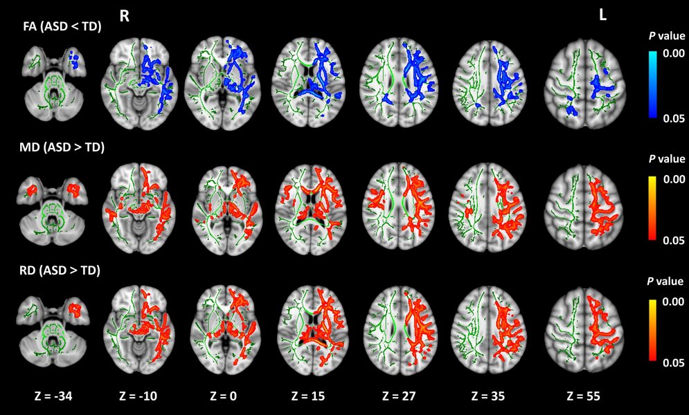
Brain Connectivity Networks
FND is increasingly understood as a disorder of brain network connectivity, where communication between different brain regions is disrupted.
Potential Intersections: PPI and FND
Shared Networks, Different Manifestations?
While studies directly investigating PPI specifically within FND populations appear limited based on current literature, there's a compelling overlap in the neural circuitry implicated in both. Both PPI (as a measure of sensorimotor gating) and FND involve disruptions in how the brain processes sensory information, regulates motor output, and integrates bodily signals with emotional and cognitive states.
Regions like the insula and TPJ are central players in both contexts. In PPI, they contribute to filtering and attentional modulation. In FND, their dysfunction is linked to altered self-awareness, emotional dysregulation, and aberrant sensorimotor control. It's plausible that underlying vulnerabilities or alterations in these shared networks could manifest differently – contributing to subtle gating deficits measurable by PPI in some conditions, and overt neurological symptoms in FND under certain circumstances (like stress or attentional shifts).
Further research is needed to explore whether PPI deficits are present in FND and how sensorimotor gating mechanisms might be specifically affected in this disorder.
Relative Involvement of Key Brain Regions
A Comparative Neuroanatomical Perspective
This chart provides a conceptual overview comparing the relative degree of involvement or alteration of key brain regions in Prepulse Inhibition (PPI) processes versus Functional Neurological Disorder (FND), based on the synthesized findings from functional neuroimaging studies. The scores reflect the prominence of these regions in the literature concerning dysfunction or core mechanism involvement for each condition.
This visualization suggests that while regions like the Insula, TPJ, and Putamen/Striatum are significant in both, the pattern of involvement may differ. For instance, the Insula might be highly implicated in the altered interoception and emotional processing of FND, while the Striatum/Thalamus form the core gating circuit heavily studied in PPI.
Mindmap: Connecting PPI, FND, and Brain Regions
Visualizing the Relationships
This mindmap illustrates the key concepts and their connections, linking Prepulse Inhibition and Functional Neurological Disorder to underlying mechanisms like sensorimotor gating and functional connectivity, and highlighting the central role of specific brain regions identified through neuroimaging.
PPI & FND"] id1["Prepulse Inhibition (PPI)"] id1a["Sensorimotor Gating"] id1b["Neuropsychiatric Endophenotype
(Schizophrenia, Tourette's)"] id1c["Core Circuit
(Striatum, Thalamus, Pons)"] id1d["Modulatory Regions"] id1d1["Insula"] id1d2["TPJ"] id1d3["Temporal Gyri (STG, Planum Temporale)"] id1d4["Parietal Gyri (Supramarginal)"] id1d5["Putamen"] id1d6["PFC"] id2["Functional Neurological Disorder (FND)"] id2a["Disorder of Brain Function/Communication"] id2b["Altered Network Connectivity"] id2c["Affected Systems"] id2c1["Sensorimotor Control"] id2c2["Emotional Regulation"] id2c3["Predictive Processing"] id2c4["Attention & Agency"] id2d["Implicated Regions"] id2d1["Insula"] id2d2["TPJ"] id2d3["Basal Ganglia (Putamen)"] id2d4["SMA, PFC"] id2d5["Limbic System"] id3["Key Brain Regions of Interest"] id3a["Insula
(Sensory Integration, Emotion, Interoception)"] id3b["TPJ
(Multisensory Integration, Attention, Agency)"] id3c["Superior Temporal Gyrus
(Auditory/Sensory Processing)"] id3d["Planum Temporale
(Auditory Processing)"] id3e["Supramarginal Gyrus
(Sensory Processing, Language)"] id3f["Putamen
(Motor Control, Gating - part of Striatum)"] id4["Research Methods"] id4a["fMRI"] id4b["Functional Connectivity Analysis"] id4c["Structural MRI"] id4d["Electromyography (EMG - for Startle)"] id5["Potential Overlap"] id5a["Shared Neural Hubs (Insula, TPJ)"] id5b["Sensorimotor Integration Dysfunction"] id5c["Limited Direct PPI studies in FND"]
Summary Table: Key Brain Regions in PPI and FND
Roles and Alterations
This table summarizes the known or hypothesized roles of the specific brain regions of interest in the mechanisms of Prepulse Inhibition and the pathophysiology of Functional Neurological Disorder, based on functional and structural neuroimaging findings.
| Brain Region | Role/Involvement in PPI | Role/Alteration in FND |
|---|---|---|
| Insula | Sensory integration, emotional modulation of gating, interoceptive awareness related to prepulse significance, functional connectivity hub. | Altered interoception, emotional dysregulation, abnormal sensory processing, connectivity changes with sensorimotor and limbic areas. |
| Temporoparietal Junction (TPJ) | Multisensory integration, attentional modulation of gating, coordination of sensory predictions, connectivity with insula. | Altered agency, body representation issues, abnormal multisensory integration, connectivity changes related to symptom generation. |
| Superior Temporal Gyrus (STG) | Auditory and complex sensory processing (especially for acoustic prepulses), early sensory filtering, functional connectivity within gating networks. | Less commonly highlighted than Insula/TPJ, but potentially involved in auditory symptoms or altered sensory processing networks. |
| Planum Temporale | Auditory processing, contributes to processing acoustic prepulses, involved in sensory filtering alongside STG. | Potential role in altered auditory processing or sensory integration, but less frequently studied in FND compared to other regions. |
| Supramarginal Gyrus | Sensory integration, phonological processing, contributes to processing prepulse information within parietal networks. | Potential involvement in sensory symptoms, body schema disturbances, or altered sensorimotor integration. |
| Putamen (part of Striatum) | Core component of striatum involved in motor control aspects of startle modulation, gating motor output, functional connectivity within basal ganglia loops. | Potential contribution to FND motor symptoms (tremor, dystonia, weakness) via altered basal ganglia network function/connectivity. |
Frequently Asked Questions
What exactly is sensorimotor gating?
Sensorimotor gating refers to a neurological process that filters or "gates" sensory information to prevent the brain and motor system from being overwhelmed. Prepulse inhibition (PPI) is the most common way to measure it. Essentially, the brain uses preceding, less intense information (the prepulse) to automatically dampen the response to a subsequent, more intense stimulus (the pulse). This allows organisms to focus on relevant stimuli while ignoring or reducing reactions to predictable or less important ones. Deficits imply the brain isn't filtering sensory input effectively.
How does fMRI help researchers study PPI and FND?
Functional Magnetic Resonance Imaging (fMRI) measures brain activity by detecting changes associated with blood flow. When a brain area is more active, it consumes more oxygen, and blood flow increases to that area. By having participants perform tasks (like a PPI paradigm) or observing brain activity at rest or during symptoms (in FND), fMRI allows researchers to:
- Identify specific brain regions that show increased or decreased activity related to the process or condition.
- Perform functional connectivity analyses, which assess how different brain regions' activity levels correlate over time, revealing functional networks and how they might be altered in PPI deficits or FND.
This helps map the neural circuits involved and understand how their function differs in various states or disorders.
Is FND caused by psychological stress alone?
While psychological factors, including stress and past trauma, are significant risk factors and can trigger or worsen symptoms, FND is not purely "psychological" in the sense of being consciously produced or solely caused by mental state. It's increasingly understood as a complex brain network disorder at the intersection of neurology and psychiatry. Biological vulnerabilities (like how an individual's brain is wired for sensorimotor control or emotional processing), triggering events (physical injury, illness, or stress), and perpetuating factors (like attentional focus on symptoms) all interact. The brain's mechanisms for movement, sensation, and awareness genuinely function abnormally, even if triggered or modulated by psychological states.
Are the brain regions involved in PPI specific only to gating?
No, the brain regions involved in PPI (like the insula, TPJ, striatum, thalamus, temporal gyri) are not dedicated solely to sensorimotor gating. These are highly versatile areas involved in a wide range of functions. For example:
- The Insula is crucial for interoception, emotion, pain perception, and consciousness.
- The TPJ is involved in attention, social cognition (theory of mind), and integrating different sensory modalities.
- The Striatum (including the putamen) is fundamental for motor control, habit formation, and reward processing.
- The Thalamus is the main relay center for almost all sensory information going to the cortex.
Their involvement in PPI reflects how sensorimotor gating integrates processes across sensory, motor, attentional, and emotional domains, requiring coordination across these multifunctional brain regions.
References
- Associations between psychosis-proneness and neuronal activity... - PubMed
- Prepulse Inhibition and Prepulse Facilitation: A Comprehensive Review... - Frontiers
- Investigating acoustic startle habituation and prepulse inhibition... - Frontiers
-
Challenges in Cohort Characterization for Brain Imaging Research... - PMC NCBI
-
Recent insights support a Bayesian model of functional neurological disorder - PMC NCBI
- Functional Neurologic Disorder | National Institute of Neurological Disorders and Stroke
-
Arbitration between the default mode network and the executive network... - ScienceDirect
-
Neural correlates of tactile prepulse inhibition: a functional MRI... - ScienceDirect
Recommended
Last updated April 16, 2025