
Bridging Brain and Machine: Unlocking the Secrets of the Neuron-Electrode Interface
Exploring the complex interactions, impedance challenges, and innovative solutions shaping the future of neural implants.
The interface where biological neurons meet artificial electrodes is a cornerstone of modern neuroscience and biomedical engineering. This critical junction enables technologies like brain-machine interfaces (BMIs), neural prosthetics, and advanced research tools that allow us to listen to and interact with the nervous system. Understanding the intricate mechanisms at play, tackling the persistent challenge of electrical impedance, and developing strategies for high-fidelity signal recording are paramount for advancing these life-changing technologies. As requested, this review delves into the neuron-electrode interface, its operational mechanisms, the problem of impedance, and the cutting-edge solutions being explored to enhance performance and longevity.
Key Insights into the Neuron-Electrode Interface
- Interface Impedance is Crucial: The electrical impedance at the neuron-electrode junction significantly impacts the quality of recorded neural signals and the efficiency of stimulation. High impedance leads to weaker signals and increased noise.
- The Foreign Body Response (FBR) is a Major Hurdle: The brain's natural immune reaction to implanted electrodes often leads to glial scarring, physically isolating the electrode and degrading performance over time.
- Material Science and Design are Key: Innovations in electrode materials (like nanomaterials), surface coatings, and flexible designs are critical strategies for reducing impedance, improving biocompatibility, and enhancing long-term stability.
Unveiling the Mechanisms: How Neurons and Electrodes Interact
The successful function of any neural implant hinges on the quality of the connection established at the neuron-electrode interface. This interaction is a dynamic process influenced by electrical, chemical, biological, and mechanical factors.
Electrochemical Processes
Charge Transfer
Communication across the interface involves the transfer of electrical charge. This can occur through two primary mechanisms:
- Capacitive Coupling: Ions in the electrolyte (brain fluid) accumulate near the electrode surface, forming an electrical double layer. Changes in neuronal voltage alter this layer, which the electrode detects non-faradaically (without direct electron transfer).
- Faradaic Reactions: Direct electron transfer occurs between the electrode material and the surrounding ionic species. While necessary for stimulation, excessive or unbalanced faradaic reactions can damage both the electrode and the tissue.
The efficiency of these processes is heavily dependent on the electrode material's properties and surface characteristics.
Electrode Polarization
When electrical current passes through the electrode (especially during stimulation), polarization effects can occur at the interface. This alters the local ionic environment and can contribute to increased impedance, potentially hindering subsequent signal recording or stimulation efficacy.
Biological Responses
The Foreign Body Response (FBR) and Glial Scarring
Perhaps the most significant biological challenge is the brain's reaction to the implanted device. Perceiving the electrode as foreign, the immune system initiates an inflammatory response:
- Acute Phase: Immediately following implantation, microglia (the brain's resident immune cells) and macrophages are activated, releasing inflammatory cytokines.
- Chronic Phase: Over weeks to months, astrocytes (support cells in the brain) migrate to the site and proliferate, forming a dense layer around the electrode. This is known as the glial scar or glial encapsulation.
This glial scar acts as an insulating barrier, increasing the physical distance between the electrode's recording site and the target neurons. This directly increases the electrical impedance and attenuates the neural signals, often leading to a decline or complete loss of recording capability over time.
Mechanical Factors
Mechanical Mismatch
Brain tissue is extremely soft and compliant, while traditional electrode materials (like silicon or metals) are rigid. This mismatch in mechanical properties causes strain and micro-motion at the interface due to natural brain pulsations or head movements. This constant mechanical stress can exacerbate the inflammatory response, damage nearby neurons and glial cells, and contribute to device failure.
Micromotion Damage
Even subtle movements between the electrode and the surrounding tissue can lead to chronic irritation and damage, further promoting gliosis and hindering stable, long-term recording.
Electrode Properties
Material Choice and Design
The material composition (e.g., platinum, iridium, gold, conductive polymers, carbon nanotubes), size, shape, and surface topography of the electrode significantly influence all the above factors. Material choice affects conductivity, charge injection capacity, biocompatibility, and susceptibility to corrosion or degradation. Electrode size impacts the magnitude of the FBR and the spatial resolution of recordings.
Electrode Failure Modes
Beyond the biological response, electrodes can fail due to material issues like delamination of layers, cracking of the substrate, dissolution of the recording site metal, or insulation failure. Improper electrical stimulation parameters can also accelerate material degradation and tissue damage.
The Impedance Challenge: Why Lower is Better
Impedance is a measure of the opposition to alternating current flow. At the neuron-electrode interface, it represents the difficulty electrical signals (like action potentials) encounter when travelling from the neuron to the electrode's recording site. High impedance is detrimental for several reasons:
- Reduced Signal-to-Noise Ratio (SNR): Higher impedance typically correlates with higher thermal noise, making it harder to distinguish faint neural signals from background electrical noise.
- Signal Attenuation: The glial scar and other interface factors act like a resistor and capacitor in parallel, filtering and weakening the high-frequency components of neural spikes before they reach the electrode.
- Stimulation Inefficiency: For stimulating electrodes, higher impedance means more voltage is required to deliver the necessary current, increasing the risk of unwanted electrochemical reactions and tissue damage.
Factors contributing to high impedance include the small size of microelectrode recording sites (necessary for spatial resolution), the formation of the insulating glial scar, electrode material properties, and polarization effects.
Strategies for Optimizing the Interface: Reducing Impedance and Enhancing Signals
Significant research efforts focus on overcoming the limitations of the neuron-electrode interface. Key strategies involve innovations in materials, surface engineering, and electrode design.
Material Innovation
Advanced Conductive Materials
Moving beyond traditional noble metals (Pt, Au, Ir), researchers are exploring materials with superior electrical properties and biocompatibility:
- Carbon Nanotubes (CNTs): Offer extremely high surface area-to-volume ratio, excellent conductivity, and mechanical robustness. CNT coatings can significantly reduce impedance.
- Graphene: A 2D carbon material with exceptional conductivity, mechanical strength, and potential for biocompatibility. Graphene-based electrodes show promise.
- Conductive Polymers (e.g., PEDOT:PSS): These organic materials exhibit mixed ionic-electronic conductivity, which can facilitate charge transfer with biological tissue. They can be coated onto electrodes to lower impedance and improve charge injection capacity.
- Nanostructured Metals: Techniques like sputtering or electroplating can create nanostructured surfaces (e.g., platinum 'grass' or iridium oxide films - SIROF) on traditional metals, drastically increasing the electrochemically active surface area and lowering impedance without increasing the geometric footprint.
- MXenes (e.g., Ti3C2): Emerging 2D materials with high conductivity and volumetric capacitance, showing potential for neural interfaces.
Surface Modification and Coatings
Increasing Surface Area
Techniques that increase the microscopic surface area of the electrode site are highly effective at reducing impedance. This includes creating nanostructured surfaces (mentioned above) or roughening the surface.
Bioactive and Biomimetic Coatings
Applying coatings designed to improve biocompatibility and reduce the FBR is a major focus:
- Hydrogels: Soft, water-absorbent polymer networks that can mimic the mechanical properties of brain tissue, potentially reducing mechanical mismatch and inflammation. Some can also incorporate bioactive molecules.
- Anti-inflammatory Drug Elution: Coatings that slowly release anti-inflammatory drugs (like dexamethasone) at the implantation site can suppress the acute FBR.
- Neurotrophic Factors/Cell Adhesion Molecules: Coatings incorporating molecules that encourage neuron growth towards the electrode or promote adhesion could improve coupling.
- Silica Nanoparticle (SiNP) Coatings: These can create biomimetic nanotopography, potentially improving neural integration and electrochemical properties.
Electrode Design and Geometry
Flexibility and Miniaturization
Addressing the mechanical mismatch is critical:
- Flexible/Ultraflexible Probes: Using polymer substrates (like polyimide or parylene) instead of rigid silicon allows electrodes to conform better to brain tissue, reducing micromotion and chronic inflammation.
- Reduced Dimensions: Smaller diameter microwires or thinner silicon shanks can cause less initial insertion trauma and potentially elicit a less severe FBR.
Electrode Shape and Arrangement
The size, shape, and spacing of recording sites influence performance:
- Microelectrode Arrays (MEAs): Feature multiple recording sites, allowing simultaneous monitoring of many neurons. Optimizing site size and density is key for spatial resolution.
- 3D Electrode Structures: Designs that extend into three dimensions may provide better integration with neuronal networks compared to planar arrays.
- Transparent Electrodes: Materials like graphene or indium tin oxide (ITO) allow for simultaneous optical imaging/stimulation and electrical recording, enabling multimodal studies.
Optimized Stimulation Parameters
For applications involving neural stimulation, carefully designing the electrical waveforms is crucial. Using charge-balanced pulses (where the net charge injected over a cycle is zero) helps prevent irreversible faradaic reactions, electrode corrosion, and tissue damage, thus preserving the interface integrity over time.
Advanced Fabrication Techniques
Techniques like microfabrication (photolithography, etching) and even 3D printing enable the creation of increasingly complex, precise, and functional electrode designs tailored for specific applications.
Enhancing High Spatial Signal Recording
Beyond simply reducing impedance, achieving high spatial resolution—the ability to distinguish signals from individual, closely spaced neurons—requires specific considerations:
- Electrode Geometry and Density: Smaller recording sites placed closer together (high-density MEAs) allow for sampling activity from more localized neuronal populations. Technologies like Neuropixels represent significant advances in this area.
- Multimodal Integration: Combining electrical recording with optical methods (like calcium imaging or optogenetics) provides complementary information about neural activity at different spatial and temporal scales. Transparent electrodes facilitate this synergy.
- Advanced Signal Processing: Sophisticated algorithms are essential for isolating the signals of individual neurons (spike sorting) from the recordings of multi-unit activity and background noise captured by dense electrode arrays.
- Long-Term Biocompatibility: Ultimately, achieving stable high-resolution recordings over extended periods relies on maintaining a healthy, minimally perturbed interface through optimized materials and designs that minimize the FBR.
Comparing Interface Optimization Strategies
Various strategies offer different balances of benefits for improving the neuron-electrode interface. The radar chart below provides a conceptual comparison of several key approaches based on factors like their potential for impedance reduction, biocompatibility, expected longevity, scalability for high-density arrays, and mechanical flexibility.
Mapping the Neuron-Electrode Interface Landscape
The diagram below provides a conceptual overview of the key elements involved in the neuron-electrode interface, including the challenges faced and the major categories of solutions being pursued.
Visualizing the Interface: Electrode Technologies
Advances in microfabrication have enabled the development of sophisticated microelectrode arrays (MEAs) for neural interfacing. These devices come in various forms, balancing rigidity for insertion with flexibility for chronic implantation, and incorporating different materials and geometries to optimize performance. Below are examples illustrating the diversity of modern neural electrode technologies.
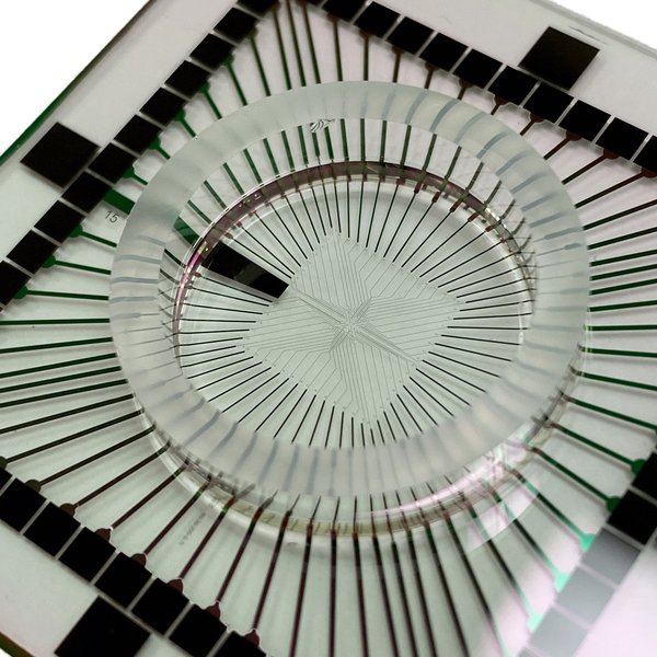
Example of a rigid glass microelectrode array (MEA), often used for acute recordings or in vitro studies due to its precise structure.
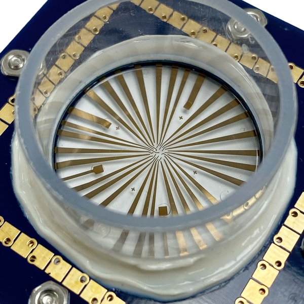
A flexible and stretchable MEA designed to better conform to brain tissue, reducing mechanical mismatch.

An innovative 3D MEA structure, potentially offering enhanced integration with neural circuits.
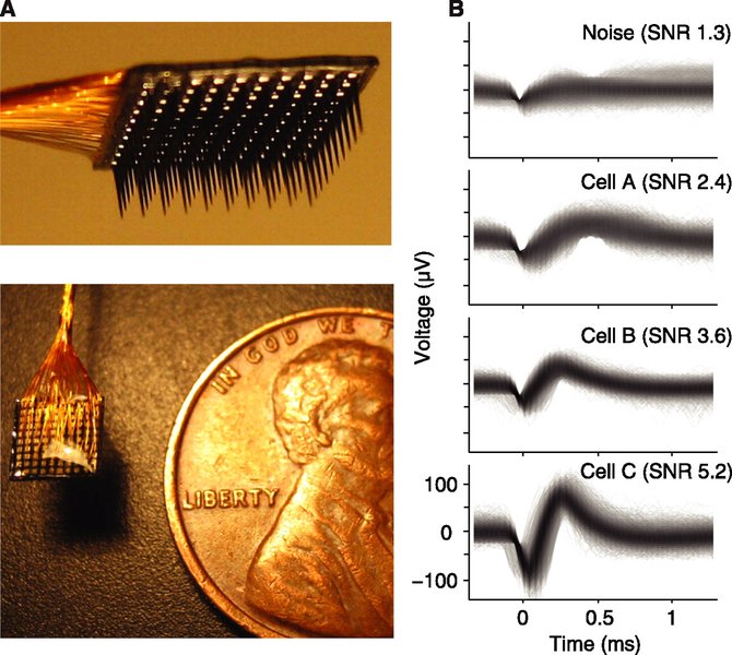
Illustration comparing recordings from a microelectrode array (MEA) with those from traditional single wires, highlighting the potential for higher density recording with MEAs.
Exploring Electrode Materials and Coatings
The choice of materials and coatings is fundamental to electrode performance. The table below summarizes some common and emerging options, highlighting their key characteristics relevant to the neuron-electrode interface.
| Material/Coating Type | Example(s) | Key Advantage(s) | Impedance Effect | Biocompatibility |
|---|---|---|---|---|
| Noble Metals (Bulk) | Platinum (Pt), Gold (Au), Iridium (Ir) | Good conductivity, relative stability | Baseline (Higher for small sites) | Generally Good |
| Nanostructured Metals | Pt-Black, Sputtered Iridium Oxide (SIROF) | Massively increased surface area, improved charge injection | Significantly Reduced | Generally Good |
| Conductive Polymers | PEDOT:PSS | Mixed ionic-electronic conductivity, processability, flexibility | Significantly Reduced | Good, can be tailored |
| Carbon Nanomaterials | Carbon Nanotubes (CNTs), Graphene | Excellent conductivity, high surface area, mechanical strength | Significantly Reduced | Promising, ongoing research |
| Hydrogels | PEG, Agarose-based | Softness (mimics tissue), potential for drug delivery | Can slightly increase (if non-conductive), but reduces FBR impact | Excellent (tunable) |
| Biomimetic/Bioactive Coatings | Laminin, Peptide sequences, SiNPs | Promote neural adhesion, reduce inflammation | Indirectly improves via reduced scarring | Designed to be High |
Expert Insights: Progress in Neuron-Electrode Interfacing
Understanding the advancements and challenges in neuron-electrode interfacing directly from researchers in the field provides valuable context. The following presentation offers insights into the progress being made in how we listen to and interpret neuronal signals through electrode technologies. It discusses the evolution of interfacing techniques and the ongoing efforts to improve the quality and longevity of neural recordings.
Frequently Asked Questions (FAQ)
What exactly is interface impedance and why is it important?
Interface impedance is the opposition to the flow of alternating electrical current (like the signals from neurons) between the electrode material and the surrounding biological tissue (electrolyte). It's typically measured across a range of frequencies. Low impedance is crucial because:
- It improves the signal-to-noise ratio (SNR), making it easier to detect weak neural signals against background noise.
- It reduces signal attenuation, ensuring more of the neuron's signal amplitude reaches the recording equipment.
- For stimulation, lower impedance requires less voltage to deliver the needed current, reducing risks of tissue damage and electrode corrosion.
Factors like electrode size (smaller = higher impedance), material properties, and the presence of the glial scar all contribute to the overall impedance.
What is the "glial scar" and why does it form?
The glial scar is a dense layer of reactive astrocytes (a type of glial cell) that encapsulates implanted objects, including neural electrodes, in the brain. It's part of the brain's chronic foreign body response (FBR).
It forms because the brain recognizes the implant as foreign and potentially damaging. Microglia (immune cells) are activated first, followed by astrocytes migrating to the site, proliferating, and changing their structure to form a physical barrier. This process aims to isolate the foreign object and protect the surrounding tissue.
However, for neural electrodes, this scar is detrimental because it physically increases the distance between the electrode and the neurons it's trying to record from or stimulate. This increases impedance and blocks signals, leading to performance degradation over time.
How do nanomaterials help reduce impedance?
Nanomaterials like carbon nanotubes (CNTs), graphene, or nanostructured metal coatings (e.g., platinum black) primarily reduce impedance by dramatically increasing the electrochemically active surface area (ECSA) of the electrode without significantly increasing its physical size (geometric footprint).
Impedance is inversely proportional to the ECSA. By creating intricate structures at the nanoscale, these materials provide vastly more sites for charge transfer (capacitive and faradaic) to occur between the electrode and the electrolyte. This increased capacity for charge exchange lowers the opposition to current flow, thereby reducing impedance, especially at the biologically relevant frequencies of neural signals.
Can flexible electrodes completely solve the problem of mechanical mismatch?
Flexible electrodes significantly mitigate, but don't completely eliminate, the problems caused by mechanical mismatch. By using soft polymer substrates that better match the low Young's modulus of brain tissue, they reduce the chronic strain and micromotion that contribute to inflammation and glial scarring.
However, challenges remain. Even flexible implants are still foreign bodies that elicit some degree of FBR. Furthermore, achieving sufficient stiffness for reliable surgical insertion while maintaining high flexibility for chronic implantation requires careful design trade-offs. While a major improvement over rigid implants, flexibility is often combined with other strategies (like bioactive coatings) to achieve optimal long-term performance.
References
- Electrode materials for brain-machine interface: A review - Wiley Online Library
- Neuron devices: emerging prospects in neural interfaces and recognition - Nature Microsystems & Nanoengineering
-
Recent advances in neural electrode–tissue interfaces - ScienceDirect Current Opinion in Biomedical Engineering
-
A Critical Review of Microelectrode Arrays and Strategies for Improving the Neural Interface - NIH National Library of Medicine
-
Micro- and nanotechnology for neural electrode-tissue interfaces - ScienceDirect Materials Today Bio
- Strategies for interface issues and challenges of neural electrodes in electrical stimulation and recording - Royal Society of Chemistry
- Optimizing the neuron-electrode interface for chronic bioelectronic interfacing - Journal of Neurosurgery: Focus
Recommended Further Exploration
Last updated April 10, 2025