
Unlocking Neural Communication: The Electrical Foundations of Brain-Spinal Cord Signaling
How understanding neuron function and electrical signaling can revolutionize flexible neural probe development for advanced brain research
Key Insights for Neural Probe Development
- Neurons communicate through precise electrical-chemical-electrical signal conversion that flexible probes must be designed to record and stimulate effectively
- The spinal cord serves as a critical hub for neural signal processing with unique anatomical and functional properties that neural probes must accommodate
- Material biocompatibility, flexibility, and electrode design are crucial considerations for developing neural probes that minimize tissue damage while maximizing signal quality
Neuronal Structure and Function: The Foundation of Neural Signaling
Neurons are specialized cells that form the building blocks of the nervous system, designed to process and transmit information through electrical and chemical signals. Understanding their structure and function is essential for developing effective neural probes that can accurately record and stimulate neural activity.
Anatomical Components of Neurons
Each neuron consists of three primary components, each serving a distinct function in neural communication:
- Soma (Cell Body): Contains the nucleus and cellular organelles that maintain the neuron's metabolic functions and protein synthesis. The soma integrates incoming signals and determines whether to generate an outgoing signal.
- Dendrites: Branching extensions that receive signals from other neurons. Human dendrites have unique electrical properties that influence signal processing, with signals weakening as they travel along the dendrite toward the cell body.
- Axon: A long, thin extension that conducts electrical signals away from the cell body to other neurons, muscles, or glands. Many axons are covered with myelin sheaths that increase the speed of signal transmission.
- Axon Terminals: The branched endings of axons where neurotransmitters are released into synapses to communicate with other cells.
Types of Neurons in the Nervous System
Neurons are classified based on their function and structure in the nervous system:
| Neuron Type | Function | Location | Key Characteristics |
|---|---|---|---|
| Sensory Neurons | Transmit information from sensory receptors to the CNS | Peripheral nervous system, with cell bodies in dorsal root ganglia | Long dendrites, unipolar or pseudounipolar structure |
| Motor Neurons | Transmit signals from the CNS to muscles and glands | Ventral horn of spinal cord, brain stem, motor cortex | Long axons, multipolar structure |
| Interneurons | Process and integrate information between neurons | CNS (brain and spinal cord) | Short axons, typically multipolar |
| Projection Neurons | Transmit signals over long distances | Throughout CNS | Very long axons, multipolar structure |
Electrical Signaling in Neurons: The Language of Neural Communication
Neurons communicate through a sophisticated electrical signaling system that allows for rapid transmission of information throughout the nervous system. This electrical activity forms the basis for all neural functions and is crucial for understanding how neural probes can interface with the nervous system.
The Neural Membrane Potential
At rest, neurons maintain a negative electrical charge inside the cell relative to the outside, known as the resting membrane potential (typically around -70 mV). This charge difference is created and maintained by the sodium-potassium pump, which actively transports sodium ions (Na⁺) out of the cell and potassium ions (K⁺) into the cell, creating an electrochemical gradient across the membrane.
Action Potentials: The Neural Electrical Signal
When a neuron receives sufficient excitatory input to reach a threshold potential (around -55 mV), it triggers an action potential—a rapid, all-or-nothing electrical event that propagates along the axon. This process involves several key steps:
- Depolarization: Voltage-gated sodium channels open, allowing Na⁺ ions to rush into the cell, making the membrane potential temporarily positive.
- Repolarization: Voltage-gated potassium channels open, allowing K⁺ ions to flow out of the cell, restoring the negative membrane potential.
- Hyperpolarization: The membrane potential becomes briefly more negative than the resting potential as potassium channels remain open momentarily.
- Return to resting potential: The sodium-potassium pump restores ion concentrations to their resting state.
Comparing Signal Transmission Properties
The radar chart above compares the signal transmission properties of myelinated and unmyelinated axons with current and potential future neural probe technologies. Understanding these properties is crucial for developing neural probes that can effectively interface with the nervous system. The chart shows that while current neural probe technology has made significant progress, there remains substantial room for improvement in areas such as energy efficiency, spatial precision, and adaptability.
Synaptic Transmission: Converting Electrical to Chemical Signals
Once an action potential reaches the axon terminals, it triggers the release of neurotransmitters into the synaptic cleft—the small gap between the presynaptic neuron and the postsynaptic cell. This process converts the electrical signal into a chemical one:
- The action potential causes voltage-gated calcium channels to open, allowing calcium ions to enter the axon terminal.
- Calcium influx triggers synaptic vesicles containing neurotransmitters to fuse with the presynaptic membrane and release their contents into the synaptic cleft.
- Neurotransmitters diffuse across the synaptic cleft and bind to receptors on the postsynaptic membrane.
- Binding can cause ion channels to open or close, generating either excitatory (depolarizing) or inhibitory (hyperpolarizing) postsynaptic potentials, converting the chemical signal back to an electrical one.
The Spinal Cord: Neural Processing Hub and Signal Relay
The spinal cord serves as a critical component of the central nervous system, functioning as both a processing center and a signal relay station between the brain and the rest of the body. Understanding its structure and function is essential for developing neural probes that can effectively interface with spinal neurons.
Anatomical Organization of the Spinal Cord
The spinal cord has a distinctive cross-sectional anatomy with gray matter (containing neuronal cell bodies) in the center, surrounded by white matter (consisting of myelinated axon tracts). The gray matter forms an H-shaped pattern with:
- Dorsal Horns: Process sensory information from the periphery.
- Ventral Horns: Contain motor neurons that innervate skeletal muscles.
- Intermediate Zone: Contains interneurons that integrate sensory and motor information.
The white matter contains ascending tracts that carry sensory information to the brain and descending tracts that carry motor commands from the brain to spinal motor neurons.
Neural Signaling Pathways in the Spinal Cord
The spinal cord processes various types of neural signals through distinct pathways:
Ascending (Sensory) Pathways
Sensory information from the periphery travels through primary sensory neurons, whose cell bodies are located in the dorsal root ganglia. These neurons synapse with second-order neurons in the spinal cord, which then send their axons up to the brain through several major pathways:
- Dorsal Column-Medial Lemniscus Pathway: Carries fine touch, vibration, and proprioception.
- Spinothalamic Tract: Transmits pain, temperature, and crude touch sensations.
- Spinocerebellar Tracts: Convey unconscious proprioceptive information to the cerebellum.
Descending (Motor) Pathways
Motor commands originate in the brain and travel down to the spinal cord through several pathways:
- Corticospinal Tract: The primary pathway for voluntary movement, originating in the motor cortex.
- Rubrospinal Tract: Influences flexor muscles of the upper limbs.
- Reticulospinal Tracts: Control posture, balance, and some autonomous functions.
- Vestibulospinal Tract: Regulates balance and posture in response to head movements.
Reflex Arcs
The spinal cord also mediates reflexes—rapid, involuntary responses to stimuli that don't require brain involvement. A typical reflex arc involves:
- A sensory receptor detects a stimulus.
- A sensory neuron transmits the signal to the spinal cord.
- Interneurons in the spinal cord process the information.
- Motor neurons send signals to effector organs (muscles or glands).
The mindmap above illustrates the complex neural signaling processes in the spinal cord that neural probes must interact with. Understanding these pathways is essential for developing probes that can effectively record from and stimulate specific neural populations within the spinal cord.
Electrical Stimuli and Neural Responses
Understanding how neurons respond to electrical stimuli is crucial for developing effective neural probes. Electrical stimulation can be used to modulate neural activity, either activating or inhibiting specific neural populations depending on the stimulation parameters.
Mechanisms of Neural Response to Electrical Stimulation
When an external electrical stimulus is applied to neural tissue, it creates an electric field that can influence the membrane potential of nearby neurons. The response depends on several factors:
- Stimulus Amplitude: Higher amplitudes recruit more neurons and can activate neurons at greater distances from the stimulation source.
- Stimulus Duration: Longer pulses allow more time for charge to accumulate across the membrane, facilitating action potential generation.
- Stimulus Frequency: Different frequencies can produce different effects, from facilitation to depression of neural activity.
- Waveform Shape: The shape of the stimulation waveform affects charge delivery and neural recruitment patterns.
- Electrode Configuration: Monopolar, bipolar, or multipolar configurations create different spatial patterns of current flow.
Neural Response Patterns
Neurons can respond to electrical stimuli in several ways:
- Direct Activation: When stimulation directly depolarizes the neuron's membrane to threshold, triggering an action potential.
- Indirect Activation: When stimulation activates presynaptic neurons, which then synaptically activate the target neuron.
- Inhibition: When stimulation activates inhibitory interneurons that suppress the activity of target neurons.
- Modulation: When stimulation alters the excitability or response properties of neurons without directly causing or preventing action potentials.
This video provides a detailed explanation of how action potentials are generated and propagated along neurons, which is fundamental to understanding how electrical stimulation interacts with neural tissue. The video illustrates the complex ionic mechanisms underlying neural electrical signaling that neural probes must detect and interact with.
Developing Novel Flexible Neural Probes: Key Considerations
As a material science and electronics engineer developing flexible neural probes, understanding the interface between electronics and neurobiology is critical. Here are key considerations for developing effective neural probes for research applications:
Material Properties and Biocompatibility
Essential Material Characteristics
- Biocompatibility: Materials must not elicit harmful immune responses or damage neural tissue. Consider using materials like PDMS, parylene-C, or polyimide that have demonstrated good biocompatibility.
- Flexibility and Elasticity: Probes should match the mechanical properties of neural tissue (Young's modulus ~1-10 kPa) to minimize tissue damage and accommodate brain/spinal cord movement.
- Durability: Materials must maintain functionality over extended periods in the physiological environment, resisting degradation from enzymes, reactive oxygen species, and pH changes.
- Permeability: Consider the diffusion of nutrients, oxygen, and waste products through probe materials to maintain tissue health.
Electrode Design for Optimal Neural Interface
Critical Electrode Parameters
- Size and Geometry: Smaller electrodes can target individual neurons but have higher impedance. Consider using nanoscale structures to increase surface area while maintaining small geometric area.
- Impedance: Lower impedance improves signal-to-noise ratio for recording and reduces power requirements for stimulation. Surface modifications like PEDOT:PSS coatings can reduce impedance.
- Charge Injection Capacity: For stimulation, electrodes must deliver sufficient charge without causing harmful electrochemical reactions. Noble metals (platinum, iridium) and conductive polymers offer good charge injection properties.
- Spatial Arrangement: The arrangement of electrodes should match the anatomical organization of the target neural tissue, with consideration for the specific pathways and functions.
Signal Processing and Electronics Integration
Electronics Design Considerations
- Amplification and Filtering: Neural signals are typically in the microvolt range and require low-noise amplification. Consider front-end amplifiers with high input impedance and low noise figure.
- Multiplexing: For multi-electrode arrays, implement efficient multiplexing strategies to reduce the number of connection wires.
- Wireless Capabilities: Consider incorporating wireless data transmission and power delivery to eliminate tethering and reduce infection risk.
- On-board Processing: Implement local signal processing to reduce data bandwidth requirements and enable closed-loop operation.
Addressing the Biological-Electronic Interface Challenges
Mitigating Interface Issues
- Foreign Body Response: Design probes to minimize the inflammatory response that can encapsulate the probe and isolate it from target neurons.
- Micro-motion: Accommodate the relative movement between probe and tissue due to respiration, heartbeat, and body movement.
- Insertion Trauma: Develop minimally invasive insertion techniques that reduce initial tissue damage.
- Long-term Stability: Address issues of electrode degradation, material breakdown, and signal drift over time.
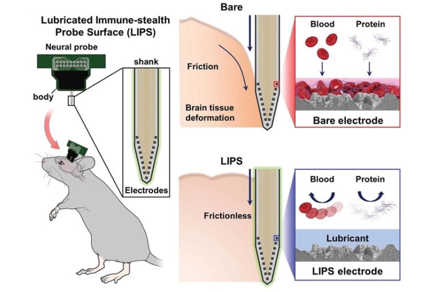
Innovative Coating Solutions for Neural Probes
The image above shows a cutting-edge lubricated coating technique being developed for brain implants. This technology uses a slippery surface that mimics the natural lubrication of the brain's tissue, potentially reducing the foreign body response and extending the functional lifespan of neural probes. Similar approaches could be adapted for spinal cord neural interfaces, where mechanical stability and reduced tissue damage are equally critical.
Frequently Asked Questions
References
- Electrical Signals in Nerve Cells - NCBI Bookshelf
- Chemical and Electrical Signals in Neurons - Georgia Tech
- Action Potentials and Synapses - Queensland Brain Institute
- Anatomy and Physiology of the Spinal Cord - NCBI Bookshelf
- Implantation of Neural Probes in the Brain - Frontiers in Bioengineering and Biotechnology
-
Ultra-Thin, Flexible Probe Provides Neural Interface - UC San Diego
- Double-Layer Flexible Neural Probe With Closely Spaced Electrodes - Frontiers in Neuroscience
Recommended Topics
Last updated April 9, 2025