
Unlocking Brain Secrets: The Cutting-Edge of Neuron-Specific EV Isolation from Plasma
Explore the advanced techniques revolutionizing neurodegenerative disease research and biomarker discovery.
Key Insights into Neuron-Specific EV Isolation
- Hybrid Approaches Dominate: The most effective current methods combine initial bulk EV isolation techniques (like Size Exclusion Chromatography or Ultracentrifugation) with highly specific immunoaffinity capture using neuron-specific markers.
- Emerging Neuronal Markers: L1CAM remains widely used, but new markers like ATP1A3 and NRXN3 are gaining prominence for their enhanced specificity in capturing neuron-derived EVs.
- Purity and Specificity are Paramount: Given the complex nature of plasma, recent advancements focus heavily on reducing contamination from non-neuronal EVs, proteins, and lipoproteins to ensure high-fidelity analyses.
The isolation of neuron-specific extracellular vesicles (nEVs) from plasma represents a significant frontier in neuroscience, offering a non-invasive window into the intricate processes occurring within the brain. These microscopic vesicles, shed by neurons, carry a cargo of proteins, lipids, and nucleic acids that reflect the physiological and pathological state of their parent cells. Their analysis holds immense potential for biomarker discovery, early disease diagnosis, and monitoring therapeutic responses in neurological disorders such as Alzheimer's, Parkinson's, and other neurodegenerative conditions.
However, isolating these neuron-specific components from the highly complex and abundant plasma matrix is a substantial challenge. Plasma contains a heterogeneous mixture of EVs from various cell types, along with high concentrations of contaminants like lipoproteins and soluble proteins, which can confound analysis. Consequently, the latest methodologies emphasize strategies that prioritize both the purity and the specificity of nEV capture, often employing multi-step protocols to achieve reliable results.
The Evolving Landscape of nEV Isolation Techniques
Modern approaches for isolating neuron-specific extracellular vesicles from plasma have moved beyond single-method reliance, embracing integrated workflows that leverage the strengths of various purification principles. These techniques aim to maximize yield while minimizing contamination, ensuring the isolated nEVs are truly representative of neuronal activity.
Foundational EV Isolation Methods: The Initial Purifiers
Before targeting neuron-specific markers, the initial step often involves a bulk isolation of total EVs from plasma to remove major contaminants. These methods lay the groundwork for subsequent specific enrichment.
Size Exclusion Chromatography (SEC): The Gentle Separator
Size Exclusion Chromatography (SEC) has emerged as a preferred initial purification step due to its ability to separate EVs based on their size, effectively distinguishing them from free proteins and lipoproteins. This gentle method preserves EV integrity and is well-suited for processing smaller plasma volumes, making it highly reproducible. Automated SEC systems, often using qEV columns, further enhance standardization and efficiency.
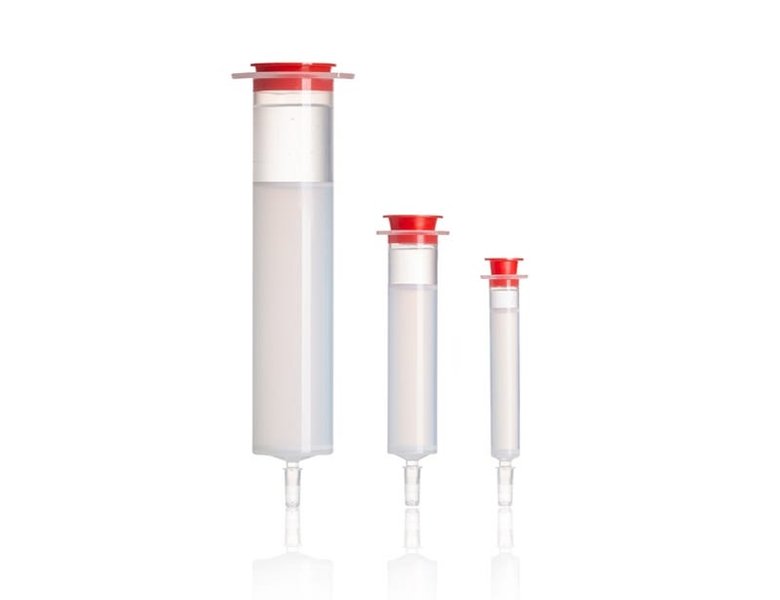
An image showing SEC columns, critical for separating extracellular vesicles from other plasma components based on size.
Ultracentrifugation (UC): The Traditional Powerhouse
Differential Ultracentrifugation (dUC) remains a foundational technique, separating EVs based on differences in their density, size, and shape. While effective for initial bulk isolation, it can be time-consuming, requires specialized equipment, and may potentially damage EVs or result in lower purity due to co-pelleting of non-EV components. Therefore, when used, UC is typically followed by additional purification steps like density gradient fractionation or immunoaffinity capture to enhance specificity and reduce contaminants.
Polymer-Based Precipitation Kits: Rapid Concentration
Kits such as ExoQuick Ultra offer a quick and straightforward way to concentrate total EVs from plasma or serum through polymer-based precipitation. While convenient and yielding higher quantities of EVs, they generally produce samples with lower purity compared to SEC or UC, often carrying over significant amounts of albumin and immunoglobulins. Consequently, these kits are often used as a preliminary concentration step, requiring further purification for neuron-specific applications.
Specialized Immunoaffinity Capture: The Neuron-Specific Key
The definitive step in isolating nEVs involves immunoaffinity capture, which selectively targets specific protein markers found on the surface of neuron-derived EVs. This step ensures high specificity and purity.
L1CAM: A Long-Standing Marker
L1CAM (L1 cell adhesion molecule) has historically been the most widely used neuron-specific marker for EV isolation. Antibodies against L1CAM are used to immunoprecipitate nEVs from the total EV pool, typically using magnetic beads or immunoplates. While effective and widely validated, recent evidence suggests that some L1CAM in biofluids might be in a soluble, non-EV associated form, necessitating rigorous validation to ensure true EV specificity.
ATP1A3: A Promising New Target
ATP1A3, a protein abundantly expressed in EVs from human neurons, brain, cerebrospinal fluid, and plasma, has emerged as a highly promising target for immunoaffinity-based isolation of nEVs. Its high specificity makes it an excellent candidate for capturing high-purity neuron-derived EVs for detailed molecular analyses.
NRXN3: An Emerging Specificity Marker
Neurexin 3 (NRXN3) is another recently identified neuron-specific protein found on EVs in cerebrospinal fluid and plasma. Research suggests NRXN3 offers high specificity, potentially surpassing L1CAM in certain applications, making it a valuable addition to the arsenal of neuron-specific EV markers.
Advanced Combined and Hybrid Methodologies
The current state-of-the-art for nEV isolation from plasma overwhelmingly favors multi-step or hybrid approaches that integrate the strengths of different techniques. This synergy addresses the challenges of both yield and purity in a complex biological fluid.
Two-Step Protocols: The Gold Standard
The most commonly recommended workflow involves a two-step approach:
- **Initial Total EV Isolation:** Utilizing methods like SEC, dUC, or polymer precipitation to reduce the overall complexity of the plasma sample and remove bulk contaminants.
- **Neuron-Specific Enrichment:** Applying immunoaffinity capture using antibodies against L1CAM, ATP1A3, NRXN3, or a combination thereof, typically linked to magnetic beads, to selectively pull down neuron-specific EVs.
This sequential approach significantly enhances the purity and specificity of the isolated nEVs, making them suitable for downstream molecular profiling.
Charge-Based Isolation: A Novel Pre-Purification
Emerging methods are exploring the use of charge differences between EVs and contaminants to achieve initial purification. By using positively charged beads to remove negatively charged impurities like lipoproteins and albumin, this rapid single-step method can yield purer EV samples, which can then be subjected to an immunoaffinity step for neuron-specific enrichment.
Comparative Analysis of Isolation Method Capabilities
The choice of isolation method significantly impacts the yield, purity, and integrity of the isolated neuron-specific EVs. The following radar chart provides an opinionated comparative analysis of the strengths of different isolation principles when applied to plasma, focusing on their utility for specific neuronal EV enrichment.
This radar chart illustrates the relative strengths of different EV isolation principles. Immunoaffinity capture excels in specificity but may have moderate yield. SEC is strong in purity and EV integrity, offering good processing time. Ultracentrifugation provides a decent yield but can be time-consuming and might compromise EV integrity. Polymer-based precipitation is fast and high-yielding but generally sacrifices purity. The chart highlights why combining methods is often the optimal strategy to leverage their complementary benefits for neuron-specific EV isolation.
Workflow for Optimal Neuron-Specific EV Isolation
A streamlined and standardized workflow is crucial for obtaining reliable and reproducible results in neuron-specific EV research. The following steps outline a generally recommended protocol:
| Step | Method | Description | Key Considerations |
|---|---|---|---|
| 1. Plasma Collection & Pre-processing | Standardized protocols, Low-speed centrifugation | Collect platelet-free plasma and remove large cellular debris by initial low-speed centrifugation to prevent contamination. | Use appropriate anticoagulants; minimize platelet contamination. |
| 2. Total EV Isolation/Purification | Size Exclusion Chromatography (SEC) or Differential Ultracentrifugation (dUC) | Purify total EVs from plasma to remove abundant plasma proteins and lipoproteins. SEC is often preferred for purity and EV integrity. | Optimize column size/speed for SEC; balance speed/purity for dUC. |
| 3. Neuron-Specific Immunoaffinity Capture | Antibodies against L1CAM, ATP1A3, or NRXN3 (on magnetic beads/plates) | Enrich neuron-derived EVs specifically by incubating the purified total EV fraction with immobilized antibodies targeting neuronal surface markers. | Choose validated and specific antibodies; optimize incubation time and washing steps. |
| 4. Elution & Concentration (Optional) | Gentle elution buffer, Ultrafiltration | Elute the captured nEVs from the beads/plates. Concentration may be performed if required for downstream analysis using ultrafiltration. | Ensure elution conditions preserve EV integrity; avoid harsh methods. |
| 5. Validation & Characterization | NTA, TEM, Western Blot, Mass Spectrometry | Confirm EV purity, size distribution, concentration, and neuronal origin by detecting specific marker proteins (e.g., neuronal vs. pan-EV markers) and morphology. | Adhere to ISEV guidelines; use orthogonal methods for robust validation. |
| 6. Downstream Molecular Profiling | Proteomics, RNA-sequencing (RNA-seq), Lipidomics | Analyze the molecular cargo of the isolated nEVs to identify biomarkers or understand pathological mechanisms. | Ensure sufficient yield for the chosen assay; maintain RNase/protease-free conditions. |
The Significance of Robust nEV Isolation
The ability to reliably isolate neuron-specific EVs from plasma is transformative for neuroscience. These vesicles serve as invaluable diagnostic tools and provide insights into fundamental brain biology and disease mechanisms.
Applications in Neurological Research and Diagnostics
Neuron-specific EVs are critical for:
- Biomarker Discovery: Identifying novel biomarkers for early diagnosis, prognosis, and monitoring disease progression in neurodegenerative disorders like Alzheimer's, Parkinson's, and ALS.
- Disease Mechanism Elucidation: Studying the molecular pathways involved in neurodegeneration by analyzing the specific cargo (proteins, RNA, lipids) within nEVs.
- Therapeutic Development: Assessing the efficacy of new treatments by monitoring changes in nEV cargo, and potentially even using nEVs themselves as therapeutic delivery vehicles.
- Non-invasive Brain Access: Providing a minimally invasive alternative to cerebrospinal fluid (CSF) or brain tissue biopsies, making repeated sampling for longitudinal studies feasible.
The Challenges of Translation to Clinical Practice
While promising, the clinical translation of nEV-based diagnostics faces challenges:
- Standardization: Ensuring consistent and reproducible isolation protocols across different laboratories and clinical settings.
- Scalability: Developing methods that can be scaled up for high-throughput processing of clinical samples.
- Cost-Effectiveness: Reducing the cost of isolation to make nEV-based diagnostics economically viable for routine clinical use.
- Validation: Rigorously validating candidate biomarkers from nEVs in large, diverse patient cohorts.
Conceptualizing the Isolation Process
The following mindmap illustrates the interconnectedness of different methods and their roles in achieving highly specific neuron-derived extracellular vesicle isolation from plasma.
This mindmap visually organizes the key components involved in isolating neuron-specific EVs. It highlights the initial bulk isolation methods, the specific neuronal markers used for capture, the preferred combined approaches, and the essential validation steps that follow to ensure the quality of the isolated vesicles. This holistic view underscores the complexity and multi-faceted nature of current best practices.
Understanding EV Isolation: A Visual Guide
To further contextualize the discussions around extracellular vesicle isolation, here is a relevant video that delves into the considerations and challenges researchers face when isolating EVs from complex biological fluids like plasma.
This video, "Isolating Extracellular Vesicles from Plasma (Blood) | Izon...", discusses the practical considerations and challenges inherent in isolating EVs from plasma. It is highly relevant to our discussion as it provides a visual and auditory explanation of the complexities of working with blood-derived samples, emphasizing the need for robust and reliable isolation techniques to ensure high-quality research outcomes. Understanding these fundamental challenges is key to appreciating the advancements in neuron-specific EV isolation.
Frequently Asked Questions
What makes isolating neuron-specific EVs from plasma challenging?
Why are two-step isolation methods preferred for neuron-specific EVs?
What are the most commonly used neuron-specific markers for EV isolation?
What are the applications of isolating neuron-specific EVs?
What are the next frontiers in neuron-specific EV isolation?
Conclusion
The isolation of neuron-specific extracellular vesicles from plasma has evolved into a sophisticated field, driven by the critical need for non-invasive diagnostic tools and deeper insights into neurological disorders. The current best practices involve multi-step strategies, typically commencing with broad EV purification using techniques like Size Exclusion Chromatography or Differential Ultracentrifugation, followed by highly specific immunoaffinity capture targeting distinct neuronal surface markers such as L1CAM, ATP1A3, and NRXN3. These hybrid approaches are designed to overcome the inherent complexities of plasma, ensuring the purity, yield, and specificity of the isolated nEVs for rigorous downstream molecular profiling. As research continues to identify novel markers and refine isolation techniques, the potential of neuron-specific EVs as diagnostic biomarkers and therapeutic targets for brain health and disease continues to expand, promising a future where neurological conditions can be detected and managed with unprecedented precision.
Recommended Further Exploration
- How to validate purity of isolated extracellular vesicles?
- Role of neuron-derived extracellular vesicles in Alzheimer's disease diagnosis
- Comparison of automated versus manual EV isolation methods
- Emerging microfluidic devices for EV isolation