
Optimal CT Protocol for Renal Cell Carcinoma (RCC) Staging
Comprehensive imaging approach for accurate staging and treatment planning
Key Takeaways
- Multiphasic Contrast-Enhanced CT: Essential for detailed assessment of tumor characteristics and extent.
- Phased Imaging: Includes precontrast, corticomedullary, nephrographic, excretory phases, and chest imaging for comprehensive evaluation.
- Technical Precision: Utilization of thin-slice acquisition, appropriate contrast dosing, and advanced reconstruction techniques enhance diagnostic accuracy.
Introduction
Accurate staging of renal cell carcinoma (RCC) is pivotal for determining appropriate treatment strategies and prognostication. Computed Tomography (CT) is the imaging modality of choice due to its high resolution and ability to provide detailed information about tumor characteristics, vascular involvement, and metastatic spread. Implementing a well-structured CT protocol ensures comprehensive evaluation, which is instrumental in surgical planning and therapeutic decision-making.
CT Protocol Phases
The optimal CT protocol for RCC staging involves a multiphasic contrast-enhanced approach, encompassing several distinct phases to capture different aspects of the renal mass and its extent. The following phases are essential:
1. Precontrast Phase
The precontrast phase serves as a baseline, allowing for the detection of intrinsic characteristics of the renal mass, such as calcifications, hemorrhage, or fat content.
Purpose
- Identify calcifications within the tumor.
- Detect hemorrhagic components.
- Assess for the presence of fat, indicative of angiomyolipomas or lipid-poor lesions.
Technical Details
- Acquisition Parameters: Axial images of the abdomen and pelvis.
- Slice Thickness: 3-5 mm for optimal resolution.
- Technique: Non-contrast axial imaging with 5-mm collimation.
2. Corticomedullary Phase (Arterial Phase)
The corticomedullary phase is timed to capture the arterial enhancement of the renal parenchyma, providing critical information about tumor vascularity and potential vascular invasion.
Purpose
- Evaluate renal vasculature, including renal arteries and veins.
- Assess tumor vascularity, which is typically hypervascular in RCC.
- Detect venous invasion, particularly in the renal vein and inferior vena cava (IVC).
Technical Details
- Timing: 25-40 seconds after intravenous contrast injection.
- Contrast Administration: Iodinated contrast media, typically 1.5 mL/kg at a rate of 3-5 mL/s.
- Acquisition Parameters: Axial images with thin slice thickness (1-3 mm).
3. Nephrographic Phase (Parenchymal Phase)
The nephrographic phase occurs during the peak enhancement of the renal parenchyma, facilitating detection of small renal masses and assessment of tumor extent within the kidney.
Purpose
- Provide uniform enhancement of the renal parenchyma.
- Detect small renal masses that may not be apparent in earlier phases.
- Assess the extent of the tumor within the renal cortex and medulla.
Technical Details
- Timing: 80-120 seconds after contrast administration.
- Technique: Axial imaging with contrast enhancement.
- Sweep Range: Entire abdomen and pelvis to evaluate nodal involvement.
4. Excretory Phase (Delayed Phase)
The excretory phase assesses the urinary collecting system, ureters, and bladder for any involvement by the tumor, such as extension into the renal pelvis or ureter.
Purpose
- Evaluate the collecting system, ureters, and bladder.
- Detect tumor involvement in the renal pelvis or ureters.
- Assess for hydronephrosis or filling defects.
Technical Details
- Timing: 5-10 minutes after contrast injection.
- Technique: Axial images with contrast enhancement.
- Slice Thickness: 3-5 mm for optimal resolution.
5. Chest Imaging
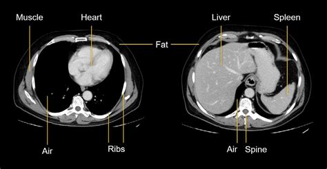
Since pulmonary metastases are common in RCC, evaluating the chest is essential for comprehensive staging.
Purpose
- Detect pulmonary metastases.
- Assess for other distant organ involvement.
Technical Details
- Technique: Non-contrast axial CT of the chest.
- Slice Thickness: 5 mm collimation.
Multiphasic CT Protocol Overview
| Phase | Timing | Purpose |
|---|---|---|
| Precontrast | Before contrast injection | Detect calcifications, hemorrhage, fat within renal mass |
| Corticomedullary | 25-40 seconds post-contrast | Evaluate renal vasculature and tumor vascularity |
| Nephrographic | 80-120 seconds post-contrast | Assess renal parenchyma and detect small renal masses |
| Excretory (Delayed) | 5-10 minutes post-contrast | Evaluate collecting system, ureters, and bladder |
| Chest Imaging | During/after abdominal phases | Detect pulmonary metastases |
Technical Parameters
To ensure high-quality imaging, the following technical parameters should be optimized:
Scanner Requirements
- Utilize a multi-detector CT (MDCT) scanner for high-resolution imaging and rapid acquisition.
- Ensure capabilities for thin-slice acquisition and advanced reconstruction algorithms.
Contrast Administration
- Use iodinated intravenous contrast agents, with dosing typically 1.5 mL/kg administered at a rate of 3-5 mL/s.
- Adjust contrast volume based on patient weight and renal function.
Slice Thickness and Reconstruction
- Employ thin-slice (1-3 mm) imaging for all phases to facilitate multiplanar reconstructions.
- Generate coronal and sagittal reconstructions to evaluate tumor extent and vascular anatomy.
Field of View (FOV)
- Cover the entire abdomen and pelvis for comprehensive evaluation.
- Include chest imaging within the scan range to assess for distant metastases.
Key Considerations
- Contrast Agent Selection: Opt for iodinated contrast media unless contraindicated, such as in patients with known allergies or significant renal impairment.
- Patient Preparation: Ensure patients are adequately hydrated prior to contrast administration to minimize the risk of contrast-induced nephropathy.
- Breath-Hold Technique: Instruct patients to hold their breath during scan acquisition to reduce motion artifacts and improve image quality.
- Radiation Dose Optimization: Employ dose-reduction strategies, including automatic exposure control and iterative reconstruction techniques, to minimize patient radiation exposure while maintaining image quality.
Advantages of the Multiphasic CT Protocol
- High Sensitivity and Specificity: Multiphasic CT scans exhibit high sensitivity (85%) and specificity (98%) for detecting venous involvement, which is critical in surgical planning.
- Comprehensive Assessment: Allows for precise evaluation of tumor size, local invasion, and detection of distant metastases, facilitating accurate staging.
- Differentiation of Lesion Types: Enables differentiation between solid and cystic renal masses, aiding in diagnostic accuracy.
- Enhanced Surgical Planning: Coronal and sagittal reconstructions aid surgeons in visualizing tumor anatomy and its relationship with surrounding structures.
Special Considerations
- Alternative Modalities: In cases where iodinated contrast agents are contraindicated, such as in patients with severe renal insufficiency or contrast allergies, a multiparametric MRI with contrast may be utilized as an alternative for RCC staging.
- Dual-Energy CT (DECT): Incorporating DECT can enhance lesion characterization, distinguishing between tumor tissue and pseudolesions, and mitigating beam hardening artifacts.
- PET-CT Utility: While PET-CT is not routinely recommended for RCC staging, it may be beneficial in specific scenarios, such as detecting soft-tissue metastases or evaluating ambiguous findings from conventional imaging.
- Vascular Invasion Assessment: If vascular invasion is suspected, including a delayed phase can improve detection of tumor thrombus or extension into the IVC.
Key Points of TNM Staging to Consider
The TNM (Tumor, Node, Metastasis) staging system is essential for categorizing RCC and guiding treatment. CT imaging plays a crucial role in accurately determining each component:
-
T (Tumor): Assess the size of the primary tumor, its local extent, involvement of sinus fat, and any direct invasion into adjacent organs or structures.
-
N (Nodes): Evaluate for the presence of regional lymphadenopathy. Enlarged lymph nodes or nodes with abnormal enhancement patterns are indicative of nodal involvement.
-
M (Metastasis): Detect and evaluate metastases in distant organs, including the lungs, liver, bones, and adrenal glands. Accurate identification of metastatic disease significantly impacts prognosis and treatment options.
Summary and Conclusion
Implementing a comprehensive multiphasic contrast-enhanced CT protocol is fundamental for the accurate staging of renal cell carcinoma. By meticulously capturing each phase—from precontrast to excretory—and integrating advanced imaging techniques, clinicians can obtain valuable insights into tumor characteristics, vascular involvement, and metastatic spread. This detailed evaluation is indispensable for effective surgical planning, therapeutic decision-making, and prognostication, ultimately enhancing patient outcomes.
References
Last updated January 18, 2025