
Preoperative Positioning for Myelomeningocele
Understanding the best positioning to protect vulnerable neural tissue
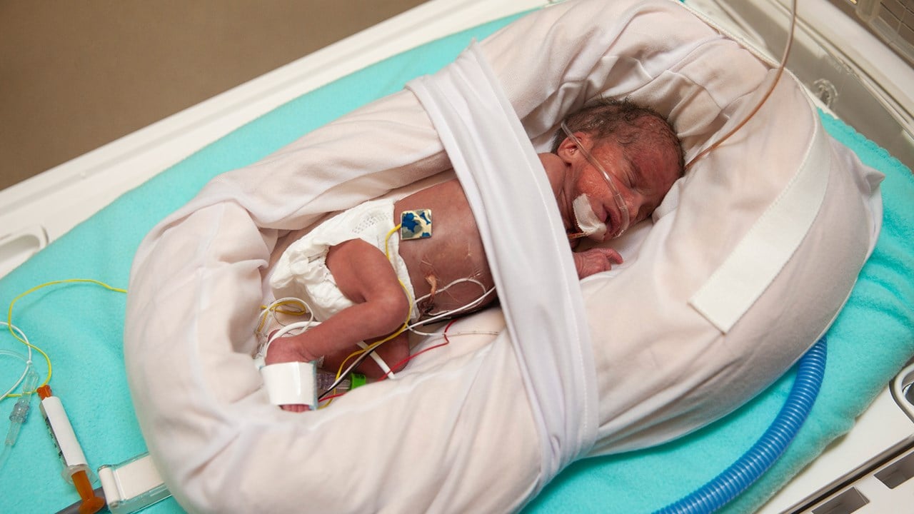
Key Takeaways
- Prone Position is Preferred: It minimizes pressure on the exposed defect and neural tissue, reducing the risk of injury and infection.
- Avoid Pressure on the Lesion: Positioning in the prone or side-lying position ensures that the neural tissue is protected until surgical repair.
- Alternatives When Necessary: Although prone is the standard, a properly supported side-lying position can be used if there are specific clinical circumstances.
Clinical Overview of Myelomeningocele Preoperative Management
Myelomeningocele is a severe form of spina bifida where the spinal canal and the backbone do not close completely during fetal development. This condition results in an exposed defect where the spinal cord and meninges can be vulnerable to trauma, infection, and further damage. The primary goal of preoperative management is to protect these exposed neural structures until surgical intervention can take place, typically within the first 24-48 hours after birth.
The preoperative positioning of infants with myelomeningocele is critical. Proper care is necessary to avoid pressure on the defect, which is the primary source of potential complications. Pressure on the exposed tissues not only increases the risk of infection but may also cause further neurologic damage. Hence, among the positioning options, the prone position is widely recommended for its ability to shield the defect from any undue pressure.
Rationale for the Prone Position
Protection of Neural Tissue
The prone position – where the infant is placed on their stomach – is the primary choice for several reasons:
Preventing Direct Pressure
When a child with myelomeningocele is placed in a prone position, the defect, usually located on the back, is not in contact with any surface. This is crucial, as direct pressure on the open lesion can lead to several complications, including:
- Risk of Infection: The exposed neural tissue is highly susceptible to bacterial contamination. By avoiding contact, the potential for infections such as meningitis is significantly lowered.
- Further Damage: Pressure on the defective area could exacerbate the injury, potentially leading to irreversible neurological deficits.
Medical guidelines and clinical studies point out that infants must be positioned in such a way as to minimize any mechanical forces exerted on the defect. This helps to ensure that the neurological tissue remains intact until corrective surgery is performed.
Enhanced Wound Integrity and Care Management
Reduced Risk of Contamination
In addition to preventing necrosis of the neural tissue, the prone position aids in keeping the surgical field relatively clean. Since the defect is not exposed to external surfaces, it is much easier for caregivers to maintain sterile conditions, which is instrumental in preoperative care.
Facilitated Nursing Interventions
The prone positioning also contributes to better management in a neonatal intensive care unit (NICU). It provides easy access for healthcare professionals to monitor respiratory function and to manage intravenous lines and other supports necessary for the infant’s survival. This thoughtful positioning supports both immediate care and the preparation process required for surgical repair.
Comparative Analysis of Positioning Options
Commonly Considered Positions
| Positioning Option | Advantages | Disadvantages |
|---|---|---|
| Prone Position |
|
|
| Side-lying Position |
|
|
| Trendelenburg Position |
|
|
| Supine Position |
|
|
Detailed Considerations in Preoperative Care
Handling and Positioning Techniques
The process of positioning a myelomeningocele infant is delicate and must be done with strict adherence to sterile techniques and established protocols. Detailed protocols involve:
Step-by-Step Positioning
1. Initial Stabilization: Immediately after birth, the infant is carefully lifted by trained neonatal staff. The aim is to stabilize the airway, breathing, and circulation while preventing inadvertent pressure on the defect.
2. Supportive Equipment: The use of specialized beds or isolettes is essential. These devices often include customized pads or cut-outs designed to accommodate the defect without applying pressure. In some cases, foam mattresses with a cut-out area are used, especially if the infant must be maintained in a supine position temporarily for other medical interventions (such as urgent intubation).
3. Prone Positioning: Once the infant is stable, the preferred positioning is accomplished by carefully turning the baby into a prone position. During this time, careful attention is paid to ensure that no part of the defect comes into contact with any surfaces.
4. Monitoring and Adjustments: Continuous observation is critical to detect any shifts in the infant's body that might place undue pressure on the defect. Nurses and doctors continuously monitor the positioning, adjusting supportive equipment as needed.
Side-lying as an Alternative
Although the prone position is the standard of care, situations may arise where a side-lying position is employed. The side-lying position involves positioning the infant on one side, thereby still keeping the defect from direct contact with any surface. However, it is considered an alternative rather than first-line management. The key considerations with side-lying include:
When Side-lying is Utilized
The side-lying position might be chosen if:
- Medical Instability: If the infant requires other interventions that make prone positioning temporarily unfeasible.
- Ease of Access for Procedures: Certain diagnostic or therapeutic maneuvers might be easier to conduct with the infant in a lateral position, provided that extensive care is taken to support the defect adequately.
Nevertheless, clinical guidelines consistently reinforce that the prone position remains the optimal choice for preoperative care in myelomeningocele due to its superior protection of vulnerable tissues.
Special Considerations and Practical Tips
Managing the Neonatal Intensive Care Environment
In neonatal intensive care settings, the multidisciplinary team, including neonatologists, nurses, and neurosurgeons, collaborates closely to ensure that every aspect of the infant’s positioning and care is optimized. Key best practices include:
Infection Prevention
Maintaining sterile techniques when handling the infant is critical. Guidelines emphasize that before and during positioning, the affected area should be protected with sterile dressings or coverings when possible. This helps curb the risk of contamination and subsequent infections.
Continuous Monitoring
The infant’s vital signs and the integrity of the lesion are continuously monitored. Monitoring devices and regular physical assessments ensure that the positioning remains effective and that any minor adjustments can be quickly executed.
Communication Among Healthcare Providers
Given the complexity and urgency of preoperative management in myelomeningocele, clear and concise communication among all members of the care team is essential. The process involves:
- Coordinated Efforts: Seamless collaboration ensures that every adjustment and decision reinforces the safety and protection of the defect. The prone position, when maintained correctly, is a cornerstone of this protective care.
- Standardized Protocols: Most institutions follow strict guidelines that delineate step-by-step procedures for infant positioning, reducing variability and minimizing complications.
Advanced Insights into Positioning Strategies
Surgical Timing and Preoperative Preparation
Surgical repair for myelomeningocele is ideally performed within 24 to 48 hours after birth. This narrow window underscores the importance of immediate and proper preoperative care. By ensuring that the infant is appropriately positioned, healthcare providers safeguard the neural tissue during the critical period before the surgical intervention.
The Impact of Timing on Outcomes
Early surgical repair has been associated with better outcomes in terms of reduced infection rates and improved protection against further neural deterioration. Hence, the immediate and careful positioning of the infant in the prone position is not merely a procedural step—it is directly linked to surgical success and long-term neurological function.
Institutional Protocols and Best Practices
Different hospitals and care centers may have nuanced protocols based on their experiences and the available resources, yet the consensus remains clear: to use the prone position preoperatively for infants with myelomeningocele. In situations where deviations from the prone positioning are necessary, such as during emergency interventions, alternative approaches like the side-lying position may be employed temporarily.
Ensuring Optimal Care through Evidence-Based Practice
Summary of Clinical Evidence
Multiple clinical studies and standard management guidelines from reputable institutions emphasize the importance of maintaining the prone position for children with myelomeningocele in the preoperative phase. This strategy stems from evidence suggesting that avoiding pressure on the defect is the single most important factor in preserving neural tissue integrity.
In addition to positioning the infant prone, efforts extend to ensuring that the often-fragile neural tissue is not compromised by external forces. The consensus derived from the literature reaffirms that while the side-lying position is a valid alternative, especially when necessary for other medical procedures, it does not offer the same level of protection as the prone position.
Practical Guidelines and Implementation
Checklist for Preoperative Positioning
The following checklist can be used by neonatal care teams to ensure that every infant with myelomeningocele is safely and correctly positioned:
- Initial Assessment: Evaluate the condition of the defect and establish a strategy for protection.
- Equipment Preparation: Use specialized isolettes or foam mattresses with cut-outs to support the infant without applying pressure.
- Sterile Handling: Ensure the defect and surrounding area are prepared using sterile techniques to prevent contamination.
- Positioning Procedure: Carefully turn the infant into the prone position with mindful adjustments to maintain pressure-free posture.
- Continuous Monitoring: Regularly check that the prone position is maintained and that there are no shifts that might compromise the defect.
- Team Coordination: Maintain clear communication among all members of the care team during the process.
Diagram of Positioning Concept
Below is a simplified diagram that illustrates the rationale behind selecting the prone position versus other alternatives:
| Characteristic | Prone Position | Side-lying Position | Supine/Trendelenburg Positions |
|---|---|---|---|
| Avoids direct pressure | ✓ High | Moderate | Low |
| Risk of contamination | ✓ Low | Low | High |
| Ease of maintaining sterile field | ✓ High | Moderate | Complex |
| Preoperative safety | ✓ Optimal | Acceptable | Suboptimal |
Integrating Best Practices into Clinical Workflows
Operational Considerations
Integrating best practices into daily neonatal operations involves multidisciplinary training sessions, standardized protocols, and quality assurance reviews. By ensuring that all medical staff are educated on the benefits of the prone positioning for myelomeningocele, hospitals can consistently safeguard the infants from complications.
Multidisciplinary Approach
A coordinated effort between neonatologists, neurosurgeons, and nursing staff is essential. The adoption of evidence-based practices such as prone positioning reflects an integrated approach to minimize risk and improve surgical outcomes.
Frequent audits and case reviews help in refining the protocols further, and sharing insights from practice-based evidence ensures that patient care remains at the highest possible standard.
Training and Simulation
Hands-on simulation training for neonatal teams is beneficial. These sessions focus on the correct technique of transferring and positioning the infant while preserving the integrity of the lesion. As practices evolve with emerging research, continuous education is necessary to keep protocols updated.
Reference Materials and Source Verification
The recommendations mentioned here are supported by multiple resources from reputable institutions that have specialized guidelines for the care of infants with myelomeningocele. Health professionals are encouraged to refer to these materials to verify the best practices.
- JSTOR Article - Academic Journal
- DB Pediatrics Guidelines - University of Washington
-
PMC Article - National Institutes of Health
- WFSA Resource Tutorial - World Federation of Societies of Anaesthesiologists
- CAHS Neonatology Guidelines - Child and Adolescent Health Service
Exploring Additional Clinical Insights
Recommendations for Further Reading
Healthcare professionals seeking to deepen their understanding of preoperative management for myelomeningocele are encouraged to explore the following related queries and topics. These additional topics provide broader context and enhance clinical decision-making in similar high-risk neonatal conditions.
Last updated March 9, 2025