
Exploring Histological Parallels Between Rat and Human Stomachs
A comprehensive comparative table and in-depth analysis of stomach histology
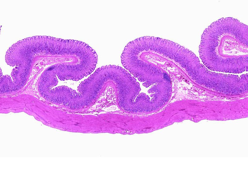
Key Highlights
- Structural Similarities: Both rat and human stomachs share many fundamental layers essential for digestion.
- Cellular and Tissue Components: Similar cell types and glandular structures enable shared functions.
- Comparative Utility: Rats offer valuable insights into human gastric physiology and pathology.
Detailed Comparative Analysis
The histological comparison between the rat and human stomach reveals remarkable similarities that underline shared functionalities despite certain anatomical differences. Researchers commonly utilize the rat as a model organism due to many overlapping features at the cellular and tissue levels. Below, we synthesize the most relevant aspects observed across multiple studies.
1. Layered Structural Organization
Both rat and human stomachs exhibit a layered organization that is crucial for their digestive roles. These layers typically include:
Mucosa, Submucosa, Muscularis Externa, and Serosa (or Adventitia)
In both species, the mucosal layer is composed of a glandular epithelium, allowing for the secretion of digestive enzymes and acid. The muscularis externa is prominently divided into distinct muscle layers that facilitate peristalsis. Although the rat stomach includes a forestomach with distinct histology (stratified squamous epithelium), the glandular portions share features with humans.
2. Gastric Glands and Cellular Components
The gastric glands are instrumental in producing digestive secretions and are found in both rat and human stomachs. Key cellular components include:
Gastric Glands
These glands contain several cell types:
- Mucous Cells: Present in both species, these cells secrete mucus to protect the epithelium from acid and digestive enzymes.
- Parietal Cells: Responsible for the secretion of hydrochloric acid (HCl) in both rats and humans.
- Chief Cells: These cells produce pepsinogen, which is converted into the digestive enzyme pepsin.
Histological staining and microscopic studies reveal that the distribution and structural appearance of these cells in the gastric mucosa show a high degree of similarity.
3. Muscular Organization and Vascularization
The muscular arrangement in the stomach is pivotal for its function. Both species demonstrate:
Muscular Layers
In humans as well as rats, the muscularis externa is classically divided into:
- Longitudinal muscle layer: The outer layer facilitating overall stomach movements.
- Circular muscle layer: The inner layer, crucial for segmental contractions and mixing.
Additionally, in certain portions of the rat stomach, an extra oblique layer exists near the gastroesophageal junction which contributes to the specialized function of that region. Both species also exhibit a well-vascularized submucosal network that supports nutrient absorption and provides a robust blood supply to the mucosa.
4. Specific Histological Observations
Detailed comparative studies have identified several points of histological commonality:
Gastric Mucosal Features
The mucosal layer in both rat and human stomachs is lined with a simple columnar epithelium and is interspersed with gastric pits, which lead to the underlying glands. Although the rat shows variation due to the non-glandular forestomach region, its glandular stomach shows an arrangement almost identical to that of humans.
Furthermore, microscopic analysis indicates that the thickness of various mucosal layers may vary; however, the fundamental histological structure, including the presence of goblet cells and specific glandular arrangements, remains consistent. This uniformity confirms the rat’s effectiveness as an experimental model for human gastric studies.
5. Comparative Table of Histological Similarities
The following table incorporates the core histological similarities between the rat and human stomach supplemented by details drawn from multiple studies:
| Feature | Rat Stomach | Human Stomach |
|---|---|---|
| General Structure | Consists of a forestomach (non-glandular), fundic and pyloric regions; clear distinction between glandular and non-glandular portions | Uniformly glandular with cardia, fundic, and pyloric regions; no forestomach |
| Mucosal Layer | Simple columnar epithelium with gastric pits; interspersed with goblet and chief cells | Similar simple columnar epithelium with multiple gastric pits and glands |
| Gastric Glands | Contain mucous cells, parietal cells (acid secretion) and chief cells (pepsinogen production) | Identical composition: mucous cells, parietal cells for acid secretion, and chief cells for pepsinogen secretion |
| Muscularis Externa | Typically organized into three layers (including unique oblique fibers at certain sites) | Consists of inner circular and outer longitudinal layers vital for peristalsis |
| Mucosal Vascularization | Rich, supporting high metabolic needs of rapidly secreting cells | Well-vascularized; ensures proper function and nutrient distribution |
| Functional Specialization | Forestomach for storage and glandular region for secretion, showing analogous digestive roles | Glandular throughout with distinct regions for acid secretion and mucus protection |
6. Visualization via Radar Chart
The radar chart below represents various aspects of histological structure observed in comparative studies, such as epithelial complexity, glandular distribution, muscular differentiation, vascularization, and cellular specialization. The datasets reflect a subjective synthesis of the key features in both models.
Supplementary Multimedia Resource
The space below includes a related video that provides additional visual and detailed explanation of the digestive system histology, with a focus on the rat model. This video reinforces the concepts discussed above and may serve as an excellent resource for educators and students alike.
FAQ Section
What are the primary histological similarities between rat and human stomachs?
How do the muscular arrangements differ between the two species?
Why is the rat used as a model for human gastric studies?
References
- Comparative Study of Digestive Systems - PubMed
- Rat Digestive System Anatomy - PW.live
- Morphology of the Rat Stomach - Wiley Online Library
- Microbial Composition in Gastric Mucosa - Nature
- Histology of Rat Stomach - ResearchGate
Recommended Further Queries
Last updated April 3, 2025