
Rat Stomach Histology: Insights into Gastrointestinal Health
Exploring the Cellular Architecture and Experimental Advantages of the Rat Model
Key Highlights
- Distinct Functional Zones: The rat stomach divides into the forestomach and glandular stomach, each playing unique roles in digestion and disease modeling.
- Research Advantages: Due to its reproducibility and anatomical similarities to the human stomach, the rat model is invaluable in studying gastric disorders such as ulcers, gastritis, and metaplasia.
- Histopathological Insights: Detailed cellular architecture analysis illuminates mechanisms behind gastrointestinal disease progression and therapeutic interventions.
Results
The rat stomach has emerged as a fundamental experimental model in gastrointestinal research. This remarkable model is leveraged due to its distinct cellular architecture and layered structure that not only facilitate digestive processes but also provide a robust framework for investigating gastric health and related diseases. Through detailed histological studies, researchers have been able to delineate functional zones within the rat stomach and correlate these with specific physiological processes as well as pathologies.
Anatomical Overview and Functional Zoning
Anatomically, the rat stomach comprises two major regions: the non-glandular forestomach and the glandular stomach. The forestomach, located proximally near the esophagus, is lined with a stratified squamous keratinized epithelium. This unique design plays a critical role in mechanically processing food and protecting against potential physical damage from ingested materials. Its primary function is storage and the initial breakdown of ingested food, setting the stage for subsequent digestive processes occurring in the glandular portion.
Non-Glandular (Forestomach) Characteristics
The forestomach is notable for its robust and protective epithelial layer that is highly resistant to wear and tear. The stratified squamous keratinized epithelium serves as a barrier to physical insults, thereby shielding the more sensitive structures of the digestive system further along the tract. Histologically, this region offers insights into cell differentiation and the mechanisms by which the body counters mechanical stress.
Glandular Stomach Structure and Function
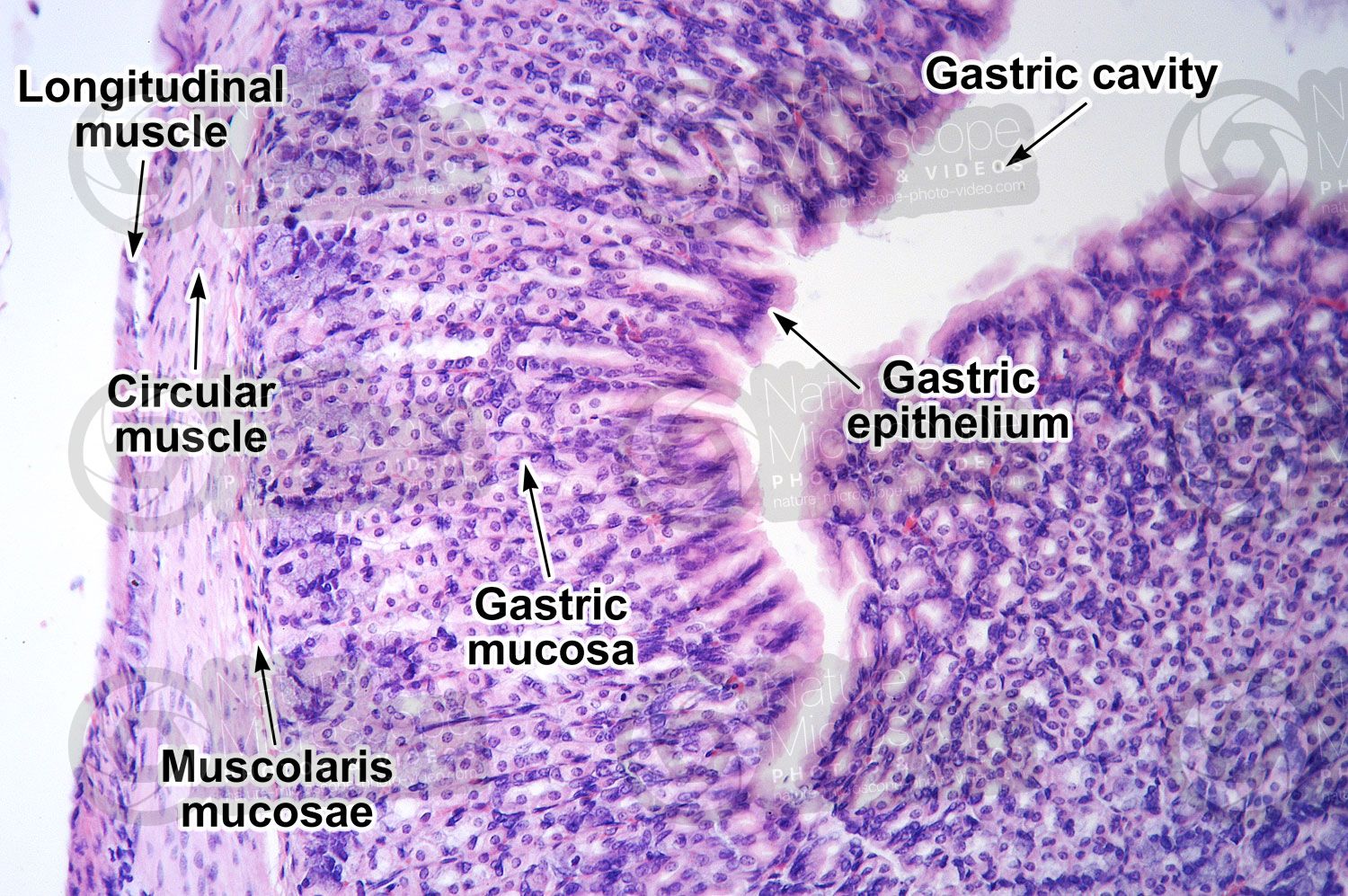
In contrast, the glandular stomach is subdivided into the fundus and pyloric regions and is primarily responsible for chemical digestion. Histological analysis of the glandular stomach shows the presence of specialized gastric glands which secrete essential digestive enzymes and acids. These secretions facilitate the breakdown of ingested food into smaller, absorbable molecules. The fundus is particularly known for its dense glandular structures responsible for the secretion of acid and pepsinogen, whereas the pylorus plays a regulatory role in gastric emptying and mucus production.
Muscular Structure and Its Implications
The rat stomach is also distinguished by its complex arrangement of muscular layers. The muscularis externa consists of three distinct muscle coats: a longitudinal coat, a circular coat, and an internal oblique (sling) muscle. This unique configuration is critical for the mechanical digestion of food, aiding in its mixing and propelling it towards the duodenum. This well-organized muscular system not only facilitates efficient peristalsis but also plays a role in maintaining the structural integrity of the gastric wall against the erosive actions of gastric acid.
Histopathological Observations and Research Applications
Detailed histological examinations have significantly advanced our understanding of the rat stomach’s cellular composition and its response to various pathological conditions. Experimental studies using rat models have successfully induced conditions such as gastric ulcers, chronic gastritis, and even precancerous lesions like metaplasia. These induced conditions mimic many aspects of human gastrointestinal diseases, thus providing a reliable experimental platform for:
- Elucidating the progression and early onset of gastric ulceration
- Understanding the cellular changes associated with chronic gastritis
- Investigating the molecular and cellular mechanisms leading to metaplasia and other dysplastic transformations
- Evaluating the efficacy of new therapeutic interventions and drugs
The reproducibility of findings in rat models is one of the foremost advantages in gastrointestinal research. It has enabled researchers to produce consistent results that are both replicable and predictable. This consistency is essential when the goal is to translate experimental data into therapeutic insights, particularly for developing interventions that can be extrapolated to human gastric diseases.
Innovative Analytical Techniques in Gastric Histology
Beyond classical histological staining and examination, recent advancements have seen the adoption of innovative analytical techniques such as fractal geometry to understand the complex structures within the gastrointestinal system. Although primarily conceptual at this stage, these techniques offer potential in enhancing diagnostic precision. Fractal geometry allows for the quantitative analysis of the intricate branching patterns in the gastric glands and cellular layers. This quantitative approach not only improves our understanding of normal histology but also helps in the early detection of pathological changes.
Fractal Analysis: Future Directions
Fractal analysis provides a novel perspective on the evaluation of complex biological structures. By applying principles of fractal mathematics, researchers have started to analyze the irregular nature of glandular architectures. These studies suggest that alterations in the fractal dimension of gastric tissues may correlate with early disease states, thus opening new avenues for non-invasive and highly detailed diagnostic methodologies. Although these techniques are still under refinement, their application underscores a shift towards more quantitative and predictive models in histopathology.
Comparative Overview: Forestomach vs. Glandular Stomach
To facilitate a better understanding of the distinctive features and functions of the two major zones in the rat stomach, we present a comparison table. This table synthesizes key histological features and functional roles that differentiate the forestomach from the glandular stomach.
| Feature | Forestomach | Glandular Stomach |
|---|---|---|
| Epithelium Type | Stratified Squamous Keratinized | Simple Columnar with Glandular Units |
| Primary Function | Food Storage and Mechanical Protection | Secretion of Digestive Enzymes and Acids |
| Histological Layers | Well-differentiated protective barrier with minimal glandular features | Multiple layers including mucosal, submucosal, muscular, and serosal layers with extensive glandular structures |
| Associated Research Models | Studies on mechanical injury resistance and epithelial differentiation | Investigations into gastric ulcers, metaplasia, and chemical digestion |
This table highlights that the forestomach and glandular stomach, while both integral to overall gastric function, contribute differently to the digestion process and respond distinctly under pathological conditions. Their unique histological structures make the rat an exemplary model for both fundamental research and the evaluation of potential therapeutic agents.
Conclusion and Final Thoughts
In summary, the rat stomach offers a comprehensive model for gastrointestinal research due to its distinct cellular architecture, functional zoning, and reproducibility in experimental settings. Its division into the forestomach and glandular stomach represents a microcosm of the complex digestive processes found in higher mammals, including humans. The forestomach, with its protective keratinized epithelium, plays a critical role in the mechanical breakdown and initial processing of food, while the glandular stomach is essential for chemical digestion, involving the secretion of digestive enzymes and acids.
The extensive histological characterization of these regions provides invaluable insights into the cellular and molecular mechanisms underlying various gastrointestinal disorders such as gastric ulcers and metaplasia. Rat models have demonstrated that, through detailed observation of structural changes and specific cellular markers, key aspects of disease progression can be elucidated. These insights support the development of more precise prevention and treatment strategies for human gastric disorders. The ability to induce and study pathologies such as chronic gastritis, epithelial dysplasia, and precancerous lesions in rats contributes to a deeper understanding of gastrointestinal disease mechanisms.
Furthermore, the incorporation of advanced techniques like fractal geometry analysis signifies the evolving landscape of histopathological research. By quantifying the irregular branching and spatial distribution of gastric glands, researchers are beginning to forge quantitative links between structural complexity and disease state progression. These developments are likely to enhance diagnostic capabilities and provide more rigorous models for therapeutic testing.
Overall, the rat stomach stands as a vital experimental tool whose detailed study not only offers a window into normal digestive functions but also mirrors pathological states with a high degree of accuracy. The reproducibility of experimental results using the rat model underpins its utility as an indispensable component of gastrointestinal research. As ongoing studies continue to refine our understanding, the insights gleaned from rat stomach histology promise to drive forward innovations in the prevention and treatment of gastrointestinal disorders.
Conclusion
The research on the rat stomach has provided a rich, detailed understanding of the gastrointestinal system's anatomy and cellular complexity. From its dual functional zoning—each with specialized roles in mechanical and chemical digestion—to the advanced analytical techniques employed for assessing structural changes, the rat stomach model epitomizes the convergence of basic science and translational research. Its histological attributes have proven essential for simulating human gastric pathologies, ranging from simple mechanical injuries to the more complex processes of metaplasia and chronic inflammation. The insights derived from these investigations not only enhance our fundamental knowledge of gastric physiology but also pave the way for developing novel therapeutic strategies aimed at mitigating gastrointestinal diseases.
Continued investment in such research models is warranted, as the reproducibility and detailed structural examination of the rat stomach offer a window into early-stage disease detection and potential avenues for treatment. In essence, by deepening our understanding of the rat stomach’s detailed histology, scientists are better equipped to decipher the multifactorial nature of gastric diseases and translate these findings into safer, more effective interventions for human health.
References
- Anatomical and Histological Insights - SpringerLink
-
Muscularis Externa in Rat Stomach - PMC
- Gastric Ulcer Models in Rats - SCIRP
-
Digestive System Morphology - PMC
- Gastric Bypass Research - PubMed
Recommended Topics
Last updated February 22, 2025