
Understanding the Dural Sheath Around the Optic Nerve
Exploring the Critical Role of the Dural Sheath in Optic Nerve Health
Key Takeaways
- Protection and Structural Integrity: The dural sheath provides essential mechanical protection and support to the optic nerve, safeguarding it from physical trauma.
- Cerebrospinal Fluid Dynamics: The presence of cerebrospinal fluid (CSF) within the sheath facilitates nutrient exchange, waste removal, and acts as a cushion against pressure changes.
- Clinical Significance: Variations in the dural sheath, such as dilation or abnormalities, are critical indicators for diagnosing intracranial pressure disorders and other optic nerve pathologies.
Anatomical Structure of the Dural Sheath
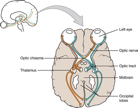
The dural sheath is a fibrous, protective layer encasing the optic nerve, which is a vital conduit transmitting visual information from the retina to the brain. Comprising three primary segments—intracranial, intracanalicular, and intraorbital—the dural sheath extends from the brain's subarachnoid space to the eye, seamlessly integrating with the periorbita. This continuity allows for the uninterrupted flow of cerebrospinal fluid (CSF), which plays a pivotal role in maintaining the optic nerve's health and functionality.
Protection and Support
The dural sheath serves as a robust barrier, shielding the delicate optic nerve fibers from external physical forces. This mechanical protection is crucial, especially given the optic nerve's vulnerability as it traverses from the ocular globe to the brain. By maintaining structural integrity, the dural sheath ensures that the optic nerve remains uncompromised, safeguarding against potential injuries that could lead to vision impairment or loss.
Cerebrospinal Fluid Dynamics
Encased within the dural sheath is cerebrospinal fluid (CSF), occupying the subarachnoid space surrounding the optic nerve. CSF serves multiple functions:
-
Cushioning: Acts as a shock absorber, mitigating the impact of sudden pressure changes and mechanical stress on the optic nerve.
-
Nutrient Exchange: Facilitates the delivery of essential nutrients and the removal of metabolic waste products, ensuring the optic nerve's optimal function.
-
Pressure Regulation: Maintains homeostasis by balancing CSF production and absorption, which is critical for proper neural function.
Intracranial Pressure Monitoring
The dural sheath's direct communication with the brain's subarachnoid space makes it sensitive to variations in intracranial pressure (ICP). Elevated ICP can lead to the expansion of the dural sheath, a phenomenon detectable through imaging modalities such as Magnetic Resonance Imaging (MRI) or ultrasound. Measuring the Optic Nerve Sheath Diameter (ONSD) has emerged as a non-invasive method to assess ICP, providing clinicians with valuable diagnostic information without the need for more invasive procedures like lumbar punctures.
Clinical Implications and Pathological Conditions
Intracranial Pressure Disorders
Elevated intracranial pressure (ICP) can result from various conditions, including hydrocephalus, traumatic brain injury, and idiopathic intracranial hypertension (IIH). The dural sheath's response to increased ICP—manifested as sheath dilation—serves as a key diagnostic indicator. Additionally, papilledema, characterized by optic disc swelling, is a direct consequence of elevated ICP, stemming from the compression of retinal veins and subsequent fluid accumulation.
Optic Nerve Sheath Meningioma (ONSM)
ONSM is a rare, benign tumor arising from the meninges surrounding the optic nerve. It leads to the thickening and potential calcification of the dural sheath, resulting in optic nerve compression. This compression can cause progressive vision loss and other ocular disturbances. Imaging techniques like MRI and CT scans are instrumental in identifying ONSM, as they reveal characteristic sheath thickening and other morphological changes.
Dural Ectasia
Dural ectasia refers to the abnormal expansion of the dural sheath, often associated with connective tissue disorders such as Marfan syndrome or Ehlers-Danlos syndrome. In the context of the optic nerve, dural ectasia can lead to the dilation of the optic nerve sheath, which may be benign or indicative of underlying pathologies like arachnoid cysts or space-occupying lesions. Clinical symptoms can range from mild visual disturbances to significant optic neuropathy, depending on the extent of sheath enlargement.
CSF Leakage and Intracranial Hypotension
CSF leakage from the dural sheath can result in intracranial hypotension, leading to symptoms such as headaches, visual disturbances, and cognitive impairments. Identifying leakage points within the optic nerve sheath is crucial for effective management and treatment, which may include surgical interventions or targeted therapies to restore CSF balance.
Neurodegenerative Diseases
Emerging research suggests that the optic nerve sheath may play a role in the clearance of interstitial fluids, potentially linking it to neurodegenerative diseases like Alzheimer's and Parkinson's. Impaired CSF circulation and drainage within the dural sheath could contribute to the accumulation of pathological proteins characteristic of these conditions, although further studies are necessary to elucidate these connections fully.
Diagnostic Imaging and Techniques
Magnetic Resonance Imaging (MRI)
MRI provides detailed images of the optic nerve sheath, allowing for the assessment of structural abnormalities, sheath thickness, and the presence of lesions such as meningiomas. T2-weighted MRI sequences are particularly useful in evaluating the fluid-filled subarachnoid space, enabling clinicians to discern between various pathological states based on imaging characteristics.
Ultrasound Measurement of Optic Nerve Sheath Diameter (ONSD)
Ultrasound offers a non-invasive, bedside method for measuring ONSD, which correlates with ICP levels. An increased ONSD is indicative of elevated ICP, making this technique valuable in emergency and critical care settings where rapid assessment is essential. The simplicity and accessibility of ultrasound make it a preferred tool for initial evaluations of suspected intracranial pressure disorders.
Computed Tomography (CT) Scans
CT scans are instrumental in identifying calcifications, sheath thickening, and other structural changes within the dural sheath. They provide rapid, high-resolution images, which are critical in acute settings such as traumatic brain injuries, where timely diagnosis can influence treatment outcomes.
Differential Diagnosis Through Imaging
Imaging studies are crucial for differentiating between various optic nerve sheath pathologies. For instance, optic neuritis, characterized by inflammation, presents differently on MRI compared to compressive lesions like meningiomas. Accurate interpretation of imaging results ensures appropriate diagnosis and management of underlying conditions affecting the optic nerve.
Functional Implications of the Dural Sheath
Nutrient and Waste Exchange
The CSF within the dural sheath facilitates the exchange of nutrients and waste products, ensuring the metabolic needs of the optic nerve are met. This exchange is vital for maintaining the health of the optic nerve fibers, supporting their ability to transmit visual information efficiently.
Protection Against Mechanical Stress
The cushioning effect of CSF absorbs mechanical shocks and mitigates the impact of movements or external forces, protecting the optic nerve from potential damage. This protective mechanism is essential for preserving the integrity of the optic nerve in various physiological and environmental conditions.
Role in CSF Circulation
The dural sheath's continuity with the brain's subarachnoid space allows it to participate actively in CSF circulation. This role is critical for the overall homeostasis of the central nervous system, as CSF circulation aids in removing metabolic waste and distributing essential molecules throughout the brain and optic nerve.
Pathological States and Their Impact
Optic Nerve Sheath Meningioma
ONSM can lead to progressive compression of the optic nerve, resulting in visual acuity loss, visual field defects, and potential blindness if left untreated. Early detection through imaging is crucial for initiating appropriate therapeutic interventions, which may include surgical resection or radiotherapy to alleviate nerve compression.
Dural Ectasia and Connective Tissue Disorders
In conditions like Marfan syndrome, dural ectasia can cause significant expansion of the optic nerve sheath, leading to visual disturbances and increased susceptibility to optic nerve injuries. Management involves regular monitoring through imaging and addressing the underlying connective tissue disorder to prevent further complications.
CSF Leakage and Intracranial Hypotension
CSF leaks can lead to a drop in intracranial pressure, manifested by severe headaches, visual disturbances, and potential cognitive impairments. Identifying the source of leakage within the dural sheath is paramount for effective treatment, which may involve epidural blood patches or surgical repair to restore normal CSF dynamics.
Clinical Monitoring and Assessment
Non-Invasive ICP Assessment
Measuring ONSD through ultrasound provides a non-invasive means to estimate intracranial pressure, especially valuable in settings where immediate assessment is necessary. This technique allows for timely interventions in cases of elevated ICP, improving patient outcomes by preventing secondary complications.
Pseudotumor Cerebri (IIH)
IIH is characterized by increased ICP without an identifiable cause, leading to symptoms like headaches, vision loss, and tinnitus. Elevated ONSD and optic disc swelling observed through imaging signify IIH, guiding clinicians to implement treatments that reduce ICP and mitigate optic nerve damage.
Differentiating Optic Neuritis from Other Pathologies
Optic neuritis presents with inflammation of the optic nerve, often associated with multiple sclerosis. Imaging differentiates it from compressive lesions by identifying features such as contrast enhancement and nerve sheath swelling, ensuring accurate diagnosis and appropriate therapeutic strategies.
Innovations in Understanding the Dural Sheath
Role in CSF and Lymphatic Drainage
Recent studies have illuminated the dural sheath's involvement in CSF and interstitial fluid clearance, linking it to the brain's glymphatic system. This connection highlights the sheath's potential role in neurodegenerative diseases, where impaired clearance mechanisms contribute to the accumulation of toxic proteins and neuronal damage.
Implications for Neurodegenerative Disease Research
Understanding the dural sheath's role in fluid dynamics opens new avenues for researching treatments targeting CSF circulation and clearance pathways. Enhancing these mechanisms could offer therapeutic benefits for conditions like Alzheimer's disease by facilitating the removal of pathological proteins from the central nervous system.
Conclusion
The dural sheath surrounding the optic nerve is a critical anatomical structure with multifaceted roles in protecting the optic nerve, facilitating CSF dynamics, and serving as a diagnostic tool for various intracranial and optic nerve pathologies. Its integrity and functionality are paramount for maintaining optimal visual function and overall neural health. Advances in imaging technologies and a deeper understanding of the sheath's physiological roles continue to enhance clinical diagnostics and open new research pathways, underscoring the dural sheath's significance in both health and disease.
References
Last updated January 20, 2025