
Comprehensive Guide to Tissue Processing for Microscopic Analysis
Understanding the Critical Steps for Optimal Histological Examination
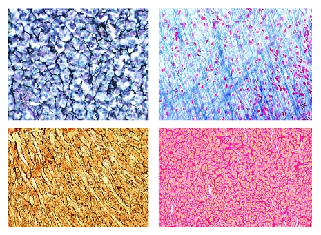
Key Takeaways
- Fixation is essential for preserving tissue structure and preventing degradation.
- Dehydration and clearing remove water and fats, preparing the tissue for embedding.
- Embedding, sectioning, and staining are crucial for creating clear, thin slices for microscopic inspection.
Introduction to Tissue Processing
Tissue processing is a fundamental procedure in histopathology, ensuring that biological tissues are adequately prepared for microscopic examination and diagnosis. The primary objective is to render the tissue specimens optically clear and sectionable at thin slices, typically ranging from 4 to 10 micrometers in thickness. This meticulous preparation allows pathologists to observe cellular and subcellular structures with high clarity, facilitating accurate diagnoses.
Step-by-Step Tissue Processing
1. Fixation
Fixation is the initial and one of the most critical steps in tissue processing. The tissue is treated with a fixative solution, commonly 10% neutral buffered formalin, which serves multiple purposes:
- Preservation of Tissue Structure: Fixatives stabilize proteins and cross-link cellular components, maintaining the integrity of the tissue architecture.
- Prevention of Degradation: Fixation halts autolysis and inhibits microbial growth, preventing tissue decay during processing.
- Facilitation of Handling: Fixed tissues become firmer, making them easier to manipulate in subsequent steps.
The duration of fixation typically ranges from 6 to 24 hours, depending on the tissue type and size. Over-fixation can obscure cellular details, while under-fixation may lead to degradation.
2. Dehydration
After fixation, the tissue undergoes dehydration to remove water, which is incompatible with the embedding medium. This process involves passing the tissue through a series of increasing concentrations of alcohol:
- 70% Ethanol: Initiates the removal of water from the tissue.
- 90% Ethanol: Further reduces water content.
- 100% Ethanol: Completely removes residual water, preparing the tissue for clearing.
The gradual increase in alcohol concentration minimizes tissue distortion and shrinkage. Dehydration is crucial for ensuring that the tissue is adequately prepared for the subsequent clearing step.
3. Clearing
Clearing removes alcohol and replaces it with a solvent compatible with the embedding medium, typically xylene or its substitutes:
- Purpose: To render the tissue transparent and prepare it for infiltration with the embedding medium.
- Method: The tissue is immersed in xylene, which is miscible with both alcohol and paraffin wax.
- Importance: Efficient clearing ensures complete removal of alcohol, preventing interference with embedding and sectioning.
Multiple exchanges of xylene may be necessary to achieve thorough clearing. This step is pivotal for the success of the embedding process.
4. Infiltration
Infiltration involves replacing the clearing agent with a medium that can solidify, such as paraffin wax:
- Embedding Medium: Paraffin wax is commonly used due to its favorable properties, including ease of handling and compatibility with most staining techniques.
- Process: The tissue is immersed in melted paraffin, which penetrates the tissue intercellular spaces.
- Outcome: The paraffin infiltrates the tissue, replacing the cleared solvent and providing structural support.
Proper infiltration is essential for the subsequent embedding and sectioning steps. Incomplete infiltration can lead to poor section quality and artifacts in microscopic analysis.
5. Embedding
Embedding encases the tissue in a solid block of paraffin, facilitating precise sectioning:
- Molding: The paraffin-infiltrated tissue is placed in a mold and surrounded by additional melted paraffin.
- Solidification: Upon cooling, the paraffin hardens, creating a solid block that immobilizes the tissue.
- Support: The rigid paraffin block provides support during the cutting of thin sections, preventing distortion.
The orientation of the tissue within the paraffin block is critical, as it determines the plane of sectioning and ultimately affects the quality of microscopic analysis.
6. Sectioning
Sectioning is the process of slicing the paraffin-embedded tissue into ultra-thin sections using a microtome:
- Microtome: A precision instrument that allows for the consistent cutting of thin tissue slices, typically 4-6 micrometers thick.
- Technique: The paraffin block is mounted on the microtome, and a sharp blade slices through the tissue, producing uniform sections.
- Quality Control: Consistent thickness is crucial for optimal light penetration and clear visualization of cellular structures under the microscope.
Proper sectioning ensures that tissues are thin enough to be transparent to light while maintaining structural integrity for accurate examination.
7. Mounting and Staining
The final steps involve mounting the sections on slides and staining them to enhance contrast:
- Mounting: Thin sections are carefully placed on glass slides and allowed to adhere.
- Staining: Dyes such as hematoxylin and eosin (H&E) are applied to highlight different cellular components, making structures like nuclei and cytoplasm more distinguishable.
- Coverslipping: A coverslip is placed over the stained tissue with a mounting medium to protect the sample and facilitate microscopy.
Staining is essential for differentiating various tissue elements, allowing for detailed examination and accurate diagnosis by pathologists.
Detailed Workflow of Tissue Processing
Below is a comprehensive workflow table summarizing each step of tissue processing for microscopic analysis:
| Step | Purpose | Method | Key Considerations |
|---|---|---|---|
| Fixation | Preserve tissue structure and prevent degradation | Immerse tissue in 10% neutral buffered formalin for 6-24 hours | Avoid over- or under-fixation to maintain cellular integrity |
| Dehydration | Remove water from tissue | Pass tissue through graded ethanol series (70%, 90%, 100%) | Ensure gradual alcohol concentration increase to prevent tissue distortion |
| Clearing | Remove alcohol and fats, making tissue transparent | Use xylene or substitutes to replace alcohol | Multiple solvent exchanges may be necessary for complete clearing |
| Infiltration | Replace clearing agent with embedding medium | Immerse tissue in melted paraffin wax | Ensure thorough paraffin penetration to prevent sectioning artifacts |
| Embedding | Create a solid matrix for sectioning | Encapsulate tissue in paraffin mold and allow solidification | Proper orientation of tissue within block is critical for effective sectioning |
| Sectioning | Produce thin tissue slices for microscopy | Use a microtome to cut sections 4-6 micrometers thick | Maintain consistent thickness and avoid compression or tearing of sections |
| Mounting & Staining | Enhance visibility of tissue structures | Mount sections on slides, apply appropriate stains, and coverslip | Select appropriate staining protocols to highlight specific cellular components |
Advanced Techniques and Considerations
Automated Tissue Processing
Modern histology laboratories utilize automated tissue processors to increase efficiency and consistency. These machines streamline the fixation, dehydration, clearing, and infiltration steps by precisely controlling temperature, duration, and reagent concentrations. Automation reduces manual handling errors and enhances throughput, especially in high-demand settings.
Specialized Staining Techniques
While hematoxylin and eosin (H&E) staining is the standard, various specialized stains are employed to highlight specific tissue components:
- Immunohistochemistry (IHC): Uses antibodies to detect specific antigens within tissues, aiding in the identification of proteins linked to diseases.
- Masson's Trichrome: Differentiates between muscle, collagen, and fibrin, useful in connective tissue studies.
- Periodic Acid-Schiff (PAS): Detects polysaccharides like glycogen, commonly used in liver and kidney biopsies.
The choice of staining technique depends on the diagnostic requirements and the nature of the tissue being examined.
Challenges in Tissue Processing
Despite standardized protocols, several challenges can arise during tissue processing:
-
Tissue Shrinkage and Expansion: Improper fixation or dehydration can lead to uneven shrinkage, distorting tissue morphology.
-
Incomplete Clearing: Residual alcohol can interfere with paraffin infiltration, resulting in poor section quality.
-
Sectioning Artifacts: Issues like tearing, folding, or compression during cutting compromise the integrity of slides.
Addressing these challenges requires meticulous adherence to protocols, regular maintenance of equipment, and ongoing training for laboratory personnel.
Applications of Tissue Processing
Diagnostic Pathology
Histological analysis is indispensable in diagnosing a wide array of diseases, including cancers, infections, and inflammatory conditions. Accurate tissue processing ensures that pathologists can observe hallmark features of diseases, such as cellular atypia, mitotic figures, and structural disruptions.
Research and Development
Beyond clinical diagnostics, tissue processing is vital in biomedical research. It facilitates the study of tissue architecture, gene expression patterns, and the effects of various treatments at the cellular level. High-quality histological samples contribute to advancements in understanding disease mechanisms and developing new therapies.
Forensic Science
In forensic investigations, tissue analysis can provide critical insights into causes of death, time since injury, and the presence of toxins. Properly processed tissues enable forensic pathologists to reconstruct events leading to death with greater accuracy.
Future Directions in Tissue Processing
Digital Pathology
The advent of digital pathology is transforming the field by enabling the digitization of histological slides. High-resolution imaging and advanced software facilitate remote diagnostics, telemedicine consultations, and the integration of artificial intelligence for automated analysis. Future advancements may include real-time processing and integration with molecular diagnostic techniques.
Sustainable Practices
Traditional tissue processing involves the use of hazardous chemicals like xylene, posing environmental and health risks. Emerging sustainable alternatives, such as water-based clearing agents and biodegradable embedding materials, are being developed to minimize ecological impact while maintaining processing efficacy.
Integration with Molecular Techniques
Combining histological processing with molecular techniques like in situ hybridization and polymerase chain reaction (PCR) allows for comprehensive analysis of tissues. This integration enhances the diagnostic capabilities by correlating morphological features with genetic and molecular profiles.
Conclusion
Tissue processing is a cornerstone of histopathological analysis, encompassing a series of meticulously orchestrated steps that preserve, prepare, and enhance tissue specimens for microscopic examination. From fixation to staining, each phase plays a vital role in ensuring the clarity and structural integrity necessary for accurate diagnosis and research. Advances in automation, staining techniques, and sustainable practices continue to refine and improve the efficacy of tissue processing, promising enhanced outcomes in both clinical and research settings.
References
Last updated February 12, 2025