
Comprehensive Strategies for Diagnosing and Evaluating Therapeutic Efficiency in Tuberculous Meningitis
A Detailed Overview of Multimodal Diagnostic and Monitoring Approaches
Key Takeaways
- Integrated Diagnostic Approaches: Combining clinical evaluation, cerebrospinal fluid analysis, advanced molecular tests, and neuroimaging ensures robust early diagnosis.
- Comprehensive Therapeutic Monitoring: Regular assessments through clinical examinations, serial CSF studies, imaging, and emerging biomarkers are crucial for evaluating treatment effectiveness.
- Emerging Technologies: Novel biomarkers, omics-based tools, and advanced imaging techniques provide promising avenues for faster and more accurate diagnosis and monitoring.
Introduction
Tuberculous meningitis (TBM) is a severe and potentially life‐threatening manifestation of extrapulmonary tuberculosis. Caused by the bacterium Mycobacterium tuberculosis, TBM presents a formidable challenge in both diagnosis and treatment evaluation due to its nonspecific clinical presentation and the complexities involved in confirming the diagnosis. Early diagnosis and continuous evaluation of therapeutic efficiency are vital not only to reduce morbidity and mortality but also to tailor treatment plans effectively and mitigate potential neurological complications.
The complexities in diagnosing TBM stem from its subacute onset and often vague clinical signs such as headache, fever, altered mental status, and neurological deficits, which can overlap with various other central nervous system conditions. The standard diagnostic approach typically employs multiple modalities including clinical assessment, analysis of cerebrospinal fluid (CSF), advanced molecular diagnostic tests, and neuroimaging techniques. A multidimensional approach is indispensable, particularly given that rapid and accurate diagnosis can be pivotal for starting treatment in a timely manner.
Likewise, evaluating the therapeutic efficacy in TBM is a dynamic process that incorporates both the immediate clinical response and the longer-term resolution of radiological and laboratory abnormalities. Treatment regimens generally extend over a period of several months to a year, making periodic assessment crucial to ensure that the prescribed therapy is effective and to allow for adjustments if complications or drug resistance are detected.
Diagnostic Strategies for Tuberculous Meningitis
Clinical Assessment and Scoring Systems
The cornerstone of TBM diagnosis begins with a detailed clinical history and physical examination. Although symptoms are often nonspecific, certain key indicators coupled with risk factors can heighten clinical suspicion:
- Symptom Duration and Presentation: Patients typically present with a subacute history of fever, headache, and a gradual decline in cognitive function spanning weeks. A deterioration in mental status accompanied by neurological deficits such as cranial nerve palsies, hemiparesis, or seizures should alert clinicians to the possibility of TBM.
- Risk Factor Assessment: A history of tuberculosis infection or exposure, particularly in endemic areas, and conditions such as HIV increase the clinical likelihood of TBM. The presence of “constitutional” symptoms related to TB elsewhere in the body further bolsters the clinical suspicion.
- Diagnostic Algorithms and Scoring Systems: Clinical scoring systems, such as the Marais criteria, integrate clinical signs, imaging findings, and laboratory test results. These systems classify cases into definite, probable, or possible TBM, aiding clinicians in decision-making, especially when diagnostic resources are limited.
Cerebrospinal Fluid Analysis
Analysis of the cerebrospinal fluid (CSF) is critical in diagnosing TBM owing to its direct contact with the infection site. The typical CSF profile in TBM includes:
- Lymphocytic Pleocytosis: A marked increase in lymphocytes forms the basis of the inflammatory response. In many cases, a lymphocyte-predominant pleocytosis is observed.
- Biochemical Markers: Elevated protein concentration is a consistent finding, while there is a concurrent reduction in glucose levels, reflecting the metabolic activity of the pathogen.
-
CSF Microbiological Testing:
- Acid-Fast Bacilli (AFB) Smear: Although simple to perform, AFB smears have limited sensitivity and may fail to detect the bacteria in low-concentration samples.
- Culture: The culture is considered the gold standard and offers definitive evidence of infection; however, it is time-consuming and may take several weeks to yield results.
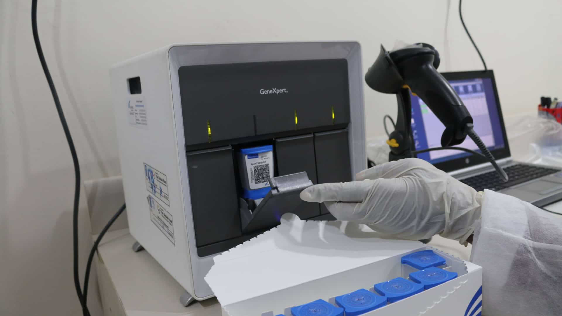
- Nucleic Acid Amplification Tests (NAATs): Techniques such as polymerase chain reaction (PCR) and the Xpert® MTB/RIF Ultra assay have transformed the diagnostic landscape by offering rapid results with high specificity. Although sensitivity may vary, these tests provide molecular confirmation of the disease.
- Additional Biomarkers: Other markers such as adenosine deaminase (ADA) levels can offer supportive evidence in the diagnosis of TBM, particularly in high-prevalence settings. While elevated ADA activity is not exclusive to TBM, its inclusion in a panel of tests improves diagnostic confidence.
Advanced Molecular and Omics-Based Technologies
Recent advances in molecular diagnostics and omics-based techniques have opened new horizons in the rapid and accurate identification of TBM. These methods include:
- Metagenomic Next-Generation Sequencing (mNGS): mNGS is capable of detecting microbial genomes directly from the CSF sample, including those of Mycobacterium tuberculosis. This technology is particularly useful when conventional tests fail to render a diagnosis.
- Proteomics and Metabolomics: The analysis of proteins and metabolic products in the CSF helps in the discovery of biomarkers that are indicative of TBM. Emerging biomarkers such as specific miRNA signatures and protein markers show promise in enhancing diagnostic accuracy.
- Emerging Biomarkers: Research has identified markers such as Delta-like 1 ligand (DLL) in CSF and serum, which has demonstrated high sensitivity and specificity in identifying TBM. Such biomarkers supplement traditional tests, thus providing an additional layer of diagnostic validation.
Neuroimaging Techniques
Neuroimaging is an indispensable element in the diagnostic pathway for TBM. Both computed tomography (CT) and magnetic resonance imaging (MRI) provide critical insights into the disease’s impact on the brain:
- Magnetic Resonance Imaging (MRI): MRI is highly sensitive in detecting meningeal contrast enhancement, tuberculomas, hydrocephalus, and cerebral infarcts. It allows clinicians to assess the extent of inflammatory exudates and complications such as infarcts.
- Computed Tomography (CT): CT scans are useful for identifying basal meningeal enhancement and may be particularly valuable when MRI is contraindicated or unavailable. CT can also detect complications like cerebral edema and infarcts.
- Adjunct Imaging Studies: Techniques such as MR angiography provide further details by assessing vascular involvement and potential circulatory compromise resulting from TBM.
Adjunctive Investigations
In addition to the primary diagnostic approaches, adjunctive investigations serve to support and refine the diagnosis:
- Chest Radiography: A chest X-ray can reveal signs of active or previous pulmonary tuberculosis, thereby indirectly supporting the diagnosis of TBM.
- Immunological Tests: Interferon-gamma release assays (IGRAs) performed on blood samples, and in some instances on CSF, offer additional evidence of tuberculosis infection when combined with other diagnostic criteria.
Evaluation of Therapeutic Efficiency
Monitoring Clinical Response
Evaluating the efficacy of therapeutic regimens in TBM patients is as critical as establishing an early diagnosis. Therapeutic monitoring comprises a blend of clinical observation, laboratory assessments, and imaging follow-ups:
- Serial Neurological Examinations: A structured schedule of neurological evaluations utilizing standardized scoring systems, such as the Glasgow Coma Scale (GCS) or the modified Rankin Scale, provides key insights into patient recovery. Improvement in cognitive function, resolution of focal neurological deficits, and stabilization of consciousness levels are indicative of a favorable therapeutic response.
- Identification of Paradoxical Reactions: In some patients, paradoxical worsening of symptoms, sometimes resembling immune reconstitution inflammatory syndrome (IRIS), may occur despite appropriate antitubercular therapy. Recognizing this phenomenon is vital for guiding adjunctive treatment strategies.
Serial Cerebrospinal Fluid Analysis
Continuous evaluation of CSF parameters provides significant insights into the biochemical and immunological response to therapy:
- Trend Analysis: Serial CSF examinations focus on trends in protein concentration, glucose levels, and cellular composition. A reduction in protein levels accompanied by a return to normal glucose concentrations underscores an improving infection status.
- Microbiological Clearance: Follow-up CSF cultures and NAATs are conducted periodically to confirm the clearance of Mycobacterium tuberculosis. Eradication of the pathogen detected by molecular techniques serves as a microbiological marker of treatment success.
- Biomarker Monitoring: Emerging biomarkers, including specific cytokines (TNF-α, IL-6) and novel molecular signatures, can be measured to provide additional evidence of the host’s response to therapy.
Follow-Up Neuroimaging
Radiological imaging is repeated at intervals to evaluate the resolution of central nervous system pathology:
- Repeat MRI/CT Scans: Serial imaging is utilized to document the resolution of meningeal enhancement, decrease in the size of tuberculomas, and management of complications such as hydrocephalus. In some cases, changes on imaging may lag behind clinical improvement, necessitating cautious interpretation.
- Advanced Imaging Techniques: Diffusion-weighted imaging (DWI) is particularly useful in distinguishing active cerebral infarcts from post-treatment sequelae. Other modalities such as positron emission tomography (PET) scans are being explored to evaluate metabolic activity related to inflammatory processes.
Evaluation of Pharmacokinetic and Pharmacodynamic Metrics
Understanding the kinetics of antitubercular agents within the cerebrospinal fluid also contributes to an effective evaluation of treatment:
- Drug Penetration and Distribution: The assessment focuses on the ability of drugs to cross the blood-brain barrier. Therapeutic efficiency is often judged by the ratio of maximum drug concentration (Cmax) to the minimal inhibitory concentration (MIC), an essential metric described by the formula:
$$\text{Cmax/MIC} = \frac{C_{\max}}{MIC}$$
A favorable Cmax/MIC ratio highlights effective drug penetration and robust antimicrobial activity within the CSF.
- Area Under the Curve (AUC): The AUC to MIC ratio further correlates with treatment efficacy. Monitoring these pharmacokinetic-pharmacodynamic (PK-PD) parameters aids in tailoring dosages, especially in patients with altered drug metabolism or blood-brain barrier integrity.
Adjunctive Therapeutic Interventions
In addition to anti-tubercular medications, supportive measures and adjunct therapies play a pivotal role in managing TBM:
- Corticosteroid Therapy: Adjunctive corticosteroids are administered to reduce the inflammatory response within the meninges. Their use has been associated with improved survival outcomes and decreased neurological complications.
- Surgical Interventions: In cases where hydrocephalus or raised intracranial pressure is a concern, neurosurgical procedures such as ventriculoperitoneal shunting or third ventriculostomy may be warranted.
- Immunomodulatory Strategies: Future approaches may include targeted immunomodulatory therapies that aim to mitigate the detrimental inflammatory cascade while preserving host defense mechanisms.
Comparative Overview of Diagnostic and Therapeutic Evaluation Modalities
The table below provides a side-by-side comparison of various diagnostic strategies and methods used to evaluate treatment efficacy in TBM:
| Method | Application | Strengths | Limitations |
|---|---|---|---|
| Clinical Assessment | Patient history, neurological exam, scoring systems | Immediate, cost-effective, comprehensive view | Nonspecific symptoms; requires high clinical suspicion |
| CSF Analysis | Cell count, protein, glucose, ADA, AFB smear, culture | Direct sampling from infection site; biochemical insights | Invasive; culture is time-consuming; low sensitivity in smear |
| Molecular Diagnostics (NAATs) | Rapid detection (PCR, Xpert® MTB/RIF Ultra, mNGS) | High specificity; faster results; simultaneous drug resistance testing | Variable sensitivity; expensive equipment |
| Omics-Based Biomarkers | Proteomics, metabolomics, miRNA profiling | Potential for early and precise diagnosis; high sensitivity | Still emerging; requires specialized laboratories |
| Neuroimaging | MRI, CT, DWI, MR angiography | Visualizes structural and inflammatory changes; guides therapy | May lag behind clinical improvement; high cost in advanced modalities |
| Pharmacokinetic/Pharmacodynamic Monitoring | Measurement of Cmax, AUC, CSF drug levels | Optimizes dosing; ensures therapeutic drug levels in CSF | Requires serial sampling and specialized assays |
Advanced Techniques and Future Perspectives
Emerging Molecular and Imaging Technologies
The field of TBM diagnostics and therapeutic monitoring is rapidly evolving. Alongside the traditional diagnostic tools, several cutting-edge techniques are being integrated into practice:
- Single-Cell Sequencing and Transcriptomics: These innovations allow for the dissection of host immune responses at the cellular level. By analyzing individual cells, researchers are unraveling the complex interplay of cytokines and inflammatory mediators in TBM, guiding more personalized treatment strategies.
- Advanced Imaging Modalities: New imaging technologies such as PET/MRI fusion not only provide structural details but also metabolic activity of lesions. These modalities help differentiate residual inflammation from active infection, aiding in the decision to taper or modify therapy.
- Quantitative Biomarker Panels: Ongoing research aims to develop composite panels that combine multiple biomarkers—including protein, RNA, and metabolite levels—to generate a diagnostic score with higher accuracy than single tests alone.
Integration of Multidisciplinary Approaches
Successful management of TBM requires seamless collaboration among various specialties. Neurologists, infectious disease experts, radiologists, and clinical laboratory scientists must work together to synthesize data from diverse diagnostic modalities. This multidisciplinary approach ensures that early signs of treatment failure, drug resistance, or complications are swiftly recognized and appropriately managed.
The integration of clinical information with laboratory and imaging findings forms the backbone of precision medicine in TBM. Case discussions and multidisciplinary team meetings contribute significantly to decision-making, ensuring both diagnostic accuracy and effective therapeutic adjustments based on individual patient responses.
The Role of Adjunctive Therapies
Adjunctive therapies have gained prominence in the management of TBM, particularly in patients who develop significant inflammatory complications. The adjunctive use of corticosteroids, for instance, serves to attenuate the inflammatory cascade within the meninges and reduce the risk of secondary neurological injuries. The following interventions are pivotal:
- Corticosteroids: Administration of steroids can short-circuit the inflammatory process and has been shown to improve survival rates in TBM. Monitoring the response to corticosteroids includes both clinical improvement and radiological regression of inflammatory markers.
- Surgical Management: In patients with severe hydrocephalus or raised intracranial pressure, neurosurgical interventions such as ventriculoperitoneal shunting or third ventriculostomy may be necessary. These procedures aim to relieve intracranial pressure and have been associated with improved neurological outcomes.
- Immunomodulatory Agents: Research into targeted immunotherapies is underway to better control the immune response, particularly in cases where paradoxical immune reconstitution inflammatory syndrome (IRIS) complicates the clinical picture.
Practical Implementation and Clinical Case Considerations
Case Study: Integrative Diagnosis and Monitoring
Consider a patient presenting with a two-week history of fever, headache, and gradual cognitive decline. The clinical suspicion of TBM leads to an initial CSF analysis that reveals a lymphocyte-predominant pleocytosis, elevated protein levels, and low glucose concentration. Given these findings, along with a history of prior tuberculosis exposure, the treating team initiates further diagnostic work-up.
The following steps are undertaken:
- Advanced Molecular Testing: A nucleic acid amplification test such as the Xpert® MTB/RIF Ultra assay is performed on the CSF sample to rapidly confirm the presence of Mycobacterium tuberculosis and to assess rifampicin resistance.
- Neuroimaging: An MRI of the brain is conducted. The imaging reveals meningeal enhancement and the presence of multiple small tuberculomas. Diffusion-weighted imaging is additionally performed to evaluate the extent of ischemic changes.
- Multidisciplinary Consultation: The infectious disease team, neurologists, and radiologists collaborate to integrate these findings. The diagnosis is classified as probable TBM based on established criteria, and a comprehensive treatment plan is instituted.
Over the following weeks, the patient is monitored closely:
-
Serial neurological examinations using standardized scores reveal gradual improvement in cognitive function and resolution of focal deficits.
-
Follow-up CSF analyses show progressive normalization of protein and glucose levels. Repeat NAATs are negative, suggesting microbiological clearance.
-
Follow-up neuroimaging demonstrates reduced meningeal enhancement and a decrease in the size of tuberculomas.
This integrative approach underscores the importance of combining clinical, laboratory, and imaging modalities in both the diagnosis and ongoing evaluation of therapeutic efficiency in TBM.
Utilizing Digital Tools and Data Analysis
In modern clinical practice, digital health tools and advanced computational methods are utilized to enhance the diagnostic and monitoring processes. For example, automated image processing algorithms can quantify the extent of meningeal enhancement from MRI scans, while machine learning models are being developed to analyze trends in serial CSF parameters. These tools assist clinicians in making data-driven decisions, often incorporating real-time analytics.
# Example of a simple Python function that calculates a PK-PD metric:
#     Cmax: Maximum drug concentration achieved in CSF
#     MIC: Minimum inhibitory concentration of the drug against Mycobacterium tuberculosis

def calculate_cmax_mic_ratio(cmax, mic):
    # Ensure that the minimal inhibitory concentration is not zero
    if mic == 0:
        raise ValueError("MIC must be greater than zero.")
    return cmax / mic

# Example usage
cmax = 10.0  # hypothetical concentration in µg/mL
mic = 1.0    # hypothetical minimum inhibitory concentration in µg/mL
ratio = calculate_cmax_mic_ratio(cmax, mic)
print("Cmax/MIC ratio:", ratio)  # Expected output: Cmax/MIC ratio: 10.0
Such computational approaches help refine treatment regimens by ensuring that drug levels remain within the therapeutic window and are adjusted as necessary over the long course of therapy.
Integration with Global Health Strategies
Public Health Implications
Tuberculous meningitis, despite being a relatively rare manifestation of tuberculosis, exerts a disproportionate burden on healthcare systems, particularly in low- to middle-income countries. Early diagnosis, prompt initiation of therapy, and regular monitoring are essential in mitigating the high morbidity and mortality associated with TBM. Incorporating advanced diagnostic techniques into resource-limited settings, along with training multidisciplinary teams, can significantly enhance patient outcomes on a global scale.
Global health initiatives continue to emphasize scaling up access to molecular diagnostics and improving the availability of imaging facilities. This is coupled with the need for standardized therapeutic protocols that accommodate variations in patient populations and healthcare resources. The integration of mobile health technologies and telemedicine also enables remote monitoring and consultation, bridging the gap between tertiary care centers and peripheral hospitals.
Research and Innovation
Ongoing research is pivotal in advancing our understanding and management of TBM. Several clinical trials and observational studies are evaluating the role of novel biomarkers and advanced neuroimaging techniques as indicators of treatment response.
Innovations in pharmacology are assessing the penetration of newer antitubercular agents into the central nervous system, thereby potentially shortening the duration of therapy. The development of fixed-dose combination therapies, capable of achieving optimal CSF concentrations, is under active investigation.
Recent studies have highlighted the potential utility of omics-based approaches — including proteomics, metabolomics, and transcriptomics — to identify unique molecular signatures of TBM. These signatures may not only assist in early and definitive diagnosis but also predict outcomes by correlating specific biomarkers with disease severity and therapeutic response.
Conclusion
In conclusion, the diagnosis and evaluation of therapeutic efficiency in tuberculous meningitis demand a multifaceted, integrated approach that leverages clinical expertise, advanced laboratory techniques, and sophisticated imaging modalities. The complexity of TBM, underscored by its nonspecific presentations and potential for rapid neurological deterioration, necessitates that clinicians adopt a pragmatic yet dynamic diagnostic strategy.
Early diagnosis using a combination of CSF analysis, nucleic acid amplification tests, and neuroimaging—complemented by emerging omics-based biomarkers—facilitates the timely initiation of antitubercular therapy. Equally, ongoing evaluation of therapeutic efficiency through serial clinical assessments, serial CSF examinations, and repeated imaging studies is vital to guide treatment adjustments, optimize drug dosing, and manage complications such as hydrocephalus and paradoxical reactions.
Multidisciplinary collaboration remains a cornerstone of effective management, ensuring that insights from neurology, infectious diseases, radiology, and clinical laboratory sciences converge to yield the best possible patient outcomes. The future of TBM management lies in embracing new technologies and digital tools that augment clinical judgment, facilitate early intervention, and ultimately reduce the disease burden worldwide.
The continued evolution of molecular diagnostics, imaging techniques, and computational analyses promises to revolutionize the way in which TBM is diagnosed and monitored, thus paving the way for more personalized and precise therapeutic regimens. With sustained research and global health initiatives, there is hope for more rapid, accurate, and cost-effective solutions for one of the most challenging forms of tuberculosis.
References
Last updated January 31, 2025