
3. Tumor Hypoxia: Mechanisms and Clinical Implications
Understanding the Critical Role of Hypoxia in Cancer Progression and Treatment Resistance
Key Takeaways
- HIF-1α is central to cellular adaptation under hypoxic conditions, driving metabolic reprogramming, angiogenesis, and survival pathways.
- Tumor hypoxia significantly contributes to therapeutic resistance, immune evasion, and enhanced metastatic potential.
- Targeting hypoxia-related pathways offers promising avenues for improving cancer treatment outcomes.
3.1 Molecular Basis of Hypoxia
Role of HIF-1α and Other Hypoxia-Inducible Factors
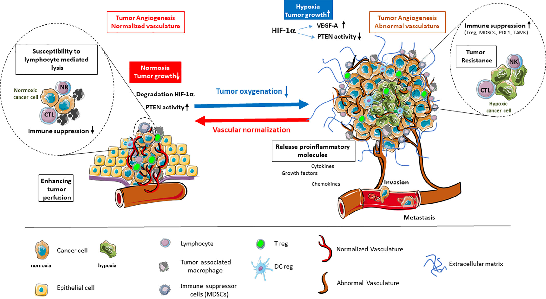
Hypoxia, characterized by reduced oxygen availability, is a common feature of the tumor microenvironment (TME) resulting from an imbalance between oxygen supply and consumption due to rapid tumor growth and aberrant vasculature. Central to the cellular response to hypoxia is the family of hypoxia-inducible factors (HIFs), particularly HIF-1α, which acts as a master regulator orchestrating a wide array of adaptive mechanisms. Under normoxic conditions, HIF-1α is hydroxylated by prolyl hydroxylase domain (PHD) enzymes, targeting it for ubiquitination and subsequent proteasomal degradation via the von Hippel-Lindau (VHL) tumor suppressor protein complex. However, in hypoxic conditions, hydroxylation is inhibited, leading to the stabilization and accumulation of HIF-1α. The stabilized HIF-1α translocates to the nucleus, where it dimerizes with HIF-1β to form a functional transcription factor complex that binds to hypoxia-responsive elements (HREs) in the promoters of target genes.
In addition to HIF-1α, other HIF isoforms such as HIF-2α and HIF-3α also play significant roles in the hypoxic response. While HIF-1α primarily regulates genes involved in glycolysis, angiogenesis, and apoptosis, HIF-2α is implicated in the regulation of genes that promote stemness and erythropoiesis, particularly in renal cell carcinoma. HIF-3α, though less studied, appears to function as a negative regulator of HIF-1α and HIF-2α, providing a feedback mechanism to fine-tune the hypoxic response. The differential expression and activity of these HIF isoforms contribute to the complexity and heterogeneity of the hypoxic response across various cancer types.
Transcriptional Reprogramming and Downstream Effects
The activation of HIF-1α initiates extensive transcriptional reprogramming that enables cancer cells to adapt and thrive under hypoxic conditions. One of the most critical adaptations is the shift in energy metabolism from oxidative phosphorylation to glycolysis, a phenomenon known as the Warburg effect. HIF-1α upregulates the expression of glucose transporters (e.g., GLUT1) and glycolytic enzymes (e.g., hexokinase 2, lactate dehydrogenase A), facilitating increased glucose uptake and anaerobic glycolysis. This metabolic reprogramming not only ensures a continuous supply of ATP but also generates metabolic intermediates crucial for biosynthetic processes essential for rapid cell proliferation.
Beyond metabolic adjustments, HIF-1α plays a pivotal role in promoting angiogenesis through the upregulation of vascular endothelial growth factor (VEGF), platelet-derived growth factor (PDGF), and angiopoietin-2 (ANGPT2). These pro-angiogenic factors stimulate the formation of new blood vessels, theoretically enhancing oxygen and nutrient delivery to the tumor. However, the newly formed vasculature is often aberrant and inefficient, perpetuating a cycle of hypoxia and further promoting aggressive tumor behavior.
HIF-1α also regulates the expression of genes involved in cell survival and proliferation by modulating pathways such as PI3K/AKT/mTOR and NF-κB. These pathways enhance cellular resistance to apoptosis and facilitate continuous cell division, contributing to tumor growth and resilience. Additionally, HIF-1α induces the expression of carbonic anhydrase IX (CAIX) and other pH-regulating proteins, maintaining intracellular pH homeostasis despite increased lactic acid production from glycolysis.
Moreover, the interplay between HIF-1α and other cellular signaling pathways under hypoxia underscores the multifaceted role of HIFs in tumor biology. For instance, HIF-1α can be activated through oxygen-independent mechanisms such as oncogenic signaling (e.g., RAS/RAF mutations) and loss of VHL function, highlighting its integral role even in tumors not traditionally associated with hypoxia. This robustness makes HIF-1α a compelling target for therapeutic intervention.
3.2 Clinical Implications
Hypoxia-Driven Resistance Mechanisms
Tumor hypoxia poses significant challenges to effective cancer treatment, primarily by fostering resistance to conventional therapies. Radiotherapy, for instance, relies on the generation of reactive oxygen species (ROS) to induce DNA damage in cancer cells. Hypoxic conditions reduce oxygen availability, thereby diminishing ROS production and attenuating the efficacy of radiotherapy. Studies have shown that hypoxic tumors require up to three times the radiation dose to achieve comparable cell kill rates as well-oxygenated tumors.
Chemotherapeutic agents also face reduced efficacy in hypoxic tumor regions. HIF-1α-mediated upregulation of drug efflux pumps such as P-glycoprotein (MDR1) facilitates the removal of chemotherapeutic drugs from cancer cells, thereby decreasing intracellular drug concentrations and therapeutic effectiveness. Additionally, hypoxia-induced activation of DNA repair pathways enhances the ability of cancer cells to repair drug-induced DNA damage, further contributing to chemoresistance.
Moreover, hypoxia promotes the expression of anti-apoptotic proteins (e.g., BCL-2, BNIP3) and inhibits pro-apoptotic pathways, rendering cancer cells more resilient to therapy-induced cell death. This multifaceted resistance mechanism underscores the critical need for therapeutic strategies that can effectively target hypoxic tumor regions.
Role in Immune Evasion, Angiogenesis, and Metastasis
Tumor hypoxia significantly influences the tumor immune microenvironment (TIME), facilitating immune evasion and suppressing anti-tumor immune responses. HIF-1α upregulates the expression of immune checkpoint molecules such as programmed death ligand-1 (PD-L1), which engages with PD-1 receptors on T cells, inhibiting their cytotoxic activity. Furthermore, hypoxia recruits immunosuppressive cell populations, including regulatory T cells (Tregs) and myeloid-derived suppressor cells (MDSCs), which further dampen immune surveillance and promote an immunosuppressive milieu conducive to tumor growth.
Angiogenesis driven by hypoxia is a double-edged sword. While it is essential for tumor growth by supplying oxygen and nutrients, the resultant vasculature is often disorganized and leaky, contributing to an immunosuppressive environment and facilitating metastatic dissemination. The induction of epithelial-mesenchymal transition (EMT) by hypoxia, mediated through HIF-1α-induced expression of transcription factors like Snail and Twist, enhances the migratory and invasive capabilities of cancer cells, thereby promoting metastasis.
The metastatic cascade is further supported by hypoxia-induced alterations in the extracellular matrix (ECM) and increased expression of matrix metalloproteinases (MMPs), which degrade ECM components and facilitate cancer cell invasion into surrounding tissues and entry into the bloodstream. Clinical evidence links hypoxic tumors with higher rates of lymph node involvement and distant metastasis, correlating with poor prognosis and reduced overall survival across various cancer types, including breast, prostate, pancreatic, and cervical cancers.
Quantitative Data on Prevalence and Significance of Hypoxia in Various Cancers
Hypoxia is a pervasive feature in solid tumors, with studies indicating that approximately 50-60% of solid tumors exhibit hypoxic regions. Quantitative assessments using techniques such as oxygen electrode measurements and hypoxia-specific imaging (e.g., 18F-fluoromisonidazole PET) have revealed that the extent and severity of hypoxia vary across different cancer types. For instance, pancreatic and cervical cancers frequently display extensive hypoxic areas, which are strongly associated with aggressive disease phenotypes and poorer clinical outcomes.
In breast cancer, particularly triple-negative subtypes, hypoxia has been linked to increased tumor grade, heightened metastatic potential, and reduced responsiveness to therapy. Prostate cancer studies have identified hypoxia as a key factor in the development of castration-resistant phenotypes, contributing to disease progression despite androgen deprivation therapy. Cervical cancers with significant hypoxic regions demonstrate higher rates of recurrence and lower disease-free survival rates, emphasizing the prognostic value of hypoxia assessment.
Meta-analyses have consistently demonstrated that hypoxic tumors correlate with reduced overall survival and increased mortality rates, underscoring the clinical significance of hypoxia as both a prognostic marker and a therapeutic target. The integration of hypoxia assessment into clinical practice, through advanced imaging techniques and molecular biomarkers, holds promise for stratifying patients, personalizing therapy, and improving clinical outcomes.
Conclusion
Tumor hypoxia is a fundamental characteristic of the tumor microenvironment that profoundly impacts cancer progression, therapeutic resistance, and patient prognosis. The central role of HIF-1α in orchestrating adaptive responses to hypoxia—ranging from metabolic reprogramming and angiogenesis to immune evasion and metastatic dissemination—highlights the complexity of the hypoxic response and its multifaceted implications in oncology. Understanding the molecular mechanisms underlying hypoxia and its clinical ramifications is essential for developing innovative therapeutic strategies aimed at targeting hypoxia-related pathways. Such approaches hold the potential to overcome therapeutic resistance, enhance the efficacy of existing treatments, and ultimately improve patient outcomes. Future research should focus on integrating nanotechnology-based solutions to specifically target hypoxic tumor regions, thereby addressing one of the most critical barriers in effective cancer therapy.
References
- https://pmc.ncbi.nlm.nih.gov/articles/PMC5607450/
- https://www.nature.com/articles/s41392-023-01332-8
- https://molecular-cancer.biomedcentral.com/articles/10.1186/s12943-019-1089-9
- https://www.nature.com/articles/cdd200821
- https://www.frontiersin.org/journals/genetics/articles/10.3389/fgene.2022.849040/full
- https://www.jci.org/articles/view/159839
- https://www.sciencedirect.com/science/article/pii/S2211383515000817
- https://cancerci.biomedcentral.com/articles/10.1186/s12935-020-01370-0
- https://www.mdpi.com/1424-8247/17/2/195
- https://jenci.springeropen.com/articles/10.1186/s43046-023-00186-z
- https://www.cureus.com/articles/295248-hypoxia-inducible-factor-1-alpha-hif-1-and-cancer-mechanisms-of-tumor-hypoxia-and-therapeutic-targeting#!/
- https://www.sciopen.com/article/10.26599/NTM.2023.9130020
- https://onlinelibrary.wiley.com/doi/10.1002/mco2.203
- https://aacrjournals.org/cancerres/article/63/19/6130/510441/Predominant-Role-of-Hypoxia-Inducible
- https://ecor.mgh.harvard.edu/content/files/sac/sac2013celebrationofscience.pdf
Last updated January 23, 2025