
Understanding Electroencephalography (EEG): A Window into Brain Activity
Exploring the principles, procedures, and applications of EEG testing.
Key Highlights of EEG
- Fundamental Principle: EEG measures the electrical activity of the brain, specifically the postsynaptic potentials of neurons.
- Primary Application: It is a crucial tool for diagnosing and evaluating neurological conditions, most notably epilepsy and seizure disorders.
- Procedure: Involves placing electrodes on the scalp to detect and record brain wave patterns.
What is an EEG?
An electroencephalogram (EEG) is a non-invasive medical test that records the electrical activity of the brain. This activity is a result of communication between brain cells, primarily neurons, which generate tiny electrical charges. These charges create electrical fields that can be detected by electrodes placed on the scalp. The recorded electrical signals are displayed as wavy lines, representing different brain wave patterns.
The Science Behind Brain Waves
Brain cells, specifically neurons, communicate through electrical impulses. This synchronized firing of neurons generates electrical signals known as brain waves or neural oscillations. EEG measures these electrical fields on the scalp. While an EEG cannot detect the activity of individual neurons due to the small electrical current each generates, it captures the summated activity of a large population of neurons, particularly the postsynaptic potentials of pyramidal neurons in the cerebral cortex.
The principle behind EEG is similar to electrocardiography (EKG) in that it relies on the volume conduction of ionic current through the extracellular space. The electrodes on the scalp detect voltage differences between different points, providing a graphical representation of the brain's electrical function over time.
How is an EEG Performed?
The EEG Procedure Explained
The EEG procedure is generally painless and involves placing small metal discs called electrodes onto the scalp. These electrodes are connected by wires to an EEG machine, which amplifies and records the electrical signals. Before electrode placement, the technologist may prepare the scalp, sometimes using a conductive gel or paste to ensure good contact.
The placement of electrodes follows a standardized system, often the 10-20 international system, which ensures consistent and accurate recording across different individuals and tests. This system designates specific points on the scalp for electrode placement based on anatomical landmarks.
During the recording, the patient is usually asked to remain still and relaxed with their eyes closed. The test typically takes between 30 to 60 minutes, but longer or specialized EEGs, such as sleep EEGs or ambulatory EEGs, may take several hours or even days. The technologist monitors the recording for any abnormalities.
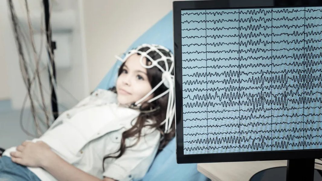
A patient with electrodes placed on their scalp for an EEG test.
Types of EEG Tests
Different types of EEG tests may be performed depending on the suspected condition and the information required:
- Routine EEG: A standard recording typically lasting 30-60 minutes.
- Sleep-Deprived EEG: Performed after the patient has had limited sleep, as sleep deprivation can sometimes trigger abnormal brain activity, particularly in individuals with epilepsy.
- Sleep EEG: Conducted while the patient is asleep to capture brain activity during different sleep stages. This is useful for diagnosing sleep disorders.
- Ambulatory EEG: A portable EEG device allows for continuous recording of brain activity over 24 hours or longer while the patient goes about their normal activities. This can help capture infrequent events.
- Video EEG Monitoring: Combines EEG recording with video recording to capture both brain activity and the patient's behavior during events like seizures. This is often done in a hospital setting.
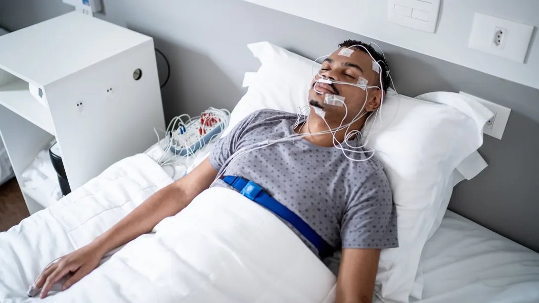
An example of a sleep EEG test.
What Does an EEG Diagnose?
EEG is a valuable diagnostic tool for a range of neurological conditions. Its primary use is in the evaluation of epilepsy and seizure disorders. By recording the electrical activity, the EEG can help identify abnormal brain wave patterns associated with seizures and classify the type of epilepsy.
Conditions Diagnosed or Evaluated with EEG
Beyond epilepsy, EEG is used to help diagnose or evaluate several other conditions:
-
Sleep Disorders: EEG is a key component of polysomnography, a comprehensive test used to diagnose sleep disorders like sleep apnea, narcolepsy, and insomnia.
-
Brain Tumors: While not directly imaging tumors, EEG can detect abnormal electrical activity around a tumor.
-
Head Injuries: Can help assess the extent of brain damage after a head injury.
-
Encephalopathy: Used to evaluate diffuse brain disease caused by various factors.
-
Inflammation of the Brain: Such as encephalitis.
-
Stroke: Can help monitor for changes in brain activity due to reduced blood flow (ischemia).
-
Creutzfeldt-Jakob disease: A rare, degenerative brain disorder.
-
Brain Death: EEG can be used as part of the evaluation to confirm brain death in critically ill patients.
-
Monitoring during Surgery: EEG can monitor the depth of anesthesia and detect potential complications like ischemia during surgical procedures.
Brain Wave Patterns and Their Significance
EEG recordings display different types of brain waves, categorized by their frequency. Changes in these patterns can indicate abnormal brain activity.
| Brain Wave Type | Frequency Range (Hz) | Associated State | Significance |
|---|---|---|---|
| Delta | 0.5 - 4 | Deep sleep, infancy, brain injury | Abnormal in awake adults (can indicate brain pathology) |
| Theta | 4 - 8 | Sleep, deep relaxation, young children | Abnormal in awake adults (can indicate brain pathology) |
| Alpha | 8 - 13 | Relaxed state, eyes closed | Disappears with open eyes or mental effort |
| Beta | 13 - 30 | Alert state, active thinking | Higher frequencies may be associated with anxiety |
| Gamma | >30 | Cognitive processing, learning | Involved in higher-level brain functions |
Common brain wave types and their characteristics.
The Technology Behind EEG
EEG technology relies on sensitive equipment to detect and amplify the very small electrical signals produced by the brain. The core components include electrodes, amplifiers, and a recording system.
Electrodes and Amplifiers
Electrodes, typically made of metal, are attached to the scalp. They serve as sensors that pick up the electrical potentials. These signals are then sent to amplifiers, which increase their amplitude to a level that can be recorded and displayed. The quality of the electrodes and amplifiers is crucial for obtaining clear and reliable recordings.

Example of EEG electrodes and recording equipment.
Digital Recording and Analysis
Modern EEG systems are digital, allowing for the recording, storage, and analysis of brain wave data on computers. This digital format offers several advantages, including easier storage, retrieval, and the ability to apply various signal processing techniques to enhance the analysis of the recordings. Analyzing EEG data requires expertise to identify normal and abnormal patterns, detect artifacts (non-brain electrical signals), and extract valuable information about brain function.
Advancements in EEG Technology
While the fundamental principles of EEG have been established for nearly a century (the first human EEG was recorded by Hans Berger in 1924), technology continues to advance. This includes the development of wireless EEG systems, dry electrodes that don't require conductive gel, and more portable devices, including wearable EEG headbands. These advancements are expanding the applications of EEG beyond traditional clinical settings into areas like research, neurofeedback, and brain-computer interfaces.
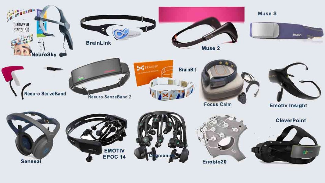
Examples of modern wearable EEG headbands.
Why is EEG Still Important?
Despite the advent of more advanced neuroimaging techniques like MRI and CT scans, EEG remains an essential tool in neurology. Its strength lies in its excellent temporal resolution, meaning it can capture changes in brain activity that occur very quickly, within milliseconds. This makes it particularly valuable for evaluating dynamic cerebral functioning and events like seizures, which are characterized by rapid electrical discharges.
Temporal Resolution vs. Spatial Resolution
While techniques like fMRI offer high spatial resolution (showing precisely where activity is occurring in the brain), EEG provides high temporal resolution (showing exactly when activity is occurring). EEG can detect changes in brain activity as they happen, which is critical for understanding the timing and progression of neurological events.
Complementary to Other Techniques
EEG is often used in conjunction with other diagnostic tools to provide a more complete picture of brain health. For example, an MRI might show a structural abnormality, while an EEG can reveal how that abnormality is affecting the brain's electrical activity.
Frequently Asked Questions about EEG
Is an EEG painful?
No, an EEG is a painless procedure. The electrodes placed on the scalp only detect electrical signals and do not deliver any electrical current.
Are there any risks associated with an EEG?
An EEG is considered a very safe procedure with no significant risks. In rare cases, the flashing lights used during photo-stimulation (a common part of the EEG to provoke brain activity) might trigger seizures in individuals with photosensitive epilepsy, but this is monitored by the technologist.
How should I prepare for an EEG?
Preparation for an EEG typically involves washing your hair the night before to ensure the electrodes can make good contact with the scalp. You may also be asked to avoid caffeine on the day of the test. If a sleep or sleep-deprived EEG is scheduled, you will receive specific instructions regarding sleep.
How long does it take to get EEG results?
The EEG recording itself is usually available immediately after the test. However, a qualified healthcare provider, typically a neurologist, needs to interpret the complex brain wave patterns. This can take several days, and the results will be shared with you during a follow-up appointment.
References
Last updated April 20, 2025