
Unlocking the Brain's Blueprint: What Exactly is a Connectome?
Delving into the intricate wiring diagram that shapes our thoughts, feelings, and actions.
Highlights
- Definition: A connectome is a comprehensive map of all neural connections within an organism's nervous system, essentially the brain's "wiring diagram."
- Types & Scales: Connectomes can map physical structures (structural), coordinated activity (functional), individual neurons (neural), or chemical signaling (chemical), ranging from macroscopic brain regions down to microscopic synapses.
- Significance: Understanding the connectome is crucial for linking brain structure to function, diagnosing diseases, tracking development, and advancing neuroscience research.
Diving Deeper into the Concept
What is a Connectome? The Brain's Intricate Network
Imagine trying to understand a complex city without a map showing its roads, power lines, and communication networks. The brain, arguably the most complex structure known, presents a similar challenge. A connectome serves as this essential map for the brain. Coined as an analogue to the "genome" (the map of an organism's genes), the connectome aims to be a complete description of the structural and functional connections within an organism's nervous system.
It's often described as the brain's "wiring diagram," detailing how billions of neurons are linked together through trillions of connections (synapses). This intricate network forms the basis for all neural activity, underpinning everything from simple reflexes to complex cognitive processes like thought, memory, and emotion. The fundamental idea is that the brain's architecture—how its parts are connected—is intrinsically linked to its function.
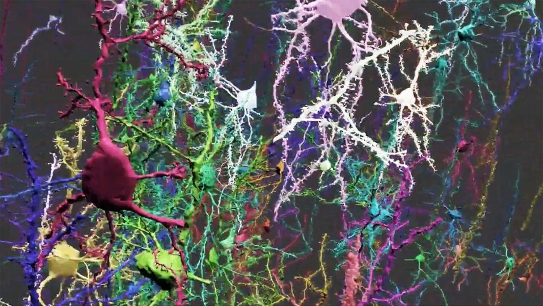
An illustration representing the dense interconnectedness of neurons in the brain.
The Different Lenses: Types of Connectomes
Mapping the brain's connections isn't a one-size-fits-all endeavor. Researchers approach it from different angles, resulting in various types of connectomes:
Structural Connectivity: The Brain's Hardware
This focuses on the physical, anatomical pathways linking different parts of the brain. It maps the tangible "wires"—bundles of axons (nerve fibers) that connect distinct brain regions or individual neurons via synapses. Structural connectomes represent the potential routes for information flow, revealing the brain's underlying architecture. Techniques like Diffusion MRI (dMRI) are often used to map large-scale white matter tracts in living humans, while electron microscopy provides ultra-high resolution maps of individual synaptic connections in smaller samples.
Functional Connectivity: Brain Regions in Sync
Instead of physical links, functional connectivity measures the statistical relationships between the activity patterns of different brain regions over time. If two areas consistently show synchronized activity (e.g., firing together during a task or even at rest), they are considered functionally connected, even if no direct structural path between them is known. Functional Magnetic Resonance Imaging (fMRI) is a key tool here, detecting changes in blood flow associated with neural activity. This reveals which brain areas tend to work together as networks.
Neural Connectomes: The Microscopic View
This is the most detailed level, aiming to map every single neuron and every single synaptic connection between them. Due to the sheer density and complexity, achieving a complete neural connectome is currently only feasible for very small organisms or tiny portions of larger brains. The classic example is the nematode worm Caenorhabditis elegans, whose nervous system of just 302 neurons was fully mapped in 1986 using laborious electron microscopy reconstruction. More recently, the complete connectome of a fruit fly larva and adult fly brain have been mapped.
Chemical Connectomes: The Neurotransmitter Network
This emerging type of map focuses on the distribution and interaction of neurochemicals, particularly neuromodulators (like dopamine, serotonin, acetylcholine). It charts which neurons produce specific neuromodulators and which neurons have receptors sensitive to them. This adds another layer of complexity, showing how chemical signaling can dynamically shape the activity and information flow across the structural network.
Mapping the Labyrinth: Scales and Techniques
From Micro to Macro: Levels of Detail
Connectomes can be described at different scales, reflecting the level of detail captured:
- Microscale: The highest resolution, mapping individual neurons and their synaptic connections. This requires techniques like electron microscopy and provides unparalleled detail but is extremely data-intensive and currently limited to very small volumes.
- Mesoscale: An intermediate level, focusing on the connectivity patterns of specific neuronal populations or local circuits, often within cortical columns or layers.
- Macroscale: The broadest view, mapping large brain regions (like the cortex, hippocampus, amygdala) and the major nerve fiber bundles (white matter tracts) that connect them. This is the scale typically studied in humans using non-invasive neuroimaging techniques like MRI and fMRI.
Tools of the Trade: How Connectomes are Built
Constructing a connectome relies on sophisticated technologies:
- Electron Microscopy (EM): Essential for microscale connectomics. Thinly slicing brain tissue and imaging each slice with an electron microscope allows researchers to trace neuronal processes and identify individual synapses. This is incredibly time-consuming and generates massive datasets.
- Magnetic Resonance Imaging (MRI):
- Diffusion MRI (dMRI): Measures the diffusion of water molecules in the brain. Since water diffuses more easily along nerve fibers than across them, dMRI can infer the orientation and pathways of white matter tracts, enabling macroscale structural connectome mapping.
- Functional MRI (fMRI): Detects changes in blood oxygen levels (BOLD signal) that correlate with neural activity. By analyzing correlations in BOLD signals across different brain regions, researchers map macroscale functional connectivity.
- Other Techniques: Light microscopy with specific stains, tract-tracing methods (injecting dyes that travel along axons, primarily in animal models), and electrophysiological recordings also contribute to understanding connectivity.
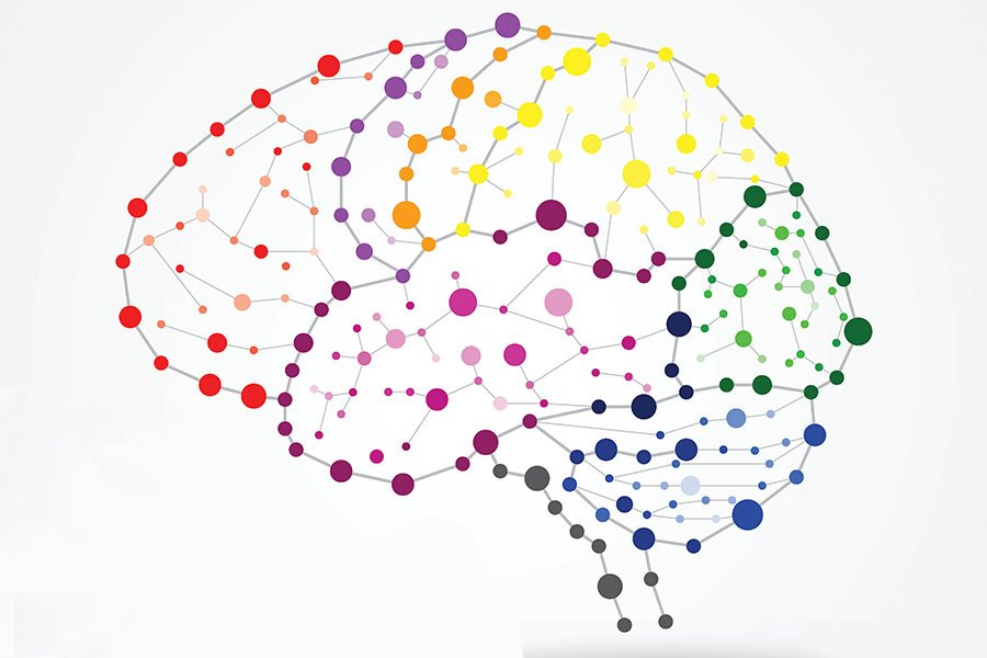
A visual representation of large-scale brain pathways derived from neuroimaging data.
The Human Connectome Project and Beyond
Charting the Human Brain
Mapping the human connectome is a monumental undertaking due to the brain's estimated 86 billion neurons and hundreds of trillions of synapses. A major initiative driving this effort is the Human Connectome Project (HCP), launched in 2009 by the U.S. National Institutes of Health (NIH). The HCP's primary goal is to build a "network map" of the healthy, living human brain by collecting vast amounts of high-quality data on structural and functional connectivity using advanced MRI techniques.
The project focuses on macroscale connectivity, mapping connections between brain regions rather than individual neurons. By studying large populations of individuals, the HCP aims to understand not only the typical patterns of brain connectivity but also how these patterns relate to behavior, cognitive abilities, and genetic factors. The data generated by the HCP and related projects is made publicly available through resources like the Connectome Coordination Facility (CCF), fostering collaboration and accelerating discovery in brain science.
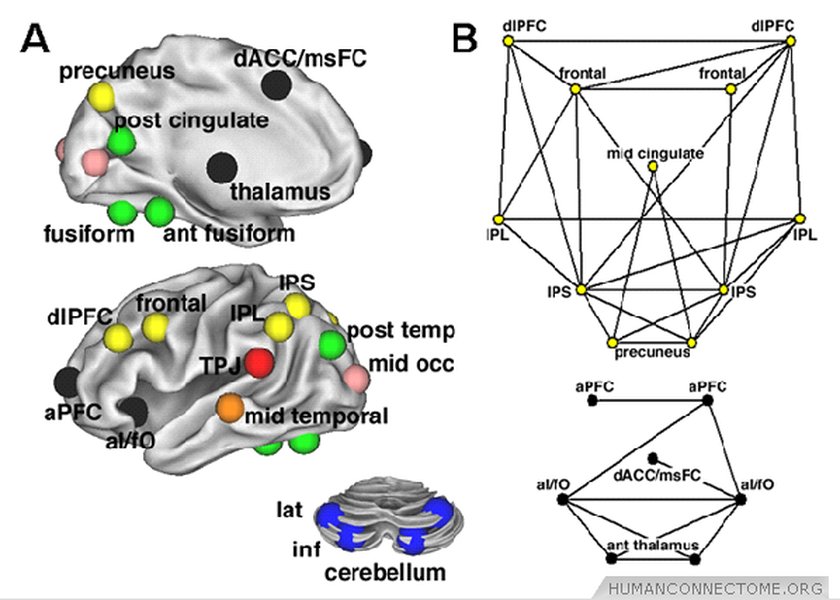
An example figure related to network analysis used in the Human Connectome Project.
Challenges and Current Status
Despite significant progress, particularly at the macroscale, mapping a complete, synapse-level neural connectome of the entire human brain remains a distant goal. The sheer scale and complexity pose immense technical and computational challenges. Current human connectomes are primarily macroscale representations derived from non-invasive imaging, providing invaluable insights but lacking the fine-grained detail of microscale maps.
Ongoing research continues to refine imaging techniques, improve data analysis methods (often leveraging artificial intelligence), and integrate different types of data (structural, functional, genetic) to create increasingly comprehensive and accurate models of brain connectivity across multiple scales.
Comparing Connectome Types
The following table summarizes the key distinctions between the main types of connectomes discussed:
| Feature | Structural Connectome (Macro) | Functional Connectome (Macro) | Neural Connectome (Micro) | Chemical Connectome |
|---|---|---|---|---|
| What it Maps | Physical pathways (axon bundles) between brain regions | Statistical correlations in activity between brain regions | Individual neurons and their synaptic connections | Distribution of neuromodulators and their receptors |
| Typical Scale | Macroscopic (regions, tracts) | Macroscopic (regions) | Microscopic (neurons, synapses) | Meso/Microscopic |
| Primary Technique(s) | Diffusion MRI (dMRI) | Functional MRI (fMRI) | Electron Microscopy (EM) | Histology, PET, advanced microscopy |
| Current Human Status Example | Mapped via HCP (regional level) | Mapped via HCP (regional correlations) | Not feasible for whole brain; small sections only | Partial maps for specific systems |
Why Does the Connectome Matter?
Linking Structure to Function
The connectome provides the structural scaffold upon which brain function unfolds. Understanding how different brain regions are wired together is fundamental to deciphering how they cooperate to produce complex cognitive functions like perception, attention, learning, memory, language, and decision-making. While the relationship isn't always simple—the same structure can support different functions at different times—the connectome defines the possible pathways of communication and constrains the dynamics of neural activity.
Insights into Brain Health and Disease
Many neurological and psychiatric disorders are increasingly understood as "connectopathies," or disorders of brain connectivity. Conditions like schizophrenia, autism spectrum disorder, Alzheimer's disease, epilepsy, and multiple sclerosis are associated with alterations in structural or functional connectivity patterns. Mapping connectomes in healthy individuals and comparing them to those with brain disorders can help identify biomarkers for diagnosis, track disease progression, and evaluate the effectiveness of treatments. Studying how the connectome changes across the lifespan also sheds light on brain development and aging.
Guiding Future Neuroscience
Connectome maps serve as essential references for neuroscience research. They inform the design of experiments, help interpret findings from brain recordings and imaging, and provide the basis for building sophisticated computational models that simulate brain activity. Ultimately, a deeper understanding of the connectome promises to guide the development of novel therapeutic strategies, including targeted brain stimulation techniques, aimed at restoring healthy connectivity patterns.
Visualizing Connectome Characteristics
Different approaches to mapping the connectome excel in different aspects. The radar chart below offers a comparative visualization of key characteristics for major connectome types, based on current capabilities, particularly regarding the human brain. Note that these are relative assessments rather than precise quantitative measures.
This chart highlights trade-offs: Microscale Neural Connectomes offer the highest resolution but are currently impossible to complete for humans and generate vast amounts of data using less accessible techniques. Macroscale Structural and Functional Connectomes provide broader coverage of the human brain using more accessible MRI methods but offer lower resolution.
Connectome Concepts Mindmap
This mindmap provides a visual overview of the core concepts related to the connectome:
The Brain's Wiring Diagram"] id1["Types"] structural["Structural
(Physical Links)"] functional["Functional
(Activity Sync)"] neural["Neural
(Neurons/Synapses)"] chemical["Chemical
(Neuromodulators)"] id2["Scales"] macro["Macroscopic
(Regions)"] meso["Mesoscopic
(Columns/Layers)"] micro["Microscopic
(Neurons/Synapses)"] id3["Mapping Techniques"] mri["MRI / dMRI
(Structural Macro)"] fmri["fMRI
(Functional Macro)"] em["Electron Microscopy
(Neural Micro)"] other["Other
(Tracing, Light Microscopy)"] id4["Significance"] understand["Understanding Function
(Cognition, Behavior)"] health["Health & Disease
(Disorders, Development, Aging)"] research["Guiding Research
(Models, Therapies)"] id5["Key Examples / Projects"] hcp["Human Connectome Project (HCP)"] celegans["*C. elegans*
(First Complete Neural Map)"] fly["*Drosophila*
(Fruit Fly Mapping)"]
Explaining the Connectome
For a dynamic explanation, neuroscientist Bobby Kasthuri breaks down the concept of the connectome in varying levels of complexity in this video:
This video provides valuable context on how scientists think about and study brain wiring, illustrating the challenges and excitement surrounding connectomics research.
Frequently Asked Questions (FAQ)
Is the connectome fixed or does it change?
The connectome is not entirely static. While the large-scale structural pathways are relatively stable in adulthood, the strength of connections, and even the number of synapses at a microscale, can change throughout life. This phenomenon, known as neuroplasticity, allows the brain to adapt based on experience, learning, development, aging, and injury. So, the connectome provides a foundational blueprint, but it's a dynamic one.
What's the difference between a connectome and a genome?
The genome is the complete set of genetic instructions (DNA) for an organism, essentially the biological blueprint inherited from parents. The connectome is the complete map of neural connections in the brain. While the genome provides the initial instructions for building the brain and influences the basic layout of the connectome, the final wiring diagram is significantly shaped by individual experiences, learning, and environmental factors throughout life. The genome dictates potential, while the connectome reflects the realized structure.
Have scientists mapped the entire human connectome?
No, not at the level of individual neurons and synapses (the microscale or neural connectome). The human brain's complexity (billions of neurons, trillions of synapses) makes this currently impossible with existing technology. However, significant progress has been made in mapping the human connectome at the macroscale, detailing the major connections between larger brain regions using techniques like MRI. Projects like the HCP have provided detailed macroscale structural and functional connectome maps.
How is connectome information useful?
Connectome data is invaluable for several reasons:
- Understanding Brain Function: It helps researchers understand how different brain areas collaborate to enable cognition, emotion, and behavior.
- Diagnosing & Treating Disorders: Identifying abnormalities in connectivity patterns can aid in diagnosing neurological and psychiatric conditions and developing targeted therapies (e.g., deep brain stimulation guided by connectivity).
- Tracking Development & Aging: It allows scientists to study how brain networks form, mature, and change over the lifespan.
- Informing AI & Models: Knowledge of brain wiring inspires new architectures for artificial intelligence and provides crucial data for building realistic computational models of the brain.
Recommended
- How does the Human Connectome Project map brain connections?
- What is the difference between structural and functional connectivity?
- How are connectomes used to study brain diseases like Alzheimer's or schizophrenia?
- What are the biggest challenges in mapping the complete human connectome?
References
Last updated May 5, 2025