
Shape-Shifting Wonders: Unveiling the World of Amoebas
Discover the fascinating life of these single-celled organisms, from their unique movement to their diverse roles in nature.
Key Insights into Amoebas
- Shape-Shifters: Amoebas are defined by their lack of a fixed shape and their ability to move and feed using temporary projections called pseudopods ("false feet").
- Diverse Lifestyles: While many are harmless inhabitants of freshwater and soil, some amoeba species are parasitic and can cause diseases in humans.
- Fundamental Biology: As simple eukaryotic cells, amoebas are crucial model organisms for studying basic cellular processes like movement, feeding, and cell division.
What Exactly is an Amoeba?
An amoeba is a type of single-celled organism, or sometimes a cell within a larger organism, renowned for its remarkable ability to alter its shape. This defining characteristic is primarily achieved through the extension and retraction of temporary cytoplasmic bulges known as pseudopods or "false feet." The term "amoeba" itself derives from the Greek word amoibe, meaning "change," perfectly capturing this fluid nature. While often used broadly to describe any cell exhibiting this "amoeboid movement," in a stricter biological sense, it refers to members of the genus Amoeba, which are protists commonly found in freshwater environments.
These microscopic organisms are eukaryotes, meaning their cells contain a true nucleus and other membrane-bound organelles, distinguishing them from simpler prokaryotic cells like bacteria. They lack a rigid cell wall, contributing to their flexibility and shape-shifting ability.
A Glimpse Inside: The Structure of an Amoeba
Despite their apparent simplicity, amoebas possess a defined cellular structure that enables all essential life functions:
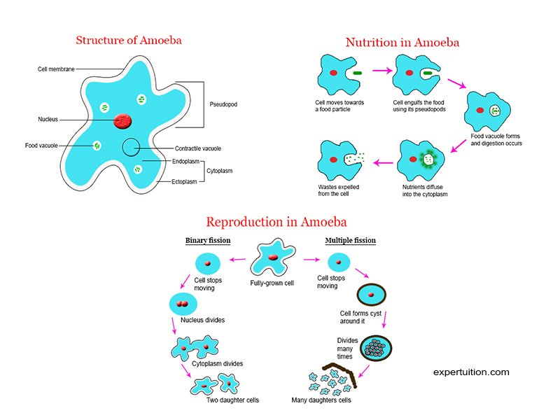
- Plasma Membrane: A thin, flexible outer boundary made of lipids and proteins. It protects the cell and controls the passage of substances in and out.
- Cytoplasm: The jelly-like substance filling the cell, differentiated into two layers:
- Ectoplasm: A clear, gel-like outer layer just beneath the plasma membrane, involved in forming pseudopods.
- Endoplasm: A more fluid, granular inner region containing the cell's organelles.
- Nucleus: Usually a single, prominent, disk-shaped or spherical organelle containing the amoeba's genetic material (DNA) and controlling cell activities, including growth and reproduction.
- Contractile Vacuole: A star-shaped, clear organelle responsible for osmoregulation – maintaining water balance by collecting excess water from the cytoplasm and periodically expelling it from the cell. This is crucial for amoebas living in freshwater environments.
- Food Vacuoles: Temporary sacs formed during feeding, containing engulfed food particles (like bacteria or smaller protists). Digestive enzymes are secreted into these vacuoles to break down the food.
- Pseudopods (False Feet): Temporary, finger-like or lobe-shaped extensions of the cytoplasm used for locomotion and capturing food. Their formation is driven by the internal cytoskeleton, particularly actin microfilaments.
- Mitochondria: Organelles responsible for cellular respiration, generating energy (ATP) for the cell's activities.
Life in Motion: Movement and Feeding
Amoeboid Movement
Amoebas move using a characteristic crawling motion known as amoeboid movement. This process involves the dynamic formation and retraction of pseudopods. The cell extends a pseudopod in the desired direction, anchors it to the substrate (surface), and then the more fluid endoplasm flows into the extension, effectively pulling the rest of the cell forward. The appearance and structure of pseudopods can vary; for instance, *Amoeba proteus* typically has broad, lobose (lobe-shaped) pseudopods, while other amoeboid organisms might have thin, thread-like (filose) or network-like (reticulose) pseudopods.

Phagocytosis: How Amoebas Eat
Amoebas are heterotrophic, meaning they obtain nutrients by consuming other organisms or organic matter. They feed using a process called phagocytosis ("cell eating"). When an amoeba detects a food particle (e.g., bacterium, alga, detritus), it extends pseudopods to surround and engulf it. The plasma membrane fuses around the particle, enclosing it within a food vacuole inside the cytoplasm. Lysosomes containing digestive enzymes then fuse with the food vacuole, breaking down the contents into smaller molecules that can be absorbed and utilized by the amoeba. Undigested waste materials are later expelled from the cell through exocytosis.
Habitat, Reproduction, and Survival
Where Do Amoebas Live?
Amoebas are found in a wide variety of environments worldwide. Many species are free-living and thrive in aquatic habitats such as ponds, lakes, streams, and damp soil, particularly where decaying organic matter is present. They often graze on bacteria and other microorganisms on surfaces like submerged vegetation or sediment. However, some amoebas are parasitic, living inside the bodies of animals, including humans.
Reproduction: The Simplicity of Binary Fission
Amoebas primarily reproduce asexually through a process called binary fission. Under favorable environmental conditions (adequate temperature, food, and moisture), a mature amoeba divides into two genetically identical daughter cells. The process begins with the replication of the nucleus (mitosis), followed by the division of the cytoplasm (cytokinesis). This simple method allows for rapid population growth when conditions are suitable. Due to this mode of reproduction, individual amoeba cells have a limited lifespan (often just a couple of days), but the lineage itself can be considered functionally immortal as it continuously divides.

Survival Strategy: Cyst Formation
When faced with unfavorable conditions, such as lack of food or water, extreme temperatures, or harmful chemical changes, many amoeba species can enter a dormant state by forming a cyst. The amoeba retracts its pseudopods, rounds up, and secretes a protective, multi-layered wall around itself. This cyst stage allows the organism to survive harsh conditions and can also aid in dispersal (e.g., through wind or water). When conditions become favorable again, the cyst wall breaks down (excystation), and the active amoeba emerges.
Diversity Within the Amoeboid World
It's important to understand that "amoeba" is not a formal taxonomic group representing a single evolutionary lineage (i.e., it's not monophyletic). Instead, the amoeboid form and mode of movement have evolved independently in several different groups of eukaryotes. Organisms traditionally called amoebas are found scattered across the eukaryotic tree of life. Historically, many were grouped under "Sarcodina," but modern molecular phylogenetics has shown this classification to be artificial.
Notable Examples of Amoebas
Here are a few well-known examples illustrating the diversity among amoeboid organisms:
- Amoeba proteus: Often considered the "classic" amoeba. It's a large, free-living species commonly found in freshwater ponds and streams, feeding on smaller protists and bacteria. Its characteristic lobose pseudopods and readily observable internal structures make it a popular subject for study in biology classrooms.
- Entamoeba histolytica: A parasitic amoeba that infects the human digestive tract. It can cause amoebic dysentery (amebiasis), a serious illness characterized by abdominal pain, diarrhea, and potentially invasive liver abscesses. It is typically transmitted through contaminated food or water.
- Naegleria fowleri: Often sensationally called the "brain-eating amoeba." This free-living amoeba thrives in warm freshwater environments like lakes, rivers, and hot springs. While infections are rare, it can cause a devastating and usually fatal brain infection called primary amoebic meningoencephalitis (PAM) if contaminated water enters the nose, allowing the amoeba to travel to the brain.
- Dictyostelium discoideum: Known as a "social amoeba" or cellular slime mold. These organisms live as individual amoeboid cells in the soil, feeding on bacteria. However, when starved, thousands of these individual cells aggregate to form a multicellular slug-like structure (pseudoplasmodium) that can migrate and eventually differentiate to form a fruiting body for spore dispersal – a fascinating example of simple multicellularity.
- Testate Amoebas: A diverse group of amoebas characterized by producing a shell or "test" around their cell body. These shells can be made of secreted organic material, cemented mineral particles collected from the environment, or silica plates. Examples include *Arcella* and *Difflugia*.
Comparing Different Amoeboid Organisms
The following table summarizes key features of some commonly discussed amoeboid organisms:
| Feature | Amoeba proteus | Entamoeba histolytica | Naegleria fowleri | Dictyostelium discoideum |
|---|---|---|---|---|
| Lifestyle | Free-living | Parasitic (Human gut) | Free-living (opportunistic parasite) | Free-living (soil), Facultative multicellular |
| Habitat | Freshwater ponds, streams | Human colon | Warm freshwater, soil | Soil, forest litter |
| Movement | Lobose pseudopods | Lobose pseudopods | Lobose pseudopods (amoeboid stage), Flagella (flagellate stage) | Lobose pseudopods (individual cells), Slug migration (multicellular stage) |
| Feeding | Phagocytosis (bacteria, protists) | Phagocytosis (host cells, bacteria) | Phagocytosis (bacteria), Cytotoxicity (brain tissue) | Phagocytosis (bacteria) |
| Reproduction | Binary fission | Binary fission (cyst formation) | Binary fission (cyst formation) | Binary fission (individual), Spore formation (multicellular) |
| Pathogenicity | Non-pathogenic | Causes Amebiasis (dysentery) | Causes Primary Amoebic Meningoencephalitis (PAM) | Non-pathogenic to humans |
Amoebas and Human Health
While most amoebas are harmless components of ecosystems, certain species pose significant threats to human health.
Amebiasis caused by Entamoeba histolytica
Entamoeba histolytica resides in the large intestine and is often transmitted via the fecal-oral route, primarily through ingestion of cysts in contaminated water or food. While many infections are asymptomatic, the amoeba can invade the intestinal lining, causing amoebic colitis (dysentery) with symptoms like bloody diarrhea, abdominal pain, and fever. In some cases, it can spread through the bloodstream to other organs, most commonly the liver, causing potentially life-threatening amoebic liver abscesses. It remains a major public health concern, particularly in developing countries with poor sanitation.
The Threat of Naegleria fowleri
Naegleria fowleri is a thermophilic (heat-loving) amoeba found globally in warm freshwater bodies and soil. It normally feeds on bacteria but can become an opportunistic human pathogen. If water containing the active amoeba form is forcefully inhaled into the nasal passages (e.g., during swimming or diving in contaminated water), the amoeba can migrate along the olfactory nerves to the brain. There, it causes PAM, a rapidly progressing and almost always fatal inflammation and destruction of brain tissue. Although extremely rare, PAM highlights the potential danger posed by some environmental amoebas.
Visualizing Amoeba Characteristics
This chart provides a comparative visualization of key characteristics across different types of amoeboid organisms based on general assessments. It helps illustrate the diversity in aspects like motility mechanisms, habitat range, potential to cause disease, and structural complexity relative to each other.
Understanding Amoeba Biology: A Mindmap
This mindmap provides a visual summary of the core concepts related to amoebas, linking their structure, behavior, types, and significance.
Visualizing the Amoeba
Videos provide a dynamic look at these fascinating organisms. The following video offers a basic introduction to what amoebas are, covering their unicellular nature and key features like pseudopods, consistent with the information discussed above. Watching amoebas in motion can truly bring their unique biology to life.
Observing their fluid movement and the process of pseudopod formation helps understand how these simple organisms navigate their environment and capture food, illustrating the concepts of amoeboid movement and phagocytosis described earlier.
Frequently Asked Questions (FAQ)
What are pseudopods?
Pseudopods, meaning "false feet," are temporary, flowing extensions of an amoeba's cytoplasm and cell membrane. They are crucial for both movement (amoeboid movement) and feeding (phagocytosis). The amoeba extends pseudopods, anchors them, and flows its cytoplasm into them to move, or uses them to surround and engulf food particles.
How big are amoebas?
Amoebas are generally microscopic. Sizes vary greatly depending on the species, but common amoebas like *Amoeba proteus* typically range from 0.1 to 0.5 millimeters (100 to 500 micrometers) in length, making them visible under a light microscope but usually not to the naked eye.
Are all amoebas dangerous to humans?
No, the vast majority of amoeba species are free-living and harmless to humans. They play important roles in ecosystems. Only a few species are known to be pathogenic, such as *Entamoeba histolytica* (causes amoebic dysentery) and *Naegleria fowleri* (causes a rare but fatal brain infection).
How do amoebas reproduce?
Amoebas primarily reproduce asexually through a process called binary fission. The parent cell divides into two genetically identical daughter cells after replicating its nucleus. Some amoebas can also form protective cysts to survive harsh conditions.
What do amoebas eat?
Free-living amoebas typically eat smaller microorganisms like bacteria, algae, other protists (like paramecia), and bits of dead organic matter (detritus). They engulf their food using pseudopods through the process of phagocytosis.
Recommended Reading
- Explore the cellular mechanisms behind amoeboid movement.
- Learn about the contrasting lifestyles of various amoeba species.
- Investigate the life cycle of the amoeba responsible for amoebic dysentery.
- Discover the fascinating social behavior and multicellularity of slime molds.
References
 davidwangblog.wordpress.com
Last updated April 29, 2025