
Decoding PSMA PET/CT: When Benign Bone Lesions Masquerade as Cancer
Unraveling the complexities of high SUVmax readings in F-18 PSMA PET/CT scans for suspected prostate cancer metastasis.
Prostate-Specific Membrane Antigen (PSMA) Positron Emission Tomography/Computed Tomography (PET/CT) using F-18 labeled tracers is a highly sensitive imaging technique for detecting prostate cancer and its spread, particularly to bones. Bone metastases from prostate cancer often appear as sclerotic (hardened or dense) lesions. While a high Standardized Uptake Value (SUVmax), such as 16.59, on a PSMA PET/CT scan can be indicative of malignancy, it's crucial to recognize that several benign (non-cancerous) conditions can also present with increased PSMA uptake and sclerotic changes, potentially mimicking metastases. This overlap necessitates careful evaluation to avoid misdiagnosis and ensure appropriate patient management.
Key Insights: Understanding Benign Mimics
- PSMA PET/CT Sensitivity vs. Specificity: While F-18 PSMA PET/CT is excellent for detecting prostate cancer cells, its specificity is not absolute; certain benign processes can also show increased tracer uptake.
- Sclerotic Lesions Aren't Always Malignant: Many benign bone conditions involve increased bone formation or turnover, leading to sclerotic appearances on CT and PSMA uptake on PET.
- High SUVmax in Benign Conditions: An SUVmax value like 16.59, while concerning, does not definitively confirm cancer, as some benign entities can also exhibit significant PSMA avidity. A comprehensive diagnostic approach is essential.
The Science Behind PSMA PET/CT and SUVmax
Why PSMA PET/CT?
PSMA is a protein found in abundance on the surface of most prostate cancer cells. PSMA PET/CT utilizes a radiotracer (like an F-18 labeled molecule) that binds to PSMA. When this tracer accumulates in areas with high PSMA expression, it emits signals detected by the PET scanner, highlighting potential cancer sites. The accompanying CT scan provides anatomical details, localizing these areas of uptake.
What is SUVmax?
The Standardized Uptake Value (SUV) is a semi-quantitative measure of radiotracer uptake in a region of interest. SUVmax refers to the maximum SUV value within that region. Generally, higher SUVmax values are associated with more metabolically active or PSMA-expressing tissue, often correlating with malignancy. However, there's an overlap in SUVmax values between malignant and some benign conditions, making interpretation complex based solely on this number.
An SUVmax of 16.59 is considerably elevated and warrants careful investigation. While often seen in true metastatic disease, understanding the potential benign mimics is crucial for accurate diagnosis.
Benign Conditions That Can Mimic Focal Sclerotic Metastases
Several benign conditions can present as focal sclerotic lesions with increased F-18 PSMA uptake, potentially leading to a false-positive interpretation of metastatic prostate cancer. These mimics often involve processes that increase bone turnover, vascularity, or non-specific tracer accumulation.
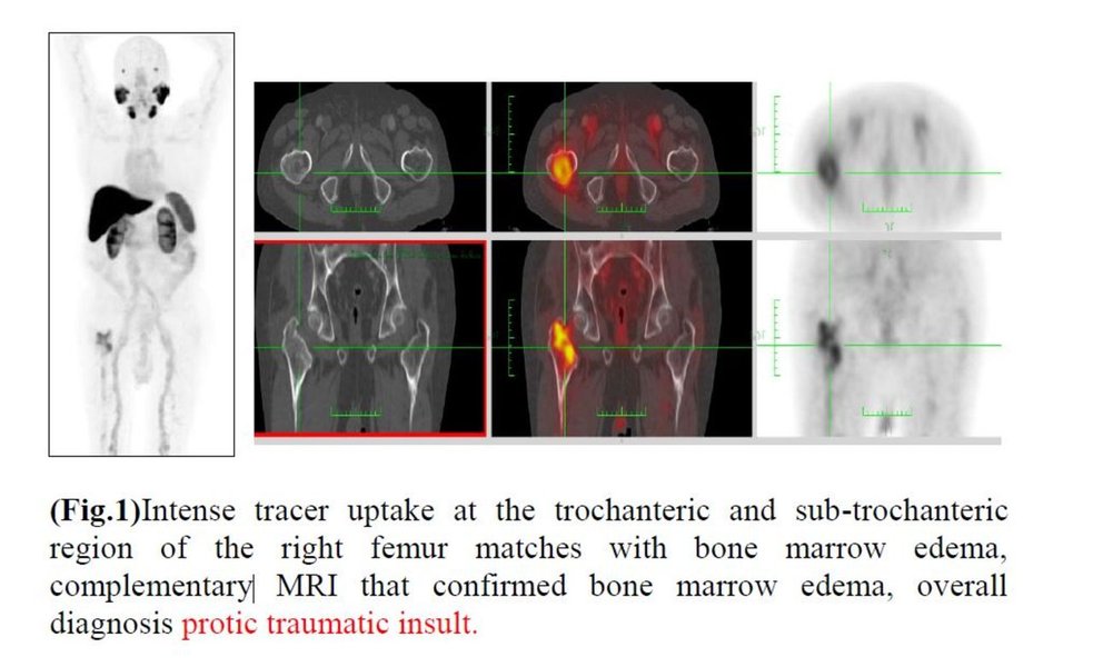
Examples of skeletal non-neoplastic lesions as seen on 18F-PSMA-1007 PET/CT, illustrating potential mimics of metastasis.
1. Degenerative and Trauma-Related Bone Changes
Osteoarthritis and Subchondral Sclerosis
Degenerative joint disease, such as osteoarthritis, can lead to increased bone turnover and reparative processes, resulting in subchondral sclerosis (hardening of bone beneath cartilage). These areas can show increased PSMA uptake. While typically located at articular surfaces, pronounced changes can sometimes be mistaken for metastases, especially in the axial skeleton.
Healing Fractures and Bone Infarcts
The bone healing process following a fracture involves significant osteoblastic activity and increased vascularity, leading to callus formation (which is sclerotic) and high PSMA uptake. This uptake can persist for months. Similarly, healing bone infarcts (areas of bone death due to lack of blood supply) can undergo sclerotic changes and show tracer uptake during the repair phase.
Post-Surgical Changes
Areas of previous bone surgery or trauma can exhibit ongoing reparative processes and inflammation, leading to sclerotic appearances and increased PSMA tracer accumulation.
2. Benign Bone Tumors and Tumor-Like Lesions
Vertebral Hemangiomas
Hemangiomas are common benign vascular tumors, frequently found in the vertebral bodies. While typically showing low PSMA uptake, atypical hemangiomas or those with reactive changes can occasionally demonstrate increased uptake and may have sclerotic components on CT, potentially mimicking metastases.
Bone Islands (Enostoses)
Bone islands are common, asymptomatic, focal areas of dense, mature compact bone within the cancellous bone. They are typically very sclerotic and usually show little to no PSMA uptake. However, larger or actively remodeling enostoses might show mild uptake, though rarely reaching very high SUVmax values like 16.59.
Fibrous Dysplasia
Fibrous dysplasia is a benign condition where normal bone is replaced by fibrous tissue. It can have a varied appearance, including sclerotic ("ground-glass") areas, and may show increased PSMA uptake depending on its metabolic activity.
Neurofibromas
Neurofibromas are benign nerve sheath tumors. While rare as bone lesion mimics, they have been reported to express PSMA and can, in some instances, particularly if adjacent to or involving bone, cause focal PSMA uptake that might be misinterpreted if associated with sclerotic bone changes.
3. Chronic Bone Disorders
Paget's Disease of Bone
Paget's disease is a chronic disorder characterized by abnormal and accelerated bone remodeling. Affected bones can become enlarged, deformed, and show mixed lytic (bone breakdown) and sclerotic (bone formation) phases. The active sclerotic phase often demonstrates very high PSMA uptake due to intense osteoblastic activity, making it a significant mimic of prostate cancer metastases, especially in older individuals. SUVmax values in Paget's disease can indeed be quite high.
4. Inflammatory and Infectious Conditions
Osteomyelitis (Chronic)
Chronic bone infections (osteomyelitis) can lead to sclerotic changes and increased vascularity and inflammation, resulting in increased PSMA uptake. While acute osteomyelitis is often lytic, chronic forms can be predominantly sclerotic.
5. Other Non-Specific Uptake
Unspecific Bone Uptake
Sometimes, focal PSMA uptake is seen in bone lesions, particularly in the ribs or axial skeleton, without a clear malignant or specific benign cause. These can be due to minor trauma, localized inflammation, or benign red marrow depositions. F-18 PSMA tracers, in particular 18F-PSMA-1007, have been noted to have a higher incidence of such non-specific bone uptake compared to Gallium-68 (Ga-68) based PSMA tracers, especially in benign rib lesions.
Mindmap of Benign Mimics
This mindmap illustrates the various categories of benign conditions that can appear as focal sclerotic metastases of prostate cancer on F-18 PSMA PET/CT scans. Understanding these categories helps in the differential diagnosis process.
Prostate Cancer Metastases
on F-18 PSMA PET/CT"] id1["Degenerative & Trauma-Related"] id1a["Osteoarthritis &
Subchondral Sclerosis"] id1b["Healing Fractures"] id1c["Bone Infarcts (healing)"] id1d["Post-Surgical Changes"] id2["Benign Bone Tumors &
Tumor-Like Lesions"] id2a["Vertebral Hemangiomas
(especially atypical)"] id2b["Bone Islands (Enostoses)
(rarely high uptake)"] id2c["Fibrous Dysplasia"] id2d["Neurofibromas
(if adjacent/involving bone)"] id3["Chronic Bone Disorders"] id3a["Paget's Disease of Bone"] id4["Inflammatory & Infectious"] id4a["Osteomyelitis (chronic)"] id5["Other Non-Specific Uptake"] id5a["Benign Red Marrow
Depositions"] id5b["Solitary Rib Lesions
(unspecific etiology with
F-18 PSMA tracers)"]
Comparative Characteristics: Metastasis vs. Benign Mimics
The radar chart below provides a conceptual comparison of how prostate cancer metastasis might differ from common benign mimics based on several imaging and clinical characteristics. The scores are illustrative (1=low, 10=high) and represent general tendencies rather than absolute values, as significant overlap can exist. An SUVmax of 16.59 would place a lesion on the higher end of "PSMA Avidity".
Diagnostic Strategies for Differentiation
Given the potential for benign mimics, a comprehensive approach is vital to accurately interpret focal sclerotic lesions with high PSMA uptake:
1. Multimodal Imaging Correlation
Correlating with CT Morphology
The CT component of the PET/CT scan provides crucial anatomical information. Benign lesions often have well-defined, sclerotic margins without cortical destruction or an associated soft tissue mass, unlike aggressive metastases. Features like osteophytes in osteoarthritis, bone enlargement in Paget's disease, or the "polka dot" sign in vertebral hemangiomas can suggest a benign etiology.
Magnetic Resonance Imaging (MRI)
MRI can be very helpful in differentiating benign from malignant bone lesions. Metastases typically replace normal fatty marrow, appearing hypointense (dark) on T1-weighted images and hyperintense (bright) on fluid-sensitive sequences like STIR, often showing restricted diffusion. Many benign sclerotic lesions, like bone islands, maintain normal fatty marrow signal or have characteristic MRI appearances (e.g., T1 hyperintensity in typical hemangiomas).
2. Clinical and Biochemical Correlation
The patient's clinical history (e.g., prior trauma, known arthritis, history of Paget's disease), symptoms (bone pain characteristics), PSA levels, and Gleason score are essential. A very high PSA or rapidly rising PSA would increase suspicion for metastasis, while a stable low PSA might favor a benign finding, though this is not absolute.
3. Biopsy and Follow-Up
In equivocal cases, especially if the finding would significantly alter treatment, a biopsy of the suspicious lesion may be necessary for definitive histological diagnosis. Alternatively, short-term follow-up imaging (e.g., in 3-6 months) can assess for stability or progression, which can help differentiate benign from malignant processes.
Summary Table of Common Benign Mimics
This table summarizes key features of some benign conditions that can mimic focal sclerotic prostate cancer metastases on F-18 PSMA PET/CT.
| Benign Condition | Typical PSMA PET/CT Appearance (Sclerotic, SUVmax potential) | Key Differentiating Features/Notes |
|---|---|---|
| Degenerative Changes (e.g., Osteoarthritis) | Sclerotic; Can show moderate PSMA uptake (SUVmax can vary, rarely as high as 16.59 but possible in very active areas) | Location (joints), CT morphology (osteophytes, joint space narrowing), clinical history. |
| Healing Fractures | Sclerotic (callus); Can show moderate to high PSMA uptake (SUVmax can reach 16.59 or higher during active healing) | History of trauma, linear/fusiform shape, gradual resolution on follow-up. |
| Paget's Disease of Bone | Sclerotic/mixed lytic-sclerotic; Can show very high PSMA uptake (SUVmax can easily exceed 16.59) | Bone enlargement/deformity, characteristic imaging features (cortical thickening, "cotton wool"), elevated alkaline phosphatase. |
| Vertebral Hemangioma (Atypical) | Can be sclerotic; Variable, often low-moderate PSMA uptake, but atypical ones can show higher uptake. | Characteristic "corduroy" or "polka dot" sign on CT/MRI, typically T1 hyperintense (fatty marrow) on MRI, stable over time. |
| Neurofibroma (osseous-related) | Variable sclerosis if bone involved; Moderate PSMA uptake reported. | Associated with neurofibromatosis, specific location, MRI characteristics of nerve sheath tumor. Rare as a primary bone mimic. |
| Fibrous Dysplasia | Sclerotic/"ground glass" appearance; Variable PSMA uptake, can be significant. | "Ground glass" matrix on CT, bone expansion, often seen in younger patients but can persist. |
| Non-specific Rib Lesions (with F-18 PSMA) | Often sclerotic or indeterminate; Can show moderate to high PSMA uptake (SUVmax potentially up to 16.59). | Common with 18F-PSMA-1007, often benign (e.g., old trauma, costochondritis), requires careful correlation. |
This video discusses various mimics of prostate cancer, providing broader context on how benign conditions can be misdiagnosed.
Frequently Asked Questions (FAQ)
Can benign conditions really show an SUVmax as high as 16.59 on PSMA PET/CT?
Yes. While an SUVmax of 16.59 is significantly elevated and raises suspicion for malignancy, several benign conditions, particularly those with active bone remodeling or inflammation (like Paget's disease or vigorously healing fractures), can sometimes exhibit high PSMA tracer uptake. It's less common for many benign lesions to reach such high values compared to aggressive metastases, but a significant overlap exists, necessitating comprehensive evaluation beyond just the SUVmax number.
Why do these benign bone conditions take up PSMA tracer if PSMA is "prostate-specific"?
PSMA, despite its name, is not exclusively expressed in prostate cancer cells. It can be found in the neovasculature (new blood vessels) of various tumors (both malignant and benign) and in some normal tissues (e.g., salivary glands, kidneys, small intestine). In benign bone conditions, increased uptake is often related to:
- Increased blood flow and vascularity: More tracer delivery to the site.
- Increased bone turnover (osteoblastic activity): Active bone formation processes can lead to non-specific tracer accumulation.
- Inflammation: Inflammatory cells and processes can contribute to uptake.
- Non-specific binding or tracer trapping: Some tracers might accumulate due to altered tissue physiology.
If a PSMA PET/CT shows a focal sclerotic lesion with high SUVmax, is a biopsy always needed?
Not always. The decision for biopsy is made on a case-by-case basis, considering all available clinical and imaging information. If correlative imaging (like the CT component of PET/CT, or a separate diagnostic CT or MRI) shows classic benign features (e.g., typical appearance of Paget's disease, a clear fracture line with callus, characteristic hemangioma), or if the clinical context strongly suggests a benign cause, biopsy might be avoided. However, if the lesion is indeterminate despite other imaging, if its confirmation as benign or malignant would significantly change the patient's treatment plan, or if there's a high suspicion of metastasis, a biopsy may be recommended for definitive diagnosis.
Are F-18 PSMA tracers more prone to these benign mimics than Ga-68 PSMA tracers?
Some studies and clinical observations suggest that certain F-18 based PSMA tracers (notably 18F-PSMA-1007) might have a slightly higher incidence of non-specific or false-positive bone lesions compared to Ga-68 based PSMA tracers. This can be due to differences in tracer biodistribution, clearance mechanisms (e.g., 18F-PSMA-1007 has less urinary excretion, potentially improving pelvic imaging, but shows different patterns of benign uptake). For example, benign rib lesions showing uptake are more commonly reported with 18F-PSMA-1007. However, both types of tracers are highly valuable for prostate cancer imaging, and awareness of potential mimics and careful interpretation in conjunction with CT morphology and clinical data are crucial regardless of the specific tracer used.
Recommended Further Insights
- How does MRI help differentiate benign sclerotic lesions from prostate cancer metastases?
- What are the typical SUVmax ranges for common benign bone lesions versus prostate cancer metastases on PSMA PET CT?
- Are there specific locations in the skeleton where benign PSMA uptake is more common?
- What is the role of follow-up imaging in managing indeterminate bone lesions found on PSMA PET CT?
References
Last updated May 7, 2025