
Comprehensive Review on Cytological and Histopathological Correlation in Palpable Breast Lesions
An in-depth exploration of diagnostic methods and their convergence in breast lesion analysis
Highlights
- Diagnostic Accuracy: FNAC combined with scoring systems improves cytological accuracy nearly to the level of histopathological analysis.
- Multimodal Approach: Integrating cytology, histopathology, and imaging-guided biopsies provides the best diagnostic pathway.
- Importance of Correlation: Coordinated assessment between cytological findings and detailed tissue analysis is essential to prevent misdiagnoses.
Introduction
In the realm of breast disease diagnostics, achieving an accurate diagnosis is a critical component in ensuring effective patient management and treatment planning. Breast lesions, whether benign or malignant, require a thorough examination to determine their nature and appropriate course of action. Two prominent diagnostic methodologies are cytological assessment – primarily using Fine Needle Aspiration Cytology (FNAC) – and histopathological analysis, which examines tissue architecture in greater detail. This review article presents an integrated discussion on the correlation between cytological and histopathological findings in palpable breast lesions, elucidating the strengths and limitations of each method while emphasizing the key role of their interplay in clinical decision-making.
Understanding the Diagnostic Techniques
Cytological Assessment: FNAC
Fine Needle Aspiration Cytology, commonly known as FNAC, is a minimally invasive technique that has become a cornerstone in the initial evaluation of palpable breast lesions. In this procedure, a fine gauge needle is used to aspirate cells from the suspected lesion, and these cells are then smeared onto a slide for microscopic examination. The process is valued for several reasons:
Advantages of FNAC
- Minimally Invasive: FNAC avoids the need for extensive surgical biopsies during the preliminary examination.
- Rapid Turnaround Time: The analysis can often be performed quickly, thereby reducing patient anxiety while informing immediate clinical decisions.
- Cost-Effective: Given its simplicity and efficiency, FNAC is an economical option in the diagnostic arsenal.
- High Sensitivity: When performed by experienced clinicians and interpreted with the aid of scoring systems, FNAC exhibits high sensitivity in differentiating benign from malignant cells.
Despite these advantages, FNAC is not without its challenges. In some instances, cells collected may be insufficient, or the sample may not adequately represent the lesion's cellular diversity. This can lead to false-negative or inconclusive results, especially in lesions with atypical features. Additionally, discerning subtle morphological differences between benign and malignant cells may occasionally require supplementary diagnostic procedures.
Histopathological Examination
Histopathology involves the microscopic analysis of the entire tissue architecture, which is usually obtained via surgical or core needle biopsy. This method is considered the gold standard for the definitive diagnosis of breast lesions. The detailed evaluation of the architecture and arrangement of cells allows pathologists to confirm the presence of malignancy or accurately classify benign conditions.
Key Advantages of Histopathology
- Detailed Tissue Analysis: Provides information on the arrangement of cells and the structural aspects that are crucial for a comprehensive diagnosis.
- Definitive Diagnosis: Often considered conclusive, especially when cytological findings are ambiguous.
- Detection of Subtle Changes: Can reveal nuanced cellular changes that might be overlooked in cytology.
Histopathological diagnosis is instrumental in guiding treatment, especially in cases where FNAC results are either suspicious or inconclusive. While its robust analysis is invaluable, the process is more invasive and generally takes longer than cytological assessments.
Correlation Between Cytology and Histopathology
Establishing a strong correlation between cytological and histopathological findings is pivotal in achieving accurate diagnosis and effective patient management. Although FNAC is highly efficient, its benefits can be significantly enhanced when integrated with histopathological analysis. This combination ensures:
Enhancing Diagnostic Accuracy
Research has consistently demonstrated that diagnostic accuracy improves markedly when FNAC findings are correlated with histopathological data. For example, the application of standardized scoring systems, such as the Modified Masood's Scoring Index (MMSI), has been shown to elevate the diagnostic accuracy of FNAC from around 80% to nearly 93.8%. This index functions by categorizing lesions into various risk groups:
Lesion Classification Using Scoring Systems
The Modified Masood's Scoring Index categorizes lesions into the following groups:
| Category | Description |
|---|---|
| Nonproliferative Breast Disease (NPBD) | Lesions with no significant cell proliferation; generally benign. |
| Proliferative Breast Disease without Atypia (PBD) | Lesions that show some proliferation; typically require monitoring. |
| Proliferative Breast Disease with Atypia | Lesions with abnormal cellular features, warranting further investigation. |
| Malignant Lesions | Lesions that exhibit clear signs of malignancy. |
These scoring systems facilitate risk stratification and help determine the need for surgical or additional diagnostic interventions. The structured categorization not only improves the correlation between cytological and histopathological findings but also streamlines the pathway for clinical management.
Challenges in Establishing a Correlation
While the integration of FNAC and histopathological analysis ideally yields more accurate diagnostic outcomes, several challenges and limitations must be acknowledged:
Potential Pitfalls
- Sampling Error: FNAC may sometimes retrieve non-representative samples from heterogeneous lesions, which can lead to discrepancies in subsequent histopathological examinations.
- Inadequate Material: In cases where only scant material is aspirated, FNAC may not provide sufficient detail to make a definitive diagnosis.
- Interpretive Variability: The interpretation of cytological features can be subjective and may vary between observers, making standardized scoring systems particularly useful for reducing such variability.
- Atypical Lesions: Lesions exhibiting atypical features pose a significant challenge; a considerable number of lesions grouped as 'atypical' on cytology may, upon more detailed tissue analysis, be reclassified as malignant.
These challenges underline the importance of a multidisciplinary approach that includes clinical evaluation, imaging, cytological analysis, and histopathology.
Role of Imaging-Guided Biopsy in Enhancing Correlation
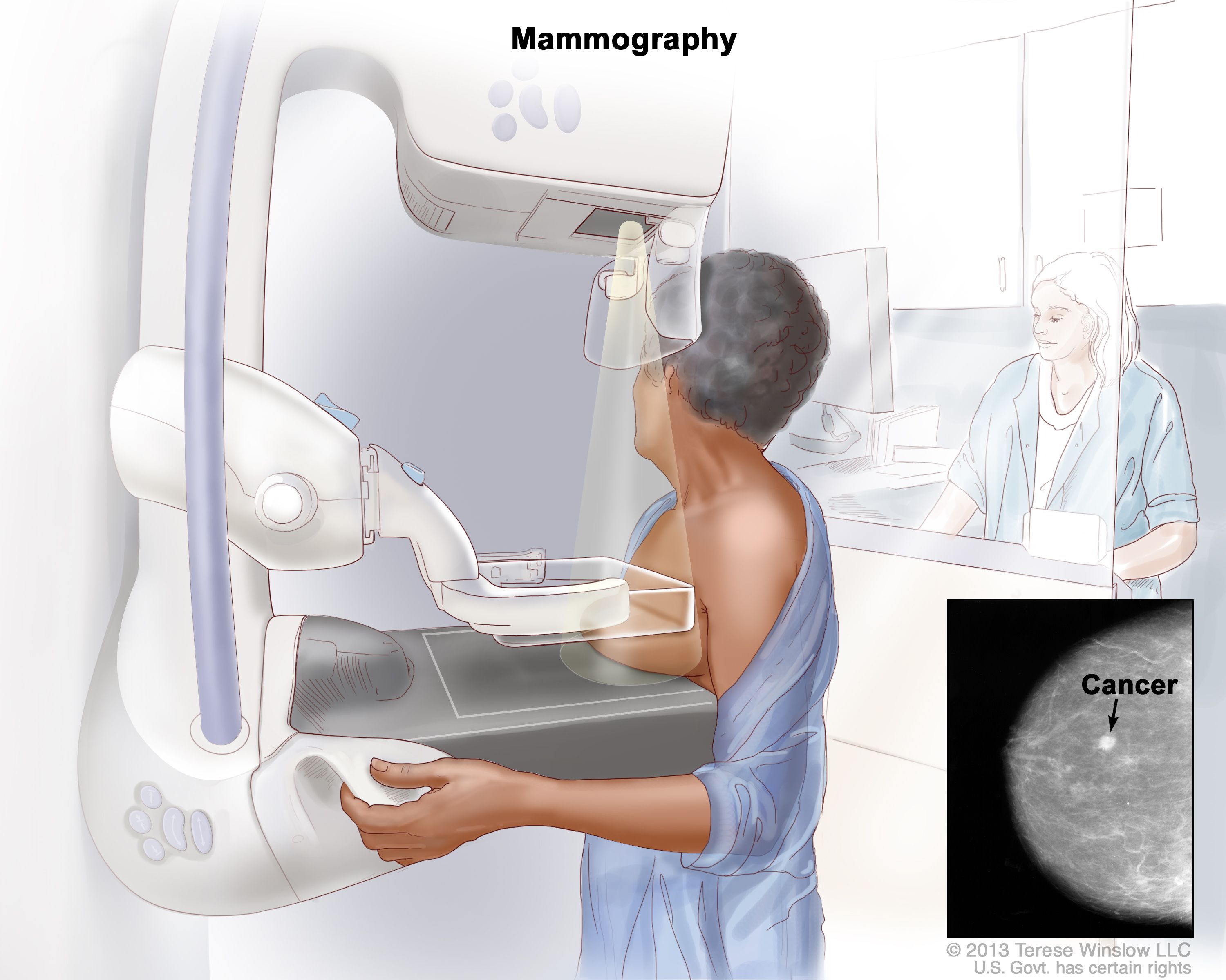
An additional aspect that bolsters the diagnostic process is the use of imaging-guided core biopsies. For lesions that are small, deep-seated, or not easily palpable, imaging techniques such as ultrasound or mammography assist in guiding the biopsy needle accurately into the lesion site. This method not only improves the quality of the tissue sample but also minimizes the risk of sampling error. When imaging-guided core biopsy is employed in tandem with FNAC, the overall diagnostic accuracy is further enhanced, ensuring that effectively ambiguous cases receive a definitive diagnosis through histopathology.
Clinical Implications and Practical Applications
Impact on Patient Management
The profound impact of accurate diagnostic correlation between cytological and histopathological assessments is most clearly seen in how it influences patient management decisions. The following points highlight the clinical implications:
Treatment Decisions
An accurate early diagnosis through FNAC can reduce patient anxiety, shorten the diagnostic timeline, and aid clinicians in making prompt treatment decisions. When FNAC results are corroborated by histopathological findings, the confidence in the diagnosis increases significantly. This is particularly reassuring in cases where imaging findings are inconclusive or when clinical symptoms suggest a high risk of malignancy.
Surgical Planning
For cases where the cytological findings hint at malignancy or atypia, the subsequent histopathological examination can provide additional clarity on the tumor’s grade, type, and extent. Such detailed information is invaluable for surgical planning and determining the necessity and scope of surgery. It is often the case that a preliminary cytological report informs the immediate management plan, while the comprehensive tissue architecture evaluated in the histopathology report confirms the need for aggressive or conservative surgical intervention.
Follow-Up and Monitoring
In benign or borderline cases, the correlation between the two diagnostic modalities can reduce unnecessary surgical interventions. Instead, patients may be managed conservatively with periodic follow-up. In contrast, discrepancies—especially in cases where cytology underestimates the severity—highlight the need for more vigilant follow-up and possibly repeat biopsy.
Role of Multidisciplinary Collaboration
Achieving the best outcomes in diagnosing breast lesions often requires coordinated efforts among several specialists. Radiologists, pathologists, cytotechnologists, and surgeons contribute their expertise to validate the findings from FNAC and histopathology. Such multidisciplinary collaboration not only enhances the accuracy of the diagnosis but also ensures that patients receive a cohesive treatment plan that aligns with the specific nature of their lesion.
Future Directions in Diagnostic Correlation
The continuous evolution of diagnostic techniques promises to further refine the correlation between cytological and histopathological findings in palpable breast lesions. Future research and development in this arena are geared toward:
Improved Sampling Techniques
Innovations in needle design, imaging guidance, and sampling protocols aim to mitigate current limitations such as sampling errors and inadequate tissue acquisition. As these advances emerge, the quality and reliability of FNAC are expected to improve, narrowing the gap with histopathological evaluations.
Enhanced Diagnostic Algorithms
The integration of automated cytological image analysis and artificial intelligence holds great promise. Such systems could assist pathologists in interpreting cellular atypia with greater objectivity and minimizing interobserver variability. By standardizing the analysis, these technologies may further solidify the diagnostic value of FNAC.
Advances in Molecular Diagnostics
Incorporating molecular markers and genetic profiling into both cytological and histopathological examinations represents another frontier for improved diagnostic accuracy. Molecular diagnostics can pinpoint specific markers associated with malignancies, thereby guiding tailored treatment strategies and offering additional layers of diagnostic clarity.
Comparative Analysis: Cytology Versus Histopathology
To facilitate a clear comparison, the following table outlines the principal features of cytological assessment using FNAC and histopathological examination:
| Aspect | FNAC (Cytological Assessment) | Histopathological Examination |
|---|---|---|
| Invasiveness | Minimally invasive | More invasive |
| Turnaround Time | Rapid results | Longer processing time |
| Diagnostic Accuracy | High with scoring systems (approx. 80-93.8%) | Gold standard for definitive diagnosis |
| Tissue Architecture | Limited cellular arrangement information | Detailed assessment of tissue structure |
| Role in Clinical Management | Initial screening and risk stratification | Confirmatory diagnosis and surgical planning |
This comparative analysis underscores the complementary nature of each diagnostic modality; while FNAC provides rapid, cost-effective preliminary insight, histopathology offers the detailed analysis necessary for a definitive diagnosis.
Conclusion and Final Thoughts
In conclusion, the correlation between cytological and histopathological findings in palpable breast lesions forms the cornerstone of effective diagnostic practice in breast pathology. FNAC, with its minimally invasive nature, rapid turnaround, and cost-effectiveness, is an indispensable tool in the initial assessment. When supported by standardized scoring systems, such as the Modified Masood's Scoring Index, its diagnostic accuracy is significantly enhanced, approaching that of histopathological evaluations.
However, histopathological examination remains the definitive standard for diagnosis due to its comprehensive analysis of tissue architecture and detailed cellular morphology. The interplay between rapid cytological screening and in-depth histological analysis facilitates a robust diagnostic algorithm that significantly improves patient management. Clinical discrepancies, potential sampling errors, and interpretive challenges highlight the necessity of adopting a multidisciplinary approach involving radiologists, pathologists, and surgeons to ensure that every patient receives optimal care.
Looking ahead, future improvements in sampling techniques, diagnostic algorithms, and the incorporation of molecular diagnostics are poised to further refine the precision of these diagnostic modalities. The ongoing evolution of technology and interdisciplinary collaboration continues to pave the way for enhanced accuracy, fewer false negatives, and ultimately, better outcomes for patients with breast lesions.
Ultimately, maintaining a high correlation between cytological and histopathological findings is essential. It minimizes diagnostic ambiguity, guides effective treatment planning, and ensures a patient-centered approach in managing breast disease.
References
- Comparative Study of Cytological and Histopathological Finding in Breast Lesion - Academia.edu
- Evaluation of Cytological Scoring and Comparison with Histopathological Finding in a Palpable Breast Lump - ResearchGate
-
Cytological Scoring of Breast Lesions - Journal of Clinical Oncology
-
Preoperative Cytological and Histological Diagnosis of Breast Lesions - ScienceDirect
- Cytological Study of Breast Lesions with Histopathological Correlation - LWW Journals
Recommended Related Queries
Last updated February 23, 2025