
Managing Chest Pain with Left Shoulder Radiation in the Australian Healthcare System
Comprehensive Protocols for Acute Coronary Syndrome Management
Key Takeaways
- Immediate Triage and Assessment: Prioritize patients presenting with chest pain and left shoulder radiation as high-risk for acute coronary syndrome (ACS).
- Rapid Diagnostic Workup: Conduct essential tests such as ECG and cardiac biomarkers within the first 10 minutes of presentation.
- Evidence-Based Management: Implement standardized pharmacological and interventional treatments based on ACS classification.
1. Initial Assessment and Triage
a. Immediate Triage
Upon arrival, patients presenting with chest pain radiating to the left shoulder must be triaged as high-priority cases due to the potential for acute coronary syndrome (ACS). Utilizing the Emergency Care Assessment and Treatment (ECAT) protocol, healthcare professionals should initiate care within 10 minutes to ensure timely intervention.
b. History and Physical Examination
A comprehensive history should be obtained, focusing on the nature, duration, and intensity of the pain, as well as associated symptoms such as shortness of breath, diaphoresis, or nausea. Evaluating risk factors for ACS, including smoking, diabetes, hypertension, and family history of heart disease, is crucial. Special attention should be given to atypical presentations, especially in elderly, diabetic, or female patients, who may present with non-classical symptoms like indigestion or syncope.
c. Vital Signs Monitoring
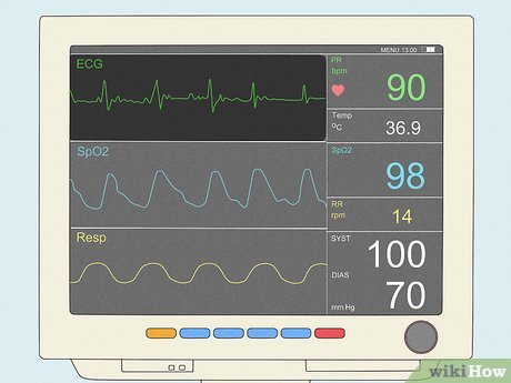
Immediately attach the patient to continuous cardiac monitoring. Measure and record vital signs including blood pressure, heart rate, respiratory rate, and oxygen saturation. Administer supplemental oxygen if SpO₂ is below 90%, adhering to the Thoracic Society of Australia and New Zealand guidelines.
2. Diagnostic Workup
a. Electrocardiogram (ECG)
A 12-lead ECG must be performed within 10 minutes of the patient's arrival to identify any ischemic changes such as ST-segment elevation or depression, T-wave inversions, which are indicative of myocardial infarction. If symptoms persist or evolve, repeat ECGs should be conducted every 15-30 minutes.
b. Cardiac Biomarkers
Order blood tests for high-sensitivity troponin levels to detect myocardial injury. Initial and subsequent troponin measurements should be taken at baseline and 2 to 3 hours post-presentation. Additional blood tests may include a complete blood count, electrolytes, renal function, and glucose levels to assess the patient's overall condition and identify any concurrent abnormalities.
c. Chest X-ray
A chest X-ray may be necessary to exclude other causes of chest pain, such as pneumothorax or aortic dissection. This imaging should be obtained promptly to facilitate differential diagnosis.
3. Immediate Management
a. Oxygen Therapy
Administer supplemental oxygen to patients who are hypoxic (SpO₂ < 90%) to ensure adequate tissue oxygenation and reduce myocardial workload.
b. Pharmacological Interventions
- Aspirin: Administer 300 mg of aspirin orally unless contraindicated by allergies or active bleeding. Aspirin acts as an antiplatelet agent, reducing the risk of thrombus formation.
- Nitroglycerin: Provide sublingual nitroglycerin for pain relief if the patient is not hypotensive. Nitroglycerin helps by dilating blood vessels, thereby reducing cardiac preload and afterload.
- Morphine: Consider intravenous morphine for pain management if nitroglycerin is ineffective. Morphine provides both analgesic and anxiolytic effects, contributing to patient comfort.
- Anticoagulation Therapy: Initiate anticoagulants such as enoxaparin or unfractionated heparin for patients with confirmed or highly suspected ACS to prevent further thrombus formation.
- P2Y12 Inhibitors: Administer P2Y12 inhibitors like clopidogrel or ticagrelor if planning for coronary intervention, enhancing the antiplatelet effect.
4. ACS-Specific Management
a. ST-Elevation Myocardial Infarction (STEMI)
For patients diagnosed with STEMI, immediate reperfusion therapy is critical. This involves:
- Percutaneous Coronary Intervention (PCI): Ideally performed within 90 minutes of hospital arrival to restore blood flow.
- Thrombolysis: Administer thrombolytic agents if PCI cannot be performed within the recommended timeframe.
b. Non-ST Elevation ACS (NSTE-ACS)
Patients with NSTE-ACS should receive dual antiplatelet therapy (e.g., aspirin and clopidogrel) and consider anticoagulation with heparin. Early invasive strategies, such as coronary angiography, should be based on risk stratification using tools like the GRACE or HEART scores to determine the necessity and timing of further interventions.
5. Risk Stratification and Further Management
a. Risk Assessment Tools
Utilize clinical tools such as the GRACE Score or HEART Score to evaluate the risk of mortality and guide management decisions. These scores consider factors like history, ECG findings, age, risk factors, and troponin levels to classify patients into low, intermediate, or high-risk categories.
b. Referral to Cardiology
Patients with confirmed or suspected ACS should be referred to a cardiologist for specialized evaluation and management. Early involvement of cardiology ensures that patients receive appropriate interventions, such as PCI, and comprehensive post-ACS care.
c. Admission or Discharge
-
Admission: High-risk patients should be admitted to a Coronary Care Unit (CCU) or a monitored bed for continuous observation and management.
-
Discharge: If ACS is ruled out and the patient remains stable, they can be discharged with appropriate follow-up instructions and outpatient evaluations if symptoms recur.
6. Supportive Care and Monitoring
a. Continuous Monitoring
Maintain continuous monitoring of vital signs, ECG, and oxygen saturation to promptly detect any changes in the patient’s condition. Regular reassessment ensures that any deterioration is identified and managed swiftly.
b. Pain Management
Ensure effective pain control through the administration of analgesics like nitroglycerin and morphine. Regularly reassess pain levels to adjust management strategies as needed.
c. Patient Education
Provide comprehensive information about the patient’s condition, the treatment plan, and necessary lifestyle modifications to reduce cardiovascular risk. Educating patients empowers them to participate actively in their recovery and prevent future cardiac events.
7. Documentation and Patient Communication
a. Standardized Pathways
Utilize a standardized Chest Pain Assessment Pathway to streamline care, ensuring all findings and interventions are consistently documented. This facilitates communication among the healthcare team and maintains a clear record of the patient’s clinical course.
b. Clear Communication
Provide clear and concise explanations to the patient regarding their condition, the rationale behind diagnostic tests and treatments, and the expected outcomes. Effective communication enhances patient understanding and satisfaction.
8. Escalation of Care
a. Clinical Emergency Response Systems (CERS)
Activate local CERS if the patient experiences deterioration, such as cardiac arrest or hemodynamic instability. Prompt activation ensures rapid deployment of advanced life support measures and specialist intervention.
b. Transfer to Tertiary Care
Consider transferring the patient to a tertiary care facility equipped with advanced cardiac intervention capabilities if necessary. This ensures that patients receive the highest level of care, especially when advanced procedures like PCI are required.
9. Special Considerations
a. Atypical Presentations
Recognize that certain populations, such as diabetic, elderly, female, or young patients, may present with atypical symptoms that do not fit the classical presentation of ACS. Healthcare providers should maintain a high index of suspicion and conduct thorough assessments in these cases.
b. Renal Impairment
Pay additional attention to patients with known renal impairment, as they may have altered pharmacokinetics and increased risk of complications from contrast agents used in diagnostic procedures. Adjust treatment protocols accordingly to minimize risks.
c. Consistent Use of Assessment Pathways
Ensure that all clinicians consistently use established assessment pathways to maintain uniformity in patient care and optimize outcomes. Regular training and protocol updates can help reinforce adherence to best practices.
Conclusion
Effective management of chest pain with radiation to the left shoulder in the Australian healthcare system necessitates a structured and evidence-based approach. From immediate triage and comprehensive diagnostic workup to targeted pharmacological and interventional treatments, adherence to standardized protocols ensures timely and high-quality care. Continuous monitoring, risk stratification, and patient education are integral to optimizing outcomes and preventing future cardiac events. By following these comprehensive guidelines, healthcare professionals can provide exemplary care to patients presenting with potential acute coronary syndrome.
References
Last updated January 22, 2025