
Exploring the Carbon Footprint of Chest X-Rays
A detailed look at imaging impacts and sustainable practices
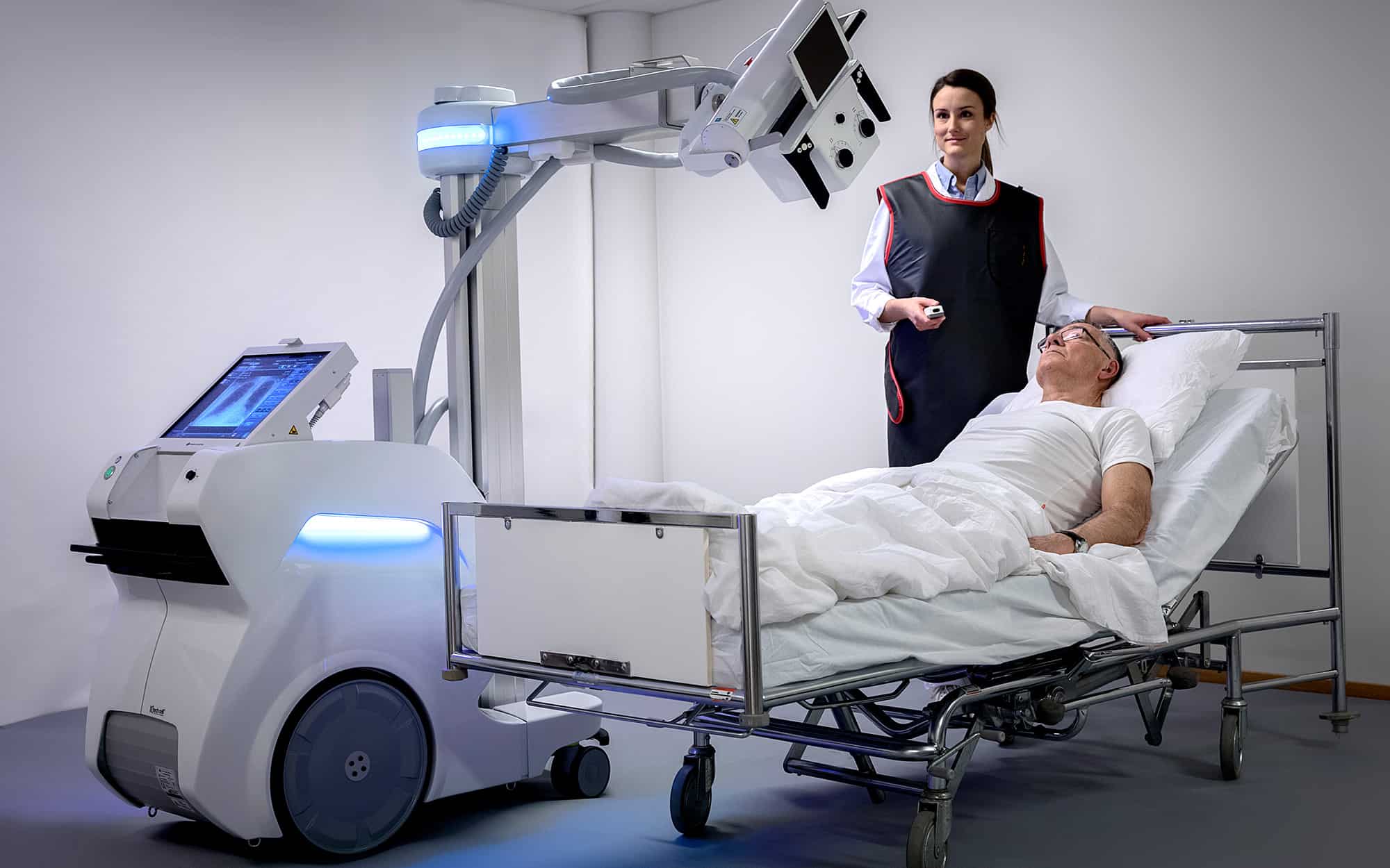
Key Highlights
- Low Emissions: Chest X-rays typically produce around 0.76–0.8 kg CO₂e per scan, substantially lower compared to CT and MRI.
- Comparative Analysis: Mobile chest X-rays can emit as little as 0.5 kg CO₂e, while diagnostics like CT and MRI have much higher footprints.
- Sustainability Opportunities: Reduction in unnecessary imaging and adoption of energy-efficient technologies offer paths to further lessen environmental impact.
Understanding Carbon Footprint in Diagnostic Imaging
Recently, concerns over environmental impacts have driven many sectors, including healthcare, to assess their carbon footprint. Radiology, a key part of medical diagnostics, is under focus for its energy consumption and greenhouse gas emissions. Amidst various imaging modalities, the chest X-ray stands out as having one of the lowest carbon footprints. Typically, the carbon footprint of a standard chest X-ray ranges between roughly 0.76 and 0.8 kg CO₂ equivalent (CO₂e) per scan. When employing a mobile chest X-ray system, which operates efficiently and sometimes with lower energy consumption, the output can be around 0.5 kg CO₂e.
Comparative Emission Values
Studies have measured and compared the energy use and resulting CO₂ emissions of various medical imaging techniques. Here is a comparative breakdown for better perspective:
| Imaging Modality | Carbon Footprint (kg CO₂e per scan) | Notes |
|---|---|---|
| Chest X-ray | 0.76–0.8 | Standard radiography with low energy requirements |
| Mobile Chest X-ray | 0.5 | Optimized for energy efficiency and lower emissions |
| CT Scan | 9.2 | High energy use due to detailed imaging requirements |
| MRI | 17.5 | Extremely energy intensive mainly because of the cooling and magnet systems |
Detailed Analysis and Environmental Context
Quantitative Assessment of Chest X-ray Emissions
The emission values for chest X-rays have been derived using Attributional Life Cycle Analysis (ALCA) and Consequential Life Cycle Analysis (CLCA). These assessments are designed to capture the overall carbon output of different imaging examinations. Typically, ALCA takes into account all parts of the imaging process, including standby power, culminating in an approximate figure of 0.76 to 0.8 kg CO₂e per scan. However, CLCA, which excludes some standby power contributions, can show a slightly lower figure around 0.6 kg CO₂e per scan for chest X-rays.
It is important to note that these figures are averages that might vary by facility, equipment age, and operational protocols. The emitted CO₂ equivalent is a critical metric for understanding the indirect environmental impacts that the healthcare sector is striving to reduce.
Contextualizing Chest X-ray Emissions with Broader Healthcare Practices
Although a chest X-ray stands as a relatively low-carbon imaging procedure, the broader implications for sustainable healthcare practices are both significant and multifaceted. With rising environmental consciousness, the healthcare industry is reviewing its entire range of operations—from imaging modalities to overall energy consumption policies.
While high-tech imaging modalities such as CT and MRI play a vital role in detailed diagnostic processes, they considerably elevate a facility's carbon footprint. For example, CT scans and MRIs require far more energy because of the intense computational, cooling, and operational demands. In the context of energy efficiency, a shift toward using lower-energy procedures when clinically appropriate can contribute to significant emissions reductions. Moreover, the deactivation of scanners during periods of inactivity—a measure to curb standby power wastage—represents one of the simple yet effective strategies toward sustainable radiology.
Environmental Strategies in Radiology
Besides choosing imaging modalities with lower energy profiles, several strategies can be integrated into radiology to further reduce the environmental impact:
1. Reducing Unnecessary Imaging
By implementing stricter guidelines and promoting the principle of “justified imaging,” clinicians can reduce the number of unnecessary scans performed. This helps in minimizing the cumulative carbon emissions across a healthcare facility.
2. Utilizing Lower Impact Modalities
When clinically appropriate, chest X-rays or ultrasound imaging can be preferred over more energy-intensive modalities such as CT scans or MRIs. This substitution can have an immediate effect in reducing the overall carbon footprint.
3. Enhancing Equipment Efficiency
Modernizing equipment to contemporary models that use energy more sparingly can further cut CO₂ emissions. Clinics and hospitals are encouraged to invest in newer technologies that are designed with energy efficiency as a priority.
4. Optimizing Operational Protocols
Beyond equipment and imaging decisions, strategies such as turning off scanners when not in use and scheduling maintenance to ensure optimal performance can offer incremental but steady improvements in reducing emissions.
5. Policy and Training Initiatives
Educating radiologists, technicians, and hospital administrators on sustainable practices can result in a cultural shift toward environmentally friendly operations. Incorporating these guidelines into regular training sessions ensures a consistent approach to minimizing energy wastage.
Integrating Sustainability into Clinical Practices
It is evident that chest X-rays, with their relatively low carbon footprint, are an ideal starting point for integrating sustainable practices into diagnostic procedures. As healthcare institutions become increasingly aware of their environmental footprint, there is a growing trend to adopt technologies and protocols that align with climate change mitigation strategies.
Continuous research and systematic reviews are essential for accurately determining the carbon footprint of various imaging modalities. They also pave the way for developing innovations aimed at reducing greenhouse gas emissions within healthcare systems. By benchmarking the performance of chest X-rays against other modalities such as CT and MRI, clinicians and policymakers can better understand which practices to emphasize in sustainability programs.
Furthermore, the competition between traditional imaging and mobile radiographic services provides additional incentives for improving environmental efficiency. Mobile chest X-rays, in particular, are proving to be a valuable asset in circumstances where reducing the infrastructure demand leads directly to lower emissions while still maintaining clinical effectiveness.
Case Study: Implementing Energy-Efficient Imaging Practices
A growing number of hospitals have documented the benefits of integrating energy-efficient practices in their radiology departments. Hospitals that have switched to modern imaging equipment with lower standby power consumption and implemented strict guidelines to avoid unnecessary imaging have observed measurable reductions in their overall CO₂ emissions.
One such approach has been the adoption of advanced scheduling software that minimizes idle time for imaging equipment. This strategy not only improves device utilization rates but also reduces the energy wasted during periods when equipment is on standby.
The methodologies commonly include analyzing the life cycle of each imaging modality. Both Attributional and Consequential Life Cycle Analyses provide insights into operational energy demands, thereby guiding better decision-making regarding technology investments and clinical protocols.
Understanding Life Cycle Analyses in Radiology
Life Cycle Analysis (LCA) is a critical tool for comprehensively understanding the environmental impacts of diagnostic imaging. Attributional LCA assigns emissions values to each stage of the imaging process, including production, operation, and disposal. As noted, this method typically results in an estimate of 0.76–0.8 kg CO₂e for a chest X-ray.
In contrast, Consequential LCA focuses more on the changes in emissions due to specific decisions such as turning off equipment when not in active use, resulting in estimates as low as 0.6 kg CO₂e per scan for chest X-rays. These analyses have become central as radiology departments work to meet rigorous sustainability goals and regulatory requirements.
Implementing Best Practices for Sustainable Radiology
To further emphasize, the implementation of sustainable practices in radiology encompasses numerous aspects including, but not limited to:
Energy Conservation Regulations
Many healthcare facilities have taken steps to integrate energy conservation into their operational guidelines. These include:
- Regular audits on energy consumption of radiology equipment
- Integration of smart energy management systems to monitor usage
- Encouragement of facility-wide initiatives to reduce wasted energy
Adopting Green Technologies
Adoption of greener technologies involves upgrading legacy equipment to models that feature energy-saving modes and utilize modern, energy-efficient components. These efforts are often supported by governmental incentives designed to reduce the environmental impact of the healthcare sector.
Staff Training and Awareness
Educating staff about the importance of energy efficiency contributes greatly to the cultural shift towards sustainable practices. Ongoing training ensures that the radiology department maintains protocols that optimize both clinical outcomes and environmental performance.
Regular Equipment Maintenance
Routine maintenance not only extends the lifespan of imaging devices but also guarantees that machines operate at their peak efficiency with minimal energy wastage. Hospitals that institute systematic maintenance schedules see a consistent reduction in their radiologic carbon footprints.
Broader Implications and Future Innovations
The discourse around the carbon footprint of chest X-rays also shifts the focus to the importance of continuous innovation in imaging technology. As research and development progress, it is expected that even the modest emissions associated with chest X-rays might be reduced further through technological advancements.
Future developments may include the design of ultra-efficient systems that use renewable energy sources, improved imaging protocols that minimize unnecessary repeat scans, and the implementation of automated systems designed to manage energy consumption in real time.
As such, the integration of sustainability into clinical practices is not merely a regulatory or ethical imperative, but also an opportunity for innovation and improved operational efficiencies across the healthcare industry. With rigorous research and continued policy support, radiology departments will likely serve as a model for other medical practices aiming to lower their environmental footprint.
References
-
The carbon footprint of hospital diagnostic imaging in Australia - PMC
- The carbon footprint of hospital diagnostic imaging in Australia - The Lancet
- Sustainable radiology: greener imaging systems - Healthcare in Europe
- Systematic review on carbon footprint in healthcare - Wiley Online Library
- Research on the environmental impact of imaging procedures - PubMed
Recommended Further Queries
Last updated March 25, 2025